Validating SPR Kinetic Data Despite Unstable Baselines: A Troubleshooting and Optimization Guide
Unstable baselines in Surface Plasmon Resonance (SPR) experiments pose a significant challenge, potentially compromising the accuracy of kinetic data.
Validating SPR Kinetic Data Despite Unstable Baselines: A Troubleshooting and Optimization Guide
Abstract
Unstable baselines in Surface Plasmon Resonance (SPR) experiments pose a significant challenge, potentially compromising the accuracy of kinetic data. This article provides researchers, scientists, and drug development professionals with a comprehensive framework for identifying, troubleshooting, and validating data collected under unstable baseline conditions. Covering foundational principles, methodological adjustments, systematic optimization, and robust validation techniques, this guide offers practical strategies to ensure data reliability and confidence in kinetic parameters for critical applications in drug discovery and biomolecular interaction analysis.
Understanding SPR Baseline Instability: Root Causes and Impact on Data Integrity
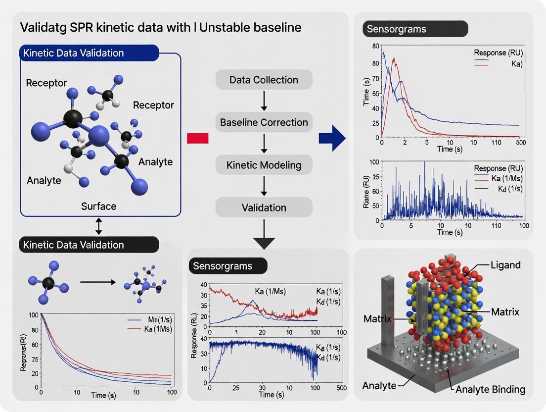
In Surface Plasmon Resonance (SPR) analysis, a sensorgram provides real-time, label-free monitoring of molecular interactions. The baseline—the initial flat portion of the sensorgram before analyte injection—serves as the critical reference point from which all binding events are measured. Baseline drift refers to the gradual increase or decrease of this signal when no analyte is present, representing a significant source of experimental artifact that can compromise data integrity [1] [2]. For researchers validating SPR kinetic data, particularly in unstable baseline conditions, recognizing and correcting for baseline drift is not merely a procedural step but a fundamental prerequisite for obtaining reliable kinetic parameters (kₐ, kₑ, and Kᴅ). Uncorrected drift can distort binding curves, leading to inaccurate calculation of association and dissociation rates, ultimately affecting conclusions about binding affinity and mechanism [3]. This guide examines the symptomatic presentation of baseline drift across SPR platforms and provides methodologies for its identification and correction within the context of rigorous kinetic data validation.
Recognizing Visual Symptoms in Sensorgrams
The manifestation of baseline drift can vary from subtle, slow deviations to pronounced, directional trends. The following table categorizes the primary visual symptoms observed in sensorgrams and their immediate implications for data quality.
Table 1: Symptom Profiles of Common Baseline Drift Types
| Symptom Profile | Visual Description | Impact on Sensorgram | Common Causes |
|---|---|---|---|
| Upward Drift | Gradual, often linear increase in Response Units (RU) before analyte injection [4]. | Overestimation of binding response and complex stability; can inflate Rmax values [3]. | Slow surface equilibration, ligand leaching from a capture surface, contamination [1] [3]. |
| Downward Drift | Gradual, often linear decrease in RU during the dissociation phase or pre-injection baseline [4]. | Underestimation of binding response and complex stability; can skew dissociation rate calculations [3]. | Surface dehydration, ligand instability, buffer mismatch, or air bubbles in the fluidic system [1] [4]. |
| Start-up Drift | A pronounced drift immediately after initiating flow or docking a new chip, which levels off after 5-30 minutes [1]. | Makes the initial baseline unreliable as a reference point, affecting all subsequent binding cycles. | Rehydration of the sensor surface, wash-out of immobilization chemicals, or temperature equilibration [1]. |
| Post-Regeneration Drift | Failure of the baseline to return to the original pre-injection level after a regeneration step [5]. | Causes carryover effects between analysis cycles, leading to inconsistent analyte binding responses. | Incomplete regeneration (leaving residual analyte) or overly harsh regeneration damaging the ligand [6] [5]. |
Quantitative Characterization of Drift
Beyond visual inspection, quantifying the rate and magnitude of drift is essential for determining its severity and for applying mathematical corrections during data analysis.
Measuring Drift Magnitude and Rate
The most straightforward metric is the drift rate, expressed in Resonance Units per minute (RU/min). This is calculated by measuring the total change in RU (ΔRU) over a defined time period (Δt) during a stable, analyte-free baseline region [3]:
Drift Rate (RU/min) = ΔRU / Δt
For kinetic analysis to be considered reliable, the total drift over the duration of a single analyte injection cycle should be negligible compared to the specific binding signal. As a general guideline, a drift rate that contributes to less than 5% of the Rmax value for that interaction is often considered acceptable, though this threshold depends on the specific affinity and signal strength of the system.
Data Analysis and Modeling Approaches
Modern SPR analysis software incorporates models to account for drift. The Langmuir with Drift model, for instance, is specifically designed for experiments using capture surfaces where ligand loss causes a linear, time-dependent signal change [3]. This model fits the baseline drift as a constant linear variable in addition to the standard kinetic parameters, thereby deconvoluting the drift artifact from the true binding signal. Judging the quality of the fit, often by examining the chi-squared (χ²) value, is crucial for validating that the model adequately accounts for the observed drift [3].
Experimental Protocols for Diagnosis and Validation
A systematic experimental approach is key to diagnosing the root cause of baseline drift and validating kinetic data acquired under potentially unstable conditions.
Baseline Stability Assessment Protocol
This protocol evaluates the intrinsic stability of the SPR system and surface prior to any kinetic experiment.
- Surface Preparation: Dock a clean sensor chip and prime the system with degassed, filtered running buffer [1] [5].
- Initial Equilibration: Flow running buffer at the intended experimental flow rate for 30-60 minutes, monitoring the baseline continuously [1].
- Data Collection: Record the baseline response without any injections. Note the drift rate (RU/min) over the final 20 minutes.
- Acceptance Criterion: A system is considered stable for high-quality kinetics if the baseline exhibits a drift of < 1.0 RU/min over this period [1].
Diagnostic Run for Kinetic Validation
When analyzing an interaction with a suspected unstable baseline, the following modified kinetic experiment is recommended.
- Start-up Cycles: Incorporate at least three "start-up" or "dummy" cycles at the beginning of the method. These cycles should use buffer injections instead of analyte over the ligand surface, including any regeneration steps. Their purpose is to condition the surface and stabilize the system; these cycles are not used in the final analysis [1].
- Extended Dissociation: For interactions with slow off-rates, include a long dissociation phase (e.g., 1-2 hours) following a mid-range analyte concentration injection.
- Blank Referencing: Intersperse regular blank (buffer) injections throughout the analyte concentration series. These are critical for double referencing—a data processing step that subtracts both signal from a reference flow cell and systemic artifacts from the buffer injection itself [1].
- Data Processing:
- Double Referencing: Subtract the reference surface data and then the average blank injection response from all analyte sensorgrams [1].
- Model Fitting: Fit the processed data to both the standard Langmuir model and the Langmuir with Drift model. Compare the chi-squared (χ²) values and the residual plots. A significantly better fit with the drift model, along with random (non-systematic) residuals, validates the use of drift correction for that data set [3].
The logical workflow for a systematic diagnosis is outlined below.
The Scientist's Toolkit: Essential Reagents and Materials
The following table details key reagents and materials essential for experiments focused on diagnosing and mitigating baseline drift.
Table 2: Key Research Reagent Solutions for Baseline Drift Management
| Tool | Function in Drift Management | Application Notes |
|---|---|---|
| Fresh Running Buffer (e.g., HBS-EP, PBS) [1] [7] | Maintains a stable refractive index; old or contaminated buffer is a primary cause of drift. | Prepare fresh daily, 0.22 µm filter and degas thoroughly before use [1]. |
| Sensor Chips (e.g., CM5, NTA, SA) [7] [6] | Provides a stable surface for ligand immobilization. Chip type impacts surface equilibration time. | Allow sufficient time for surface hydration and equilibration with running buffer after docking [1]. |
| Regeneration Buffers (e.g., Glycine-HCl pH 1.5-3.0, 10-50 mM NaOH) [7] [6] | Resets the baseline by removing bound analyte without damaging the ligand. | Must be optimized for each ligand-analyte pair to balance completeness of regeneration with ligand activity preservation [6]. |
| Blocking Agents (e.g., Ethanolamine, BSA) [7] [5] | Reduces non-specific binding (NSB) to the sensor surface, a potential source of drift. | Apply after ligand immobilization to block unreacted functional groups on the sensor surface [5]. |
| Detergents & Additives (e.g., Surfactant P20, Tween-20) [7] [6] | Minimizes NSB by disrupting hydrophobic and charge-based interactions. | Use at low concentrations (e.g., 0.005% P20) in running buffer to reduce drift from NSB [7] [6]. |
| Software with Drift Correction (e.g., ProteOn Manager) [3] | Mathematically corrects for linear drift during data analysis, validating kinetic parameters. | Use models like "Langmuir with Drift" for a more accurate fit when residual drift is present post-referencing [3]. |
Cross-Platform Comparison of Drift Artifacts
The manifestation and impact of baseline drift can be influenced by the specific SPR instrument and its fluidics. The table below provides a generalized comparison of how drift management is approached across different experimental setups.
Table 3: Approach to Baseline Drift Across SPR Contexts
| Experimental Context | Typical Drift Profile | Recommended Correction Strategy | Data Validation Insight |
|---|---|---|---|
| Biacore-style Systems (Multi-channel) | Start-up drift; differences in drift rates between active and reference surfaces [1]. | Double referencing; use of a dedicated, untreated reference flow cell [1]. | After referencing, residual systematic errors in residuals indicate inadequate drift compensation. |
| Capture-Based Assays (e.g., His-tag / NTA) | Linear, continuous downward drift due to ligand loss from the capture surface [3]. | Use of "Langmuir with Drift" kinetic model in data fitting [3]. | The fitted drift rate should be consistent across all analyte concentrations for the model to be valid. |
| Single-Cycle Kinetics | Potential for progressive drift to accumulate over multiple, sequential analyte injections. | Inclusion of blank injections within the cycle and careful pre-equilibration [1]. | Compare the pre-injection baseline for each injection to quantify accumulated drift. |
| High-Throughput Screening | Variability in drift between different ligand spots or channels. | Robust referencing and normalization to internal controls are critical. | The z'-factor for the assay should be calculated incorporating baseline noise and drift to assess quality. |
In Surface Plasmon Resonance (SPR) biosensing, the integrity of kinetic data is paramount for accurate determination of biomolecular interactions in drug discovery and basic research. A stable baseline is the foundational prerequisite for reliable data, serving as the indicator of system equilibrium before analyte introduction. Instabilities in the baseline—manifesting as drift, injection spikes, or high buffer noise—directly compromise the accuracy of extracted kinetic parameters (association rate k_on, dissociation rate k_off, and equilibrium dissociation constant K_D). This guide provides a systematic framework for researchers to diagnose the root causes of baseline instability, categorizing them into surface, buffer, and instrumental origins, and offers validated experimental protocols for effective troubleshooting and data validation [8].
The SPR Sensorgram: A Diagnostic Blueprint
A well-defined sensorgram provides the real-time signature of a binding interaction and the first clues for diagnosing issues. The initial phase is the baseline, established by flowing a running buffer over the sensor surface. A flat, stable baseline confirms system equilibrium. Any deviation indicates potential problems requiring investigation. The subsequent phases—association (analyte binding), steady-state (binding equilibrium), dissociation (analyte wash-off), and regeneration (surface preparation for a new cycle)—are all interpreted relative to this initial baseline [8].
The following diagram illustrates the ideal sensorgram phases and common instability signatures that point to specific problem categories.
Systematic Diagnostic Framework
A systematic approach to diagnosing baseline issues efficiently isolates the root cause. The following workflow guides users from initial observation to targeted resolution.
Experimental Protocols for Cause Isolation
Protocol 1: Buffer Mismatch and Purity Test
- Objective: To isolate buffer-related causes from surface or instrumental factors.
- Method: Perform multiple, consecutive injections of the running buffer alone (with no analyte) over a freshly prepared and stabilized sensor surface [8].
- Expected Result: A flat, stable baseline with minimal noise (<1-2 Resonance Units (RU) deviation).
- Diagnostic Interpretation:
- If the baseline is stable: The buffer system and instrument are not the primary causes. Underlying surface chemistry or analyte-specific issues (e.g., non-specific binding) are more likely.
- If instability persists: The problem lies with the buffer or the instrument. Proceed to degas and filter all buffers (0.22 µm filter) to remove air bubbles and particulates. Ensure the running buffer and sample buffer are perfectly matched in composition, pH, and salt concentration.
Protocol 2: Surface Integrity and Immobilization Stability Test
- Objective: To evaluate the stability of the ligand immobilization and the sensor surface itself.
- Method: Dock a new sensor chip with a bare surface or a freshly immobilized ligand. Condition the surface with multiple short injections of a regeneration solution (e.g., 10 mM glycine-HCl, pH 2.0-2.5), followed by extensive re-equilibration in running buffer [8] [9].
- Expected Result: The baseline should return to its original position after each regeneration cycle, demonstrating a stable and reusable surface.
- Diagnostic Interpretation:
- If the baseline is stable and recovers fully: The surface chemistry is robust.
- If the baseline shows irreversible drift or poor recovery: The ligand may be degrading, the coupling chemistry may be unstable, or the sensor surface may be contaminated. Consider optimizing the immobilization level, using a different coupling chemistry, or employing a more stringent blocking agent to reduce non-specific binding.
Protocol 3: Instrumental and Fluidic System Diagnostic
- Objective: To identify hardware-related issues, such as clogging, bubbles, or temperature fluctuations.
- Method: Run the instrument's built-in priming and cleaning procedure. Then, test the system with a blank buffer flow across multiple independent flow cells or channels [10].
- Expected Result: All flow cells should exhibit identical, stable baseline responses.
- Diagnostic Interpretation:
- If instability is localized to one flow cell: A clog or a bubble is likely present in that specific microfluidic path.
- If instability is consistent across all flow cells: A systemic instrumental issue is probable, such as a failing pump, a leak in the fluidic system, or an unstable temperature controller. Consult the instrument's service manual.
Quantitative Data Comparison of Common Issues
The table below summarizes the characteristic signatures, diagnostic tests, and solutions for the three primary categories of baseline instability.
Table 1: Systematic Diagnosis of Baseline Instability Causes
| Problem Category | Characteristic Sensorgram Signature | Key Diagnostic Test | Most Effective Solution |
|---|---|---|---|
| Surface Causes | Gradual, irreversible downward drift after regeneration; slow stabilization; high binding in reference cell [8] [9] | Surface Integrity Test (Protocol 2) | Optimize ligand density; use different coupling chemistry; improve surface blocking with inert proteins [9]. |
| Buffer Causes | Sharp injection spikes; increased baseline noise (>"chatter"); steady upward/drift during buffer flow [8] | Buffer Mismatch Test (Protocol 1) | Degas and filter all buffers; ensure perfect match between running and sample buffer; use high-purity reagents. |
| Instrumental Causes | Consistent drift across all flow cells; large, sudden signal jumps (bubbles); complete signal dropout [10] | Instrumental Diagnostic Test (Protocol 3) | Execute system prime and purge; inspect for fluidic leaks or clogs; verify instrument temperature stability. |
The Scientist's Toolkit: Essential Reagents and Materials
Successful SPR experimentation and troubleshooting rely on a set of core reagents and materials. The following table details key items for surface preparation, analysis, and regeneration.
Table 2: Essential Research Reagent Solutions for SPR
| Reagent/Material | Function & Application | Key Considerations |
|---|---|---|
| CM5 Sensor Chip (or equivalent) | A gold sensor surface with a carboxymethylated dextran matrix that facilitates ligand immobilization via amine coupling [9]. | The standard choice for most applications; other chips (e.g., lipophilic, nitrilotriacetic acid) are available for specific needs like membrane protein studies [11]. |
| HEPES-NaCl Buffer (HBS-EP) | A standard running buffer (e.g., 10 mM HEPES, 150 mM NaCl, 3 mM EDTA, 0.05% surfactant P20, pH 7.4) for conditioning and maintaining the sensor surface [8]. | The surfactant reduces non-specific binding. Buffer must be degassed and filtered (0.22 µm) before use to prevent air bubbles and particulates. |
| NHS/EDC Mixture | A mixture of N-hydroxysuccinimide (NHS) and N-ethyl-N'-(3-dimethylaminopropyl)carbodiimide (EDC) used to activate carboxyl groups on the sensor chip surface for covalent amine coupling of ligands [9]. | Fresh preparation is recommended for optimal activation efficiency. |
| Ethanolamine Hydrochloride | A blocking reagent used to deactivate and quench any remaining activated ester groups on the sensor surface after ligand immobilization, minimizing non-specific binding [9]. | A critical step to ensure a stable, non-reactive surface post-immobilization. |
| Glycine-HCl (pH 2.0-2.5) | A low-ppH regeneration solution used to break the binding interaction between the ligand and analyte, effectively "resetting" the sensor surface for a new analysis cycle [8]. | The exact pH and composition must be optimized for each specific ligand-analyte pair to ensure complete regeneration without damaging the immobilized ligand. |
A stable baseline is the cornerstone of valid SPR kinetic data. By applying this systematic diagnostic framework—differentiating between surface, buffer, and instrumental origins through targeted experimental protocols—researchers can efficiently troubleshoot their systems, minimize experimental artifacts, and significantly enhance the reliability of their biomolecular interaction data. This rigorous approach to data validation is indispensable for accelerating drug discovery and ensuring the accuracy of scientific conclusions.
Surface Plasmon Resonance (SPR) is a powerful, label-free technique for determining the kinetic parameters of biomolecular interactions, including the association rate constant (ka), dissociation rate constant (kd), and the equilibrium dissociation constant (KD). However, the accuracy of these measurements is critically dependent on the stability of the baseline signal. Baseline drift, a gradual shift in the signal when no binding occurs, is a common artifact that can significantly skew derived kinetic parameters. Within the context of validating SPR kinetic data, understanding, identifying, and mitigating drift is essential for producing reliable and publication-quality data. This guide explores the impact of drift, provides protocols for its identification and correction, and compares these strategies against alternative experimental approaches.
What is Baseline Drift and What Causes It?
In SPR, the baseline is the signal recorded from the sensor surface when only the running buffer is flowing over it, representing a state of no binding activity. A stable baseline is fundamental for accurately measuring the response units (RU) change upon analyte injection.
Baseline drift is the phenomenon where this baseline signal unstablely increases or decreases over time instead of remaining constant. A good fit is characterized by a drift contribution of less than ± 0.05 RU s⁻¹ [12].
The primary causes of baseline drift include:
- Insufficient System Equilibration: The instrument and sensor surface are not fully stabilized before the experiment begins. This is a frequent cause of initial drift [12] [4].
- Buffer Incompatibility: Differences in temperature, composition, or degassing between the running buffer and the sample buffer can cause refractive index shifts and drift [5] [4].
- Surface Contamination or Incomplete Regeneration: Residual material buildup on the sensor surface over multiple injection cycles can alter the baseline [5].
- Environmental Factors: Fluctuations in ambient temperature or vibrations can introduce instrumental noise and drift [4].
How Drift Skews Kinetic Parameters
Baseline drift introduces a non-random error that the fitting algorithms for standard binding models (like the 1:1 Langmuir model) cannot account for. This leads to systematic inaccuracies in the calculated kinetic constants, as outlined in the table below.
Table 1: Impact of Baseline Drift on Key SPR Kinetic Parameters
| Kinetic Parameter | Impact of Upward Drift | Impact of Downward Drift |
|---|---|---|
| Association Rate Constant (ka) | Artificially inflated; the binding appears faster as the drifting baseline adds to the binding signal. | Artificially lowered; the binding appears slower as the drift subtracts from the binding signal. |
| Dissociation Rate Constant (kd) | Artificially lowered; the complex appears more stable because the upward drift counteracts the signal decrease from dissociation. | Artificially inflated; the complex appears less stable because the downward drift accelerates the apparent signal loss. |
| Equilibrium Dissociation Constant (KD) | Inaccurate; the overall affinity (KD = kd/ka) is skewed, typically resulting in an underestimated KD (falsely high affinity). | Inaccurate; the overall affinity is skewed, typically resulting in an overestimated KD (falsely low affinity). |
The following diagram illustrates the causal pathway of how drift originates and ultimately compromises data integrity.
Experimental Protocols for Detection and Mitigation
Protocol 1: Visual and Statistical Detection of Drift
A robust workflow for detecting drift combines visual inspection of sensorgrams with quantitative goodness-of-fit metrics.
Detailed Methodology:
- Visual Inspection: Before any data correction or fitting, examine the raw sensorgrams. Focus on the baseline regions immediately before analyte injection and during the final dissociation phase. A sloping line, rather than a flat one, indicates drift [13].
- Residuals Analysis: After fitting the data to a kinetic model (e.g., 1:1 binding), plot the residuals—the difference between the experimental data and the fitted curve.
- Interpretation: A good fit with no significant drift will show residuals randomly scattered around zero. A systematic pattern (e.g., a U-shape or a slope) in the residuals indicates that the model cannot account for the drift, and the fit is poor [12].
- Chi² (Chi-Squared) Value: The Chi² value is a statistical measure of the accuracy of the fit. While it increases with the number of curves fitted, a high Chi² value suggests a poor fit, which can be caused by significant drift or other artifacts [12].
Protocol 2: Mitigation and Correction Strategies
The most effective approach to drift is to prevent it through careful experimental design. The following protocol outlines key steps.
Detailed Methodology:
- Extended System Equilibration:
- Buffer Matching:
- Prepare the analyte samples in the exact same running buffer used in the instrument. This is critical to avoid bulk refractive index (RI) shifts and associated drift [6] [14].
- For analytes dissolved in DMSO, ensure the DMSO concentration is identical in all analyte samples and the running buffer [14].
- Instrument and Surface Maintenance:
- Degas Buffers: Always degas buffers to prevent the formation of air bubbles in the microfluidics, a common cause of baseline noise and drift [4].
- Proper Regeneration: Develop a regeneration scouting protocol to find conditions that completely remove bound analyte without damaging the ligand. Incomplete regeneration leads to carryover and a drifting baseline over multiple cycles [6] [5].
- Environmental Control: Place the instrument in a stable environment with minimal temperature fluctuations and vibrations [4].
- Data Processing:
- Double Referencing: This standard data processing technique involves subtracting both the signal from a reference flow cell (with no ligand or an irrelevant ligand) and the signal from a blank injection (buffer alone). This effectively corrects for systemic drift and bulk effects [12].
- Drift Correction in Software: Some analysis software allows for the inclusion of a drift parameter in the fitting model. This should be used as a last resort, and its contribution should be minimal. As one source states, "Fit the curves first without a drift component. Then add a drift component in the final fitting" [12].
Table 2: Research Reagent Solutions for Drift Mitigation
| Reagent/Solution | Function in Drift Mitigation | Application Notes |
|---|---|---|
| Matched Running Buffer | Prevents bulk refractive index shifts and associated drift by ensuring analyte and buffer composition are identical. | Use for dissolving/diluting the analyte and as the system running buffer [6] [14]. |
| Ethanolamine | Blocks unused active sites on the sensor surface after ligand immobilization, reducing non-specific binding that can cause drift. | Standard blocking agent for carboxymethylated dextran chips (e.g., CM5) after NHS/EDC coupling [5]. |
| Regeneration Buffer (e.g., Glycine pH 2.0) | Removes all bound analyte from the ligand surface between cycles, preventing carryover and baseline drift. | Must be optimized for each ligand-analyte pair to be effective without damaging the ligand [6] [14]. |
| Bovine Serum Albumin (BSA) | Acts as a blocking agent to reduce non-specific binding (NSB) to the sensor chip, a potential source of drift. | Typically used at 0.1-1% concentration in running buffer during analyte injections only [6]. |
| High-Salt Regeneration (e.g., 2 M NaCl) | A mild regeneration solution that disrupts electrostatic interactions to remove bound analyte. | An alternative to low-pH regeneration for sensitive ligands [14]. |
Comparison of Kinetic Methods Under Drift-Prone Conditions
The choice of kinetic method can influence a study's resilience to drift. The two primary methods, Multi-Cycle Kinetics (MCK) and Single-Cycle Kinetics (SCK), offer different advantages and vulnerabilities.
Table 3: Method Comparison: MCK vs. SCK in the Context of Drift
| Feature | Multi-Cycle Kinetics (MCK) | Single-Cycle Kinetics (SCK) |
|---|---|---|
| Principle | Each analyte concentration is injected in a separate cycle, with a regeneration step in between [15]. | Increasing analyte concentrations are injected sequentially in a single, continuous cycle without intermediate regeneration [15]. |
| Advantages for Drift Management | - Individual fitting of cycles allows for diagnosis of drift in specific segments.- A buffer blank can be injected and subtracted from each cycle to correct for baseline drift [15]. | - Fewer regeneration steps reduce the risk of surface-based drift caused by incomplete regeneration or ligand damage [15]. |
| Vulnerabilities to Drift | - Cumulative surface damage or contamination over many regeneration cycles can cause progressive baseline drift [5]. | - A single, long run is more susceptible to system-wide drift (e.g., from temperature changes) affecting the entire dataset [15]. |
| Best Suited For | Interactions where a robust regeneration condition is available and the ligand surface is stable over many cycles. | Interactions where regeneration is difficult or damages the ligand, or for capture-based immobilization [15]. |
Baseline drift is not merely a cosmetic issue in SPR data but a significant source of systematic error that directly compromises the accuracy of kinetic parameters. Unchecked drift leads to miscalculated ka and kd values, resulting in a fundamentally skewed understanding of molecular affinity (KD). Through rigorous experimental practice—including thorough system equilibration, meticulous buffer matching, and proper surface regeneration—researchers can effectively mitigate drift. Furthermore, selecting the appropriate kinetic method and diligently using referencing techniques are critical for validating SPR data. In the broader context of a thesis on data validation, establishing and adhering to a strict protocol for identifying and correcting for baseline instability is a cornerstone of generating reliable, reproducible, and scientifically defensible kinetic data.
In Surface Plasmon Resonance (SPR) studies, particularly those focused on validating kinetic data amidst unstable baselines, the experimental setup phase is paramount. The integrity of pre-experimental choices—specifically in sensor chip selection, buffer formulation, and sample quality—directly dictates the robustness, reproducibility, and ultimate validity of the derived kinetic constants (association rate constant, ka; dissociation rate constant, kd; and equilibrium constant, KD). An unstable baseline is a frequent challenge that can stem from inadequate buffer compatibility, poor surface preparation, or sample impurities, leading to significant drift and compromising the accuracy of kinetic measurements [5] [16]. This guide objectively compares available options and provides foundational protocols to safeguard your data from these common pitfalls, ensuring that your kinetic analysis rests on a solid experimental foundation.
Sensor Chip Selection: A Comparative Guide
The sensor chip is the stage upon which biomolecular interactions occur. Its surface chemistry must be meticulously chosen to ensure proper ligand orientation, stability, and minimal non-specific binding, all of which are critical for obtaining clean data with a stable baseline [6] [5].
Comparative Analysis of Common Sensor Chips
Table 1: Comparison of key sensor chip types for SPR kinetics.
| Chip Type | Immobilization Chemistry | Ideal Ligand Type | Key Advantages | Limitations & Baseline Risks |
|---|---|---|---|---|
| CM5 (Dextran) | Covalent (amine coupling) | Proteins, antibodies | High binding capacity; widely applicable | Prone to non-specific binding; dextran matrix can cause mass transport limitations and baseline drift if not properly blocked [6] [5] |
| NTA | Capture (His-tag) | His-tagged proteins | Controlled orientation; surface regenerable | Requires low imidazole; ligand leaching during runs can cause instability and inaccurate dissociation rates [6] [16] |
| SA (Streptavidin) | Capture (biotin) | Biotinylated molecules | High-affinity, stable capture; excellent orientation | High surface density can lead to avidity effects for multivalent analytes, distorting kinetic fits [6] |
| C1 / Flat Carboxyl | Covalent (amine coupling) | Large particles, cells | Minimal steric hindrance; no hydrogel | Lower binding capacity; more susceptible to non-specific binding on the flat surface, increasing noise [5] |
Experimental Protocol: Chip Surface Preparation and Validation
A standardized protocol for surface preparation is essential for minimizing baseline drift from the outset.
- Surface Cleaning/Activation: For covalent chips like CM5, inject a 1:1 mixture of EDC (N-ethyl-N'-(dimethylaminopropyl)carbodiimide) and NHS (N-hydroxysuccinimide) for 5-7 minutes at a flow rate of 10 µL/min to activate the carboxyl groups [5].
- Ligand Immobilization: Dilute the ligand in a low-salt buffer at a pH 0.5 units below its isoelectric point (pI) to ensure a positive charge for efficient coupling to the activated surface. Inject until the desired immobilization level (Response Units, RU) is achieved. For kinetic studies, lower ligand densities (e.g., 50-100 RU for a 50 kDa protein) are recommended to minimize mass transport effects [6] [16].
- Surface Blocking: Inject 1 M ethanolamine-HCl (pH 8.5) for 5-7 minutes to deactivate and block any remaining activated ester groups. This is a critical step to reduce non-specific binding and stabilize the baseline [5].
- Validation: Perform a buffer blank injection (0 nM analyte). A stable, flat baseline with minimal drift (< 5 RU over 5 minutes) and a small, square bulk shift indicates a well-prepared surface. Significant drift suggests incomplete blocking or buffer incompatibility [16].
Buffer Formulation and Optimization
The running buffer serves as the solvent environment for the interaction, and its composition is a frequent source of baseline instability and experimental artifacts [6] [5].
Key Buffer Components and Their Impact on Baseline Stability
Table 2: Critical buffer components, their functions, and optimization strategies to ensure stability.
| Component | Primary Function | Risk to Baseline & Kinetics | Optimization Strategy |
|---|---|---|---|
| Buffering Agent | pH Stability (e.g., HEPES, PBS) | Inadequate buffering can cause pH drift, altering binding kinetics and ligand activity. | Use 10-20 mM buffer concentration. Match the pH of analyte samples exactly to the running buffer [6]. |
| Salts (e.g., NaCl) | Maintain Ionic Strength | High salt can promote non-specific hydrophobic binding; low salt can increase electrostatic non-specific binding. | Titrate salt concentration (typically 150 mM NaCl). Use additives to shield charges if NSB is charge-based [6]. |
| Detergents (e.g., Tween-20) | Reduce Non-Specific Binding | Can coat the sensor surface or form micelles, causing baseline drift and signal suppression. | Use low, consistent concentrations (e.g., 0.005-0.01% v/v). Ensure it is present in both running buffer and analyte samples [6] [5]. |
| Carrier Proteins (e.g., BSA) | Blocking Agent | Can bind to the sensor surface or the ligand, increasing signal and drift. Inconsistent use between cycles causes major instability. | Use sparingly (e.g., 0.1 mg/mL BSA) and only in analyte samples, not during immobilization. Avoid if possible by optimizing other parameters [6]. |
Experimental Protocol: Buffer Scouting and Bulk Shift Correction
- Buffer Scouting: Prepare running buffer and analyte samples using the same stock solutions of all components to ensure perfect matching. A tell-tale "square" shape in the sensorgram at the start and end of injection indicates a bulk refractive index (RI) difference, often due to mismatched buffer composition [6].
- Reference Surface Subtraction: Always use a reference flow cell immobilized with an irrelevant ligand or a mock-coupled surface. The signal from this channel is automatically subtracted to correct for bulk RI shifts and instrument noise [6] [16].
- Additive Titration: If non-specific binding (NSB) is observed on the reference surface, systematically titrate additives like Tween-20 or BSA into the running buffer and analyte samples, starting from low concentrations (0.002% Tween-20, 0.1 mg/mL BSA) [6].
Sample Quality and Preparation
The quality of the interacting molecules is non-negotiable for reliable kinetics. Impurities or aggregates are a major source of instability and complex binding artifacts [5].
Essential Reagent Solutions for SPR Kinetics
Table 3: Key research reagent solutions and materials required for high-quality SPR experiments.
| Reagent / Material | Function in SPR Experiment | Critical Specification for Kinetics |
|---|---|---|
| Ultra-Pure Water | Solvent for all buffers and samples | ≥18 MΩ·cm resistivity to minimize particulate and organic contaminants. |
| Chromatography System | Post-purification of analyte and ligand | For size-exclusion chromatography (SEC) to remove aggregates immediately before the experiment. |
| EDC/NHS Crosslinkers | Activation of carboxylated sensor surfaces | Freshly prepared or single-use aliquots to ensure efficient coupling. |
- Ethanolamine-HCl: Used to block the activated sensor surface after ligand immobilization, quenching remaining esters.
- Regeneration Solutions: Harsh buffers (e.g., low pH glycine, high salt, mild detergent) used to remove bound analyte without damaging the ligand. Must be empirically scouted [6].
- Serial Dilution Tools: Automated liquid handlers or reverse pipetting techniques are recommended to prepare an accurate analyte dilution series for kinetics, minimizing pipetting errors [6].
Experimental Protocol: Sample Quality Control and Concentration Series
- Sample Purification and Clarification: Purify the analyte using size-exclusion chromatography (SEC) immediately prior to the SPR experiment to remove aggregates. Centrifuge all samples at >14,000 × g for 10 minutes and use the supernatant to remove particulates that can clog the microfluidics [5].
- Analyte Concentration Series: For kinetic analysis, a minimum of five analyte concentrations is recommended. The concentrations should span a range from 0.1 to 10 times the expected KD value to adequately define the association and dissociation phases [6] [16]. A serial dilution method should be used to maintain constant buffer composition across all concentrations.
- Positive Control: Include a well-characterized interaction (e.g., a known antibody-antigen pair) in the experimental run to verify instrument performance and surface functionality.
Integrated Workflow for Pre-Experimental Safeguards
The following diagram synthesizes the critical pre-experimental decisions and their interrelationships into a single, logical workflow designed to preemptively address unstable baselines and ensure kinetic data validity.
SPR Pre-Experimental Safeguards Workflow
This integrated approach ensures that the foundational elements of your SPR experiment are aligned to produce the most reliable kinetic data, providing a robust defense against the confounding effects of an unstable baseline.
Methodological Adjustments for Stable Baselines and Reliable Data Acquisition
Surface Plasmon Resonance (SPR) technology has established itself as a gold-standard technique for directly measuring the kinetics of molecular interactions in real-time, providing crucial data on association rates (ka), dissociation rates (kd), and equilibrium dissociation constants (KD) [17]. These parameters are particularly vital in drug discovery and development, where understanding the precise binding characteristics of therapeutic candidates can determine success in clinical trials. The validation of SPR kinetic data becomes especially critical when working with systems exhibiting unstable baselines, which can compromise data integrity and lead to erroneous conclusions about binding behavior [18].
Unstable baselines in SPR experiments may arise from various sources, including instrumental drift, temperature fluctuations, buffer mismatches, or improper surface conditioning. Within the context of a broader thesis on validating SPR kinetic data with unstable baseline research, this guide objectively compares experimental design strategies that incorporate start-up cycles and blank injections to enhance data reliability. These methodological elements serve not merely as procedural formalities but as critical components for distinguishing specific binding signals from experimental artifacts, particularly when investigating challenging molecular interactions with fast kinetics that might otherwise yield false-negative results in traditional endpoint assays [17].
Theoretical Foundation: The Role of Start-up Cycles and Blank Injections
Start-up Cycles: Stabilizing the Measurement System
Start-up cycles, often referred to as system conditioning cycles, constitute the initial series of buffer injections performed before sample analysis. These cycles serve multiple essential functions in SPR experimental design:
- System Equilibration: Start-up cycles allow the instrument fluidics and sensor surface to reach thermal and chemical equilibrium with the running buffer, minimizing baseline drift during subsequent analyte injections [18].
- Surface Validation: Initial cycles verify the integrity and functionality of the immobilized ligand before valuable samples are introduced.
- Signal Stabilization: Modern SPR instruments, particularly high-throughput systems like those described by Carterra, require stable baselines for accurate kinetic measurements across hundreds or even thousands of interactions simultaneously [19].
The implementation of start-up cycles becomes particularly crucial when working with unstable baselines, as these preliminary cycles help identify whether instability originates from the experimental system itself rather than the molecular interaction under investigation.
Blank Injections: The Cornerstone of Signal Referencing
Blank injections, comprising running buffer or sample buffer without analyte, provide the reference signals necessary for proper data interpretation through a process termed "double referencing" [18]. This approach offers two critical functions:
- Bulk Refractive Index Correction: Blank injections account for signal contributions arising from minor differences in composition between running buffer and sample buffers [18].
- Non-specific Binding Assessment: Control injections help identify and quantify non-specific binding to the sensor surface or reference regions.
The strategic incorporation of blank injections throughout the experimental run, not merely at the beginning, enables researchers to account for temporal changes in baseline behavior, which is especially valuable when working with extended run times or complex sample matrices.
Table 1: Strategic Implementation of Start-up Cycles and Blank Injections
| Experimental Phase | Purpose | Recommended Practice | Impact on Data Quality |
|---|---|---|---|
| Initial Start-up Cycles | System equilibration & stabilization | 3-5 buffer injections before first sample | Reduces baseline drift; validates surface functionality |
| Interspersed Blank Injections | Double referencing & baseline correction | Regular intervals throughout experiment | Corrects for bulk shift; identifies non-specific binding |
| Pre-concentration Blank | Analyte-specific background | Blank matching sample buffer composition | Accounts for buffer-specific refractive index effects |
| Post-regeneration Blank | Surface integrity verification | After regeneration steps | Confirms successful regeneration without ligand damage |
Comparative Analysis of Experimental Strategies
Multi-Cycle Kinetics vs. Single-Cycle Kinetics
SPR experimental design offers several injection strategies, each with distinct advantages and limitations concerning baseline management and data validation:
Multi-Cycle Kinetics represents the most common approach, where each analyte concentration is injected in a separate cycle with regeneration steps between injections [20] [18]. This method provides several advantages for unstable baseline scenarios:
- Individual reference subtraction for each concentration
- Opportunity for baseline re-equilibration between injections
- Capacity to identify and exclude outliers without losing entire dataset
Single-Cycle Kinetics (kinetic titration) involves injecting increasing analyte concentrations sequentially without regeneration between steps [20] [18]. This approach offers particular benefits for systems with challenging regeneration requirements or ligand instability but presents different baseline considerations:
- Reduced total experiment time minimizes long-term drift
- Single baseline reference point for multiple concentrations
- Potential for cumulative baseline effects during consecutive injections
Table 2: Strategic Comparison of Injection Approaches for Unstable Baselines
| Parameter | Multi-Cycle Kinetics | Single-Cycle Kinetics | Steady-State Approach |
|---|---|---|---|
| Baseline Management | Individual correction per concentration | Global correction for all concentrations | Requires stable baseline throughout |
| Regeneration Impact | Frequent potential for baseline shifts | Minimal regeneration requirements | Dependent on dissociation characteristics |
| Data Validation | Internal replication through multiple cycles | Limited validation within single experiment | Direct measurement at equilibrium |
| Optimal Application | Interactions with stable regeneration | Difficult-to-regenerate ligands [20] | Fast-dissociating interactions (kd > 10-³ s⁻¹) [18] |
High-Throughput SPR Implementation
Advanced SPR platforms like Carterra's LSAXT instrument and SPOC technology enable unprecedented throughput, with capacity for up to 1,152 binding interactions in a single automated run [17] [19]. These systems present unique baseline challenges and solutions:
- Parallel Processing: High-throughput systems perform simultaneous measurements across multiple flow cells, requiring careful normalization of baseline behavior across all channels [19].
- Integrated Referencing: Modern SPR software incorporates automated referencing protocols that systematically employ blank injections throughout extended runs [19].
- Data Quality Metrics: Advanced analytics provide quantitative assessment of baseline stability as a key parameter for validating individual binding curves within large datasets.
Experimental Protocols for Enhanced Data Validation
Comprehensive Start-up Cycle Protocol
The following detailed methodology ensures optimal system stabilization before sample analysis:
Initial System Preparation:
- Clean instrument according to manufacturer specifications using recommended solutions [18]
- Filter (0.22 µm) and degas all buffers to minimize air spikes and particulate contamination
- Equilibrate all solutions to experimental temperature
Surface Conditioning:
- Prime system with running buffer until stable baseline is achieved (±5 RU/min)
- Perform 3-5 initial start-up injections of running buffer using planned experimental flow rate and contact time
- Monitor baseline return after each injection; consistent performance indicates proper equilibration
Ligand Validation:
- Confirm immobilization level matches experimental design parameters
- Verify consistent ligand activity across all spots/channels through control analyte injection
Strategic Blank Injection Implementation
Incorporate blank injections systematically throughout the experimental workflow:
Pre-Analyte Blank Sequence:
- Inject running buffer as initial reference
- Follow with sample matrix buffer to identify buffer-specific effects
- Include at least two replicate blanks to establish reproducibility
Intra-Run Blank Monitoring:
- Insert blank injections after every 3-5 sample injections to monitor baseline stability
- Vary blank placement to avoid rhythmic patterns that might confound data interpretation
Data Processing Integration:
- Apply double referencing using both interspersed blanks and reference surface data [18]
- Validate referencing by confirming blank injections yield flat, response-free sensorgrams
Research Reagent Solutions for Enhanced Baseline Stability
Table 3: Essential Research Reagents for SPR Experiments with Unstable Baselines
| Reagent/Chemical | Specification | Function in Experimental Design | Considerations for Unstable Baselines |
|---|---|---|---|
| CM5 Sensor Chip | Carboxymethylated dextran matrix [9] | Standard immobilization surface for amine coupling | Batch variability can affect baseline stability; pre-screening recommended |
| HBS-EP Buffer | 10mM HEPES, 150mM NaCl, 3mM EDTA, 0.05% surfactant P20 [9] | Standard running buffer for reduced non-specific binding | Surfactant concentration critical for minimizing drift; filter before use |
| NHS/EDC Mixture | 1:1 mixture of 0.4M NHS and 0.1M EDC [9] | Carboxyl group activation for covalent immobilization | Fresh preparation required; degradation increases baseline noise |
| Ethanolamine HCl | 1.0M, pH 8.5 [9] | Quenching reagent after immobilization | Proper pH essential for complete quenching without surface damage |
| Glycine-HCl | 10-100mM, pH 1.5-3.0 | Regeneration solution for surface stripping | Concentration optimization required to maintain ligand activity over cycles |
| Fatty Acid-Free BSA | 1-5% in running buffer | Blocking agent for reduced non-specific binding | Quality varies by supplier; impurities contribute to baseline instability |
Data Analysis and Interpretation Framework
Quality Assessment Metrics for Baseline Validation
Implement systematic quality control measures to validate kinetic data derived from experiments with initial baseline instability:
Baseline Stability Quantification:
- Calculate baseline drift rate (RU/min) during pre-analyte phase
- Establish acceptance criteria (typically <5 RU/min for kinetic analysis)
- Document stabilization time required after start-up cycles
Reference Channel Performance:
- Verify reference surface responses remain <5% of active surface signals
- Confirm consistent reference behavior throughout experimental run
- Identify and exclude data segments with reference channel anomalies
Blank Injection Validation:
- Quantify response variability between replicate blank injections
- Establish threshold for maximum acceptable blank response (<3 RU)
- Confirm blank sensorgrams lack characteristic binding shapes
Advanced Analytical Approaches for Challenging Data
When working with data affected by residual baseline instability despite optimized experimental design, several advanced analytical approaches can enhance data interpretation:
- Global Fitting Analysis: Simultaneously fit multiple concentrations with shared kinetic parameters to improve parameter confidence [19]
- Mass Transport Evaluation: Incorporate mass transport limitations into kinetic models when rapid binding kinetics are observed [21]
- Markov Chain Monte Carlo (MCMC) Methods: Implement Bayesian approaches for robust parameter estimation with uncertainty quantification [21]
The integration of start-up cycles and strategic blank injections represents a foundational element in validating SPR kinetic data, particularly within research contexts addressing unstable baselines. By implementing the systematic approaches and comparative strategies outlined in this guide, researchers can significantly enhance the reliability of kinetic parameters essential for informed decision-making in therapeutic development programs.
Surface Plasmon Resonance (SPR) is a label-free, real-time technology for monitoring biomolecular interactions, widely used in drug discovery and basic research [22] [17]. A fundamental challenge in SPR biosensing is distinguishing the specific binding signal from non-specific signals caused by instrumental drift, buffer mismatches, and non-specific binding to the sensor matrix [1] [23]. Effective referencing is not merely a data processing step but is critical for validating kinetic data, especially in experiments characterized by unstable baselines. Without proper compensation, these artifacts can lead to significant errors in the determination of kinetic parameters (ka, kd) and equilibrium constants (KD), undermining the validity of the research [24] [16].
Drift, often observed as a gradual baseline shift, is frequently a sign of a non-optimally equilibrated sensor surface. This can occur after docking a new sensor chip, following immobilization procedures, or after a change in running buffer [1]. In the context of kinetic data validation, uncompensated drift directly compromises the integrity of the dissociation phase, leading to inaccurate calculation of the dissociation rate constant (kd) [24] [16]. Similarly, bulk refractive index (RI) effects from buffer mismatches can mask the true association kinetics. Therefore, implementing robust referencing techniques is a foundational prerequisite for generating reliable and kinetically meaningful SPR data.
Comparing Referencing Techniques for Drift Compensation
Several referencing strategies are employed in SPR to compensate for non-specific effects. The choice of technique directly impacts the quality of the resulting sensorgrams and the confidence in the fitted kinetic parameters. The table below summarizes the core principles and limitations of common methods.
Table 1: Comparison of SPR Referencing and Drift Compensation Techniques
| Technique | Core Principle | Key Advantages | Primary Limitations | Impact on Kinetic Data Validation |
|---|---|---|---|---|
| Single Reference Subtraction | Subtract signal from a separate reference flow cell. | Compensates for bulk refractive index shifts and system noise [23]. | Does not correct for baseline drift or differences between channels [1]. | Limited utility for validating data with unstable baselines; kd values susceptible to drift artifacts. |
| Blank Buffer Subtraction | Subtract sensorgram from a blank (buffer) injection. | Simple to implement. | Ineffective for drift occurring between injections; does not account for bulk effects during analyte injection. | Does not reliably improve kinetic parameter confidence. |
| Double Referencing | 1. Subtract reference flow cell signal.2. Subtract blank injection sensorgram [25]. | Comprehensively compensates for bulk RI, drift, and channel differences [25] [1]. | Requires careful experimental design with evenly spaced blank injections. | Gold standard for producing clean data; essential for accurate ka and kd determination in drift-prone systems [16]. |
| In-Line Referencing | Use a reference surface with an immobilized, non-interacting ligand. | Better matches the physicochemical properties of the active surface. | Challenging to find a suitable ligand and immobilize it at a matched density [23]. | Can reduce but not eliminate all non-specific signals and drift. |
As illustrated, double referencing stands out as the most comprehensive strategy. It is a two-step process that first removes the bulk effect via a reference surface and then compensates for residual drift and inter-channel differences using blank injections [25]. This technique is highly recommended for rigorous kinetic analysis as it directly addresses the sources of noise that can invalidate a kinetic model fit.
Experimental Protocol: Implementing Double Referencing
A successful double referencing experiment requires careful planning in both the experimental setup and the data processing workflow. The following section provides a detailed methodology.
Experimental Design and Setup
- Surface Preparation: Immobilize your ligand on the active flow cell. Create a reference surface that closely mimics the active surface. For a carboxylated dextran chip, this typically involves activating and then deactivating the surface with ethanolamine, resulting in a surface with hydroxyl groups that is less negatively charged [23]. For more advanced in-line referencing, immobilize a non-interacting protein (e.g., BSA or a non-specific IgG) at a density similar to the ligand of interest to match volume exclusion effects [23].
- Buffer Matching: Ensure the running buffer and the analyte dilution buffer are perfectly matched to minimize bulk refractive index shifts. Dialyzing the analyte into the running buffer is the most effective method [23].
- System Equilibration: After docking the chip or changing buffers, prime the system extensively and allow the baseline to stabilize. Drift can be minimized by flowing running buffer until a stable baseline is obtained, which can take 5-30 minutes or even overnight for new surfaces [1].
- Incorporating Blank and Start-up Cycles:
- Add at least three start-up cycles at the beginning of the method. These cycles should inject buffer instead of analyte but include any regeneration steps. Their purpose is to "prime" the surface and stabilize the system; they are not used in the final analysis [1].
- Incorporate blank injections (running buffer only) evenly throughout the experiment. It is recommended to have one blank cycle for every five to six analyte cycles, including one at the end. These blanks are crucial for the second step of double referencing [1].
Data Processing Workflow
The data processing procedure for double referencing follows a sequential path to yield a fully referenced sensorgram ready for kinetic analysis. The workflow can be visualized as follows:
Diagram 1: Double referencing data workflow.
The steps outlined in the diagram are executed as follows [25]:
- Zero in Y: Select a small timeframe just before the injection start and set the response to zero for all curves. This overlays the curves relative to a common baseline.
- Align to Injection Start (Zero in X): Align the sensorgrams so that the injection start is defined as t=0. This corrects for small phase differences between flow cells.
- Crop Data: Remove all unwanted parts of the sensorgram, such as stabilization periods, washing, or regeneration steps, focusing only on the relevant association and dissociation phases.
- Reference Subtraction: Subtract the sensorgram from the reference flow cell from the sensorgram of the active ligand flow cell. This is the first critical step that removes the bulk refractive index signal and some system noise.
- Blank Subtraction: Subtract the sensorgram from a blank (buffer) injection from all analyte injection sensorgrams. This second step compensates for residual baseline drift and differences between the reference and active channels, completing the double referencing process.
Validating Kinetic Data After Referencing
After processing data with double referencing, the resulting sensorgrams must be rigorously validated before trusting the fitted kinetic parameters.
Visual and Residual Inspection
The most effective way to assess the quality of a fit is through visual inspection [16].
- Fit Overlay: The fitted curve should closely follow the measured data throughout the association and dissociation phases [24].
- Residuals Plot: The residuals (difference between measured and fitted data) should be small and randomly distributed, indicating they are due to instrument noise and not a systematic deviation of the model. The noise level should not exceed the instrument's normal noise level, typically resulting in residuals below 1-2 RU [16]. Systematic patterns in the residuals indicate an inadequate model or poorly processed data.
Parameter Consistency Checks
After a visually acceptable fit is obtained, the calculated parameters should be checked for biological and experimental sense [16].
- Rmax: The calculated maximum response should be consistent with the theoretical Rmax based on the immobilized ligand level and the molecular weights of the ligand and analyte. A fitted Rmax that is very high compared to the actual responses can indicate a wrong model.
- Kinetic Constants: The association rate constant (ka) and dissociation rate constant (kd) should fall within the instrument's valid range (e.g., for a Biacore T200, ka is typically between 10³ and 10⁷ M⁻¹s⁻¹, and kd between 10⁻⁵ and 10⁻¹ s⁻¹) [24] [16].
- Self-Consistency: The equilibrium constant calculated from the ratio KD = kd/ka should be comparable to the value obtained from an equilibrium analysis of the steady-state response (Req) versus concentration [16].
The Scientist's Toolkit: Essential Reagents and Materials
Table 2: Key Research Reagents and Materials for SPR Referencing
| Item | Function in Experiment | Application in Drift Compensation |
|---|---|---|
| Carboxylated Dextran Sensor Chip (e.g., CM5) | Standard matrix for ligand immobilization. | The reference surface is created by activating and deactivating a flow cell without ligand attachment. |
| Ethanolamine-HCl | Standard reagent for blocking remaining activated ester groups after amine coupling. | Used to prepare a deactivated reference surface, providing a chemically matched control. |
| BSA or non-specific IgG | Inert proteins. | Used to create an in-line reference surface with matched physical properties to the ligand surface, helping to compensate for volume exclusion effects [23]. |
| HBS-EP Buffer | Common running buffer (HEPES, Saline, EDTA, Polysorbate). | A well-defined, filtered, and degassed buffer is essential for minimizing baseline drift and bulk effects [1]. |
| Glycine-HCl (pH 1.5-3.0) | Regeneration solution. | Removes bound analyte from the ligand surface without damaging activity, allowing for multiple cycles and blank injections on the same surface [26]. |
In the pursuit of validated and reliable SPR kinetic data, particularly when dealing with unstable baselines, double referencing is an indispensable technique. It provides a robust methodological framework for compensating for the primary non-specific signals that plague SPR biosensing: bulk refractive index changes and instrumental drift. As demonstrated, its implementation requires careful experimental design, including the use of a matched reference surface and strategic blank injections, followed by a systematic data processing workflow. When combined with rigorous post-fitting validation through residual analysis and parameter checks, double referencing allows researchers to place a high degree of confidence in their reported kinetic parameters, thereby strengthening the conclusions drawn from SPR-based research in drug development and molecular interaction studies.
In Surface Plasmon Resonance (SPR) studies, particularly those focused on validating kinetic data with unstable baselines, the critical role of buffer management is often underappreciated. SPR is a powerful, label-free technology that measures biomolecular interactions in real-time by detecting changes in the refractive index at a metal surface [27]. It has become indispensable in drug discovery and basic research for determining the affinity and kinetics of molecular interactions [17] [9]. The sensitivity of this technique, however, makes it exceptionally vulnerable to experimental artifacts caused by improper buffer preparation. Air bubbles formed in non-degassed buffers can disrupt fluidics and create signal noise, while particulate matter can clog delicate microfluidic systems. More subtly, mismatches in buffer composition between running and sample buffers can cause profound baseline shifts due to bulk refractive index effects, potentially obscuring true binding signals and compromising kinetic data [28]. For researchers investigating complex systems with inherent instability, such as G Protein-Coupled Receptors (GPCRs) or other membrane proteins, rigorous buffer protocols are not merely a best practice but a fundamental prerequisite for obtaining reliable, publishable data [11]. This guide objectively compares the performance impact of various buffer management strategies, providing the experimental data and methodologies needed to establish a robust foundation for SPR research.
The Critical Role of Buffer Management in SPR Data Quality
The exquisite sensitivity of SPR biosensors to changes in the refractive index (RI) is the very property that enables the detection of biomolecular binding events without labels. This same sensitivity, however, renders the technique susceptible to signal noise and drift stemming from inadequate buffer management. Three primary buffer-related issues can corrupt SPR data: bubbles, particulates, and refractive index mismatch.
Bubble Formation: Air bubbles precipitating within the microfluidic system or fluid cell of an SPR instrument cause sudden, massive spikes in the sensorgram due to the drastic difference in RI between liquid and gas phases. These events can permanently disrupt an experiment by introducing air-liquid interfaces that denature proteins or by blocking flow channels [28].
Particulate Contamination: Unfiltered buffers contain microscopic particles that can accumulate and clog the instrument's fluidic path, leading to increased backpressure, inconsistent flow rates, and unstable baselines. This compromises the delivery of analyte to the sensor surface and the accuracy of kinetic measurements.
Refractive Index Shifts: The most insidious problem is the bulk refractive index shift. This occurs when the composition of the buffer in the sample plug differs from that of the running buffer flowing through the instrument. Even minor differences in salt concentration, DMSO content, or other additives between the two buffers create a sharp, square-wave "injection peak" as the sample passes over the sensor surface. This artifact can mask the beginning of a binding reaction, complicate data analysis, and for weak binders, make accurate kinetic determination impossible. In the context of validating data from unstable proteins like GPCRs, where the baseline itself may be inherently drift-prone, such artifacts can render an experiment uninterpretable.
The following diagram illustrates how these buffer-related issues directly interfere with the SPR signal and the binding events under investigation.
Figure 1: Impact of Buffer Protocols on SPR Data Quality. This workflow illustrates how failures in degassing, filtration, or composition matching introduce artifacts that compromise kinetic data validation, a critical concern in studies with unstable baselines.
Experimental Protocols for Buffer Management
Standardized protocols are essential for minimizing experimental variability in SPR. The following sections detail the core methodologies for proper buffer preparation.
Buffer Filtration Protocol
Filtration removes particulate contaminants that can clog fluidic systems and increase noise.
- Materials: Cellulose acetate or polyethersulfone (PES) membrane filters with a 0.22 µm pore size are recommended for their low protein binding characteristics [28].
- Procedure: For small volumes (less than 50 mL), use a disposable syringe and an attached syringe filter. For larger volumes, use a vacuum filtration unit with a bottle-top filter. Filter the buffer directly into a clean, sterile container. The choice between cellulose acetate and PES may depend on the specific additives in the buffer; cellulose acetate is generally preferred for its broad compatibility.
Buffer Degassing Protocol
Degassing prevents bubble formation within the microfluidic cartridges and flow cells of the SPR instrument.
- Methods: Two primary methods are effective:
- Vacuum Degassing: Place the filtered buffer in a sealed vessel connected to a vacuum source. Apply a vacuum for approximately 15 minutes while gently stirring. This method reduces the dissolved oxygen content efficiently [28].
- Ultrasonic Bath Degassing: Submerge the sealed container of filtered buffer in an ultrasonic bath for approximately 15 minutes. The ultrasonic energy encourages microbubbles to coalesce and escape from the solution.
- Timing: For optimal results, buffers should be freshly filtered and degassed daily before use. Storing degassed buffers for extended periods allows gas to slowly re-dissolve [28].
Composition Matching Protocol
Matching the composition of the running buffer and the sample buffer (including the analyte dilution buffer) is critical to avoid bulk refractive index shifts.
- Procedure: The sample containing the analyte must be prepared through serial dilution or buffer exchange into the same running buffer that is flowing through the instrument. Dialysis or desalting columns can be used for buffer exchange if the analyte is stored in a different buffer.
- Critical Consideration: Pay close attention to the concentration of all components, including salts, detergents, and DMSO. For instance, when testing small molecule drugs dissolved in DMSO, the DMSO concentration in the running buffer must be precisely matched to that in the sample dilutions. Even a 0.5% difference can cause a significant injection peak.
Performance Comparison of Buffer Management Strategies
The following table summarizes the experimental outcomes and performance impact of implementing versus neglecting key buffer management protocols.
Table 1: Comparative Performance of Buffer Management Strategies in SPR Analysis
| Management Factor | Experimental Outcome with Proper Protocol | Experimental Outcome with Neglected Protocol | Impact on Kinetic Data (ka, kd, KD) | Suitability for Unstable Baseline Research |
|---|---|---|---|---|
| Degassing | Stable baseline with minimal spike artifacts [28]. | Sudden, large signal spikes; erratic fluidics and potential protein denaturation from air-liquid interfaces [28]. | High Impact. Bubbles render specific sensorgrams unusable, reducing data points for kinetic fitting. | Unsuitable. Introduces unpredictable noise that confounds intrinsic baseline instability. |
| Filtration (0.22 µm) | Consistent flow rates and reduced non-specific binding [28]. | Increased backpressure, clogged fluidics, and drift from accumulated particulates. | Medium Impact. Clogging causes gradual signal drift, affecting equilibrium and steady-state analysis. | Unsuitable. Particulates compound baseline drift, complicating data validation. |
| Composition Matching | Clean injection profiles with minimal bulk RI shift, enabling clear observation of binding onset [17]. | Large injection peaks at start and end of sample injection, obscuring the initial binding phase. | Critical Impact. RI shifts mask early association (ka) and dissociation (kd) phases, corrupting kinetic fitting. | Essential. Separates buffer artifact from genuine signal drift, which is mandatory for validation. |
The Scientist's Toolkit: Research Reagent Solutions
The following table lists key materials and reagents essential for implementing the buffer management protocols described in this guide.
Table 2: Essential Research Reagents for SPR Buffer Management
| Item | Function/Application | Key Consideration |
|---|---|---|
| 0.22 µm Cellulose Acetate Filter | Removal of particulate matter from buffers to prevent fluidic clogging. | Preferred for low protein binding, preserving the concentration of sensitive protein additives. |
| Vacuum Filtration Unit | Sterile filtration of large-volume (>100 mL) running buffers. | Enables efficient preparation of a single, consistent buffer batch for multi-cycle experiments. |
| Vacuum Degassing Apparatus | Removal of dissolved gases to prevent bubble formation in microfluidics. | More consistent and controllable than ultrasonic baths for a wide range of buffer compositions. |
| DMSO (Hybridoma Grade) | Solvent for small molecule analytes; requires precise matching in running buffer. | High-purity grade ensures low UV absorbance and contaminant levels that can foul sensor surfaces. |
| High-Purity Detergents | Maintaining solubility of membrane proteins like GPCRs in SPR analysis [11]. | Critical for stabilizing immobilized receptors; type and concentration must be matched exactly in all buffers. |
| CM5 Sensor Chip | A widely used SPR sensor chip with a carboxymethylated dextran matrix for ligand immobilization. | The quality and consistency of the gold film and surface functionalization are critical for reproducible results [27]. |
Integrated Workflow for Reliable SPR Kinetics
To achieve reliable kinetic data, especially with challenging targets, all buffer management steps must be integrated into a single, standardized workflow. The following diagram outlines this comprehensive experimental process, from buffer preparation to data acquisition, highlighting the points where specific artifacts are mitigated.
Figure 2: Integrated SPR Buffer Management Workflow. This comprehensive protocol ensures the preparation of high-quality buffers to minimize artifacts, forming the foundation for reliable kinetic analysis, particularly with unstable proteins.
Within the rigorous framework of validating SPR kinetic data, particularly for systems with unstable baselines such as those involving GPCRs [11] or transient biomolecular interactions [17], buffer management transcends routine preparation to become a critical experimental variable. As the comparative data in this guide demonstrates, neglecting protocols for degassing, filtration, and composition matching directly introduces artifacts that corrupt the primary kinetic measurements of association (ka) and dissociation (kd). The implementation of these protocols is a non-negotiable prerequisite for data integrity. By adopting the standardized methodologies and workflows outlined here—filtering with 0.22 µm membranes, rigorously degassing buffers, and exactly matching buffer compositions—researchers can eliminate significant sources of noise and error. This establishes a stable experimental foundation, enabling them to confidently distinguish true binding kinetics from experimental artifact and thereby accelerate reliable drug discovery and biological research.
In the rigorous field of drug discovery, the validity of surface plasmon resonance (SPR) kinetic data is foundational to candidate selection. SPR technology has established itself as a gold standard for directly measuring the association (ka) and dissociation (kd) rates of molecular interactions, providing essential parameters such as bound complex half-life (t1/2) and equilibrium dissociation constant (KD) [17]. However, a pervasive challenge in generating publication-quality data is the validation of results against artifacts introduced by an unstable instrument baseline, a pre-conditioning variable often relegated to the periphery of method sections. An unstable baseline can obscure true binding events, lead to inaccurate fitting of kinetic parameters, and ultimately compromise the selection of therapeutic candidates.
This guide objectively compares the performance of contemporary SPR platforms and experimental approaches, with a focused lens on their capacity to facilitate and maintain stable system equilibration. The broader thesis posits that without standardized start-up and conditioning procedures, even advanced high-throughput systems risk generating kinetic data that is not robust, particularly for the detection of weak or transient interactions that are critical for profiling therapeutic specificity [17]. We present experimental data and detailed protocols to equip researchers with the framework necessary to validate their SPR kinetic data, starting from the moment of system initiation.
Performance Comparison: Throughput, Sensitivity, and Stability
The following table summarizes the key performance characteristics of several SPR-related technologies and configurations, highlighting aspects relevant to system stability and data quality.
Table 1: Comparison of SPR Technologies and Configurations
| Technology / Configuration | Key Feature | Reported Sensitivity | Throughput / Sample Consumption | Stability & Conditioning Considerations |
|---|---|---|---|---|
| Carterra LSA [19] | High-throughput kinetics via microfluidic printing | Not Specified | Up to 1,152 clones per run; minimal sample (e.g., 200 ng/clone) [19] | Microfluidic flow cells require precise priming and buffer equilibration; high flow rates for efficient capture. |
| SPOC Technology [17] | Cell-free protein synthesis directly on biosensor | Not Specified | ~864 protein ligand spots (high multiplex capacity) [17] | On-chip protein production minimizes surface handling variability; in-situ capture standardizes surface density. |
| ZnO/Ag/Si3N4/WS2 Sensor [29] | Novel nanomaterial architecture for cancer detection | 342.14 deg/RIU (Blood Cancer) [29] | N/A | Material layers (e.g., Si3N4) can protect the plasmonic metal (Ag), potentially improving baseline drift from oxidation. |
| Copper/MXene Sensor [30] | Cost-effective copper enhanced with MXene | 312° RIU−1 (Breast T2 model) [30] | N/A | Dielectric-MXene coatings impede copper oxidation, a key factor in long-term baseline stability. |
| Spectral Shaping Method [31] | Optical method to improve signal-to-noise ratio (SNR) | N/A | N/A | Reduces SNR difference at resonance wavelengths by ~70%, directly enhancing baseline consistency and measurement accuracy [31]. |
Experimental Protocols for System Validation
Protocol 1: Ligand Immobilization and Surface Stability Assessment
This protocol is foundational for ensuring a stable sensor surface, a prerequisite for reliable kinetic analysis. It is adapted from general SPR practices and specific studies documenting immobilization [9].
1. Surface Activation:
- Immobilize the CB1 receptor protein onto a CM5 sensor chip using a standard amine-coupling kit.
- Inject a 1:1 mixture of N-hydroxysuccinimide (NHS) and N-ethyl-N'-(3-dimethylaminopropyl)carbodiimide (EDC) over the dextran matrix. A successful activation is confirmed by an immediate response unit (RU) increase of 100-200 RU [9].
2. Ligand Coupling:
- Dilute the protein ligand into a suitable sodium acetate buffer (e.g., pH 5.0).
- Inject the ligand solution over the activated surface. Monitor the RU increase in real-time to achieve the desired immobilization level (e.g., 2500 RU, as used in a study of synthetic cannabinoids [9]).
3. Surface Blocking:
- Inject 1M ethanolamine hydrochloride (pH 8.5) to deactivate and block any remaining activated ester groups. This step is critical to minimize non-specific binding in subsequent analysis cycles [9].
4. Stability Validation:
- After blocking, run the system with a continuous flow of running buffer (e.g., HBS-EP+) for a minimum of 30 minutes at the operational flow rate (e.g., 30 µL/min).
- A stable baseline is characterized by a drift of less than 5 RU over a 5-minute period before proceeding to analyte injection.
Protocol 2: High-Throughput Capture Kinetic Assay with Baseline Verification
This protocol leverages high-throughput systems like the Carterra LSA platform and includes specific steps to verify baseline stability across hundreds of spots simultaneously [19].
1. Capture Surface Preparation:
- Use a proprietary microfluidic printer to flow an anti-Fc antibody (or other capture molecule) across the sensor surface, creating an array of up to 384 individual capture spots [19].
2. System and Baseline Equilibration:
- Prime the instrument and flow channels extensively with running buffer.
- Initiate a continuous flow of buffer over the entire array. Monitor the baseline for all spots in real-time using the instrument's software (e.g., Carterra's Kinetics Software V.2.0.0.5078 [19]).
- The system is considered equilibrated only when >95% of the spots show a baseline drift of less than 5 RU over a 10-minute window.
3. Analyte Binding Kinetics:
- Capture the molecule of interest (e.g., antibodies from crude supernatant) onto individual spots. The platform allows for extended contact time under high flow rates for efficient capture from dilute samples [19].
- Titrate several concentrations of the antigen (analyte) over the entire array in parallel. Each concentration is flowed from a single 200 µL sample volume.
- The real-time sensorgrams for all 384 spots are recorded and analyzed globally to obtain ka, kd, and KD values [19].
Visualizing Workflows and Signaling Pathways
SPR Assay Workflow for Stable Kinetics
The following diagram illustrates the critical control points in a standard SPR assay workflow that ensure system equilibration and data validity.
SPR Signal Generation Pathway
This diagram outlines the fundamental signaling pathway in SPR biosensing, from light interaction to data interpretation, highlighting where instability can introduce noise.
The Scientist's Toolkit: Essential Research Reagent Solutions
The following table details key reagents and materials critical for executing the equilibration and kinetic experiments described above.
Table 2: Essential Research Reagents and Materials for SPR Kinetics
| Item | Function / Explanation | Example in Protocol |
|---|---|---|
| CM5 Sensor Chip | A carboxymethylated dextran hydrogel surface for covalent ligand immobilization via amine coupling. | Used for immobilizing CB1 receptor proteins in synthetic cannabinoid studies [9]. |
| Amine-Coupling Kit (NHS/EDC) | Contains N-hydroxysuccinimide (NHS) and N-ethyl-N'-(3-dimethylaminopropyl)carbodiimide (EDC) to activate carboxyl groups on the sensor chip surface. | Essential for Protocol 1, Surface Activation [9]. |
| Ethanolamine HCl | A blocking agent used to deactivate excess reactive ester groups after ligand coupling, minimizing non-specific binding. | Used in Protocol 1 to quench the surface after protein immobilization [9]. |
| HBS-EP+ Buffer | A standard running buffer (HEPES buffered saline with EDTA and polysorbate) that maintains pH and ionic strength, and reduces non-specific binding. | Used for system priming, equilibration, and as a running buffer in analyte dilution [9]. |
| Anti-Fc Capture Antibody | An antibody immobilized on the sensor surface to capture analyte molecules (e.g., therapeutic mAbs) in a uniform orientation. | Enables the capture kinetics assay in Protocol 2 on high-throughput platforms [19]. |
| MXene (Ti₃C₂Tx) Nanosheets | A 2D nanomaterial used to enhance sensor sensitivity and stability by intensifying surface charge oscillations and protecting the plasmonic metal. | Can be incorporated into sensor design to improve performance and reduce baseline drift from oxidation [30]. |
Troubleshooting and Optimization Strategies for Baseline Correction
Surface Plasmon Resonance (SPR) is a powerful, label-free technique widely used for real-time analysis of biomolecular interactions, providing critical data on binding kinetics, affinity, and specificity [32]. However, the reliability of SPR-derived kinetic data is highly dependent on the stability of the baseline signal. Baseline instability, often manifested as drift or excessive noise, can severely compromise data quality, leading to inaccurate determination of kinetic parameters such as association (ka) and dissociation (kd) rate constants [16] [4] [5]. This guide provides a systematic, step-by-step troubleshooting methodology to diagnose and resolve baseline instability, ensuring the generation of robust and validated SPR kinetic data.
Diagnosing Baseline Instability
Before initiating corrective actions, it is essential to characterize the nature of the baseline problem. The flowchart below outlines a systematic diagnostic pathway.
Troubleshooting Protocols: A Step-by-Step Guide
Step 1: Surface Re-equilibration and Conditioning
A poorly conditioned sensor surface is a primary cause of baseline drift. This process stabilizes the surface before ligand immobilization or analysis.
- Detailed Protocol:
- Initial Wash: Inject 50-100 µL of a strong desorbing solution (e.g., 0.5% SDS) at a low flow rate (5-10 µL/min) [16].
- Follow-up Wash: Inject 50-100 µL of a second solution (e.g., 50 mM glycine-NaOH, pH 9.5) [16].
- Final Equilibration: Flush the system extensively with the running buffer (e.g., HBS-EP or PBS) for at least 15-30 minutes, or until the baseline signal stabilizes [5]. Monitor the baseline in real-time; a stable signal indicates successful re-equilibration.
Step 2: Buffer Compatibility and Degassing
Buffer-related issues are frequent culprits behind baseline noise and drift.
- Detailed Protocol:
- Degassing: Always degas the running buffer and all samples by vacuum filtration for ~15-20 minutes before introduction into the SPR instrument [4].
- Compatibility Check: Ensure the running buffer is compatible with the sensor chip chemistry. Verify that the pH, ionic strength, and additives (e.g., DMSO) do not cause precipitation or non-specific binding. For organic solvents, match the DMSO concentration exactly between the running buffer and analyte samples to prevent refractive index artifacts [14].
- Baseline Stabilization Test: Flow the degassed running buffer alone and observe the baseline for at least 10 minutes. A stable baseline confirms properly prepared buffer.
Step 3: Fluidic System and Leak Checks
Air bubbles and leaks within the fluidic path introduce significant noise and drift.
- Detailed Protocol:
- Visual Inspection: Check all tubing, connections, and the sample injection port for signs of moisture, which indicates a leak.
- Leak Test: Run the system with buffer at a standard flow rate (e.g., 30 µL/min) while monitoring pressure sensors (if available). An unstable or low pressure reading can indicate a leak.
- Bubble Clearance: If bubbles are suspected, use the instrument's "prime" or "purge" function according to the manufacturer's instructions. Manually inspect the flow cell and tubing for trapped air [4].
- Seal Integrity: Ensure the sensor chip is correctly installed and sealed against the flow cell gasket. A misaligned or damaged gasket can cause minor leaks that lead to major baseline disturbances.
Comparative Analysis of Troubleshooting Solutions
The table below summarizes the primary causes and solutions for baseline instability, helping to compare the effectiveness of different interventions.
Table 1: Troubleshooting Solutions for Baseline Instability
| Problem | Root Cause | Solution | Expected Outcome |
|---|---|---|---|
| Baseline Drift | Buffer mismatch/improper degassing [4] | Degas buffer; ensure DMSO/concentration consistency [14] | Stable baseline within < 1-2 RU/min drift |
| Inefficient surface regeneration [5] | Optimize regeneration buffer & protocol; use stronger conditions if needed [16] | Consistent baseline return post-regeneration | |
| Fluidic system leak [4] | Inspect & seal all connections; replace damaged tubing | Restored stable system pressure & baseline | |
| Baseline Noise | Air bubbles in fluidics [4] | Prime/purge fluidic system; degas all solutions [4] | Significant reduction in high-frequency signal noise |
| Electrical or environmental interference [4] | Ground instrument; reduce vibrations & temperature fluctuations [4] | Clean, low-noise signal | |
| Contaminated sensor chip surface [5] | Clean/condition surface with desorbing solutions (e.g., SDS, glycine) [16] | Reduced non-specific binding and noise |
The Scientist's Toolkit: Essential Research Reagents
Successful SPR troubleshooting and experimentation rely on a set of key reagents. The following table details their critical functions.
Table 2: Essential Reagents for SPR Experimental Troubleshooting
| Reagent / Material | Function | Application Context |
|---|---|---|
| HBS-EP Buffer [7] | Standard running buffer; provides stable pH and ionic strength, while surfactant P20 reduces non-specific binding. | General use running buffer for most biomolecular interaction studies. |
| SDS & Glycine Solutions [16] | Strong detergents and pH extremes for stripping residual bound material from the sensor chip surface. | Surface conditioning and cleaning to resolve drift from carryover or contamination. |
| EDC/NHS Chemistry [7] [14] | Activates carboxyl groups on sensor chips (e.g., CM5) for covalent amine coupling of protein ligands. | Standard covalent immobilization of proteins, peptides, or other amine-containing molecules. |
| Ethanolamine [7] | Blocks unreacted ester groups on the sensor surface after covalent immobilization. | Post-coupling quenching to minimize non-specific binding by deactivating the surface. |
| Regeneration Buffers(e.g., Low pH, High Salt) [16] [14] | Disrupts ligand-analyte complexes without damaging the immobilized ligand. | Surface regeneration between analysis cycles in Multi-Cycle Kinetics (MCK). |
Validating Kinetic Data Post-Troubleshooting
Once baseline stability is achieved, the validity of the kinetic data must be confirmed.
- Visual Inspection: Always inspect the fitted curves and residual plots. The fit should closely follow the measured data, and residuals should be randomly scattered within a narrow band, showing no systematic deviations [16].
- Parameter Consistency: The calculated dissociation rate (kd) should be consistent when derived from the association phase versus the dissociation phase. Similarly, the equilibrium constant (KD) calculated from kinetics (kd/ka) should match the value derived from steady-state (Req) analysis [16].
- Biological Relevance: Assess whether the calculated parameters (Rmax, ka, kd, KD) make biological sense. For instance, an unexpectedly high Rmax may indicate a faulty fitting model or non-specific binding [16].
A stable baseline is the cornerstone of reliable SPR kinetic data. By methodically implementing these troubleshooting protocols—from surface re-equilibration and buffer management to rigorous leak checks—researchers can effectively diagnose and resolve the most common sources of baseline instability. This systematic approach ensures the generation of high-quality, reproducible data, thereby reinforcing the validity of conclusions drawn in drug development and basic research.
In Surface Plasmon Resonance (SPR) biosensing, the process of surface regeneration—removing bound analyte to reuse the ligand-functionalized sensor chip—is a critical step that directly impacts data quality and reliability. Effective regeneration must strike a delicate balance: completely dissociating the analyte-ligand complex while maintaining full ligand activity for subsequent analysis cycles. This balance becomes particularly crucial when validating SPR kinetic data within research contexts characterized by unstable baselines, where regeneration artifacts can compromise kinetic parameter accuracy. Incomplete regeneration leaves residual analyte that contributes to non-specific binding and baseline drift, while overly harsh conditions cause irreversible ligand denaturation, progressively diminishing binding capacity over multiple cycles. For researchers and drug development professionals, optimizing this process is essential for generating reproducible, high-quality binding data across diverse molecular systems, from antibody-antigen interactions to membrane protein-ligand studies.
Comparative Analysis of Surface Regeneration Approaches
The following table summarizes the primary regeneration strategies, their applications, and their relative impact on ligand integrity and data completeness.
Table 1: Comparison of Surface Regeneration Strategies in SPR
| Regeneration Strategy | Typical Solutions | Best For Dissolving | Impact on Ligand Integrity | Key Advantages | Key Limitations |
|---|---|---|---|---|---|
| Acidic Conditions | 10 mM Glycine/HCl (pH 1.5-3.0), 1-10 mM HCl, 0.5 M Formic Acid [33] | Electrostatic, Hydrogen Bonds [33] | Moderate to High Risk (Protein unfolding at low pH) [33] | Highly effective for many protein-protein interactions [33] | Can cause irreversible ligand denaturation; requires careful pH optimization [33] [5] |
| Basic Conditions | 10-100 mM NaOH, 10 mM Glycine/NaOH (pH 9-10) [33] | Hydrogen Bonds, Hydrophobic Interactions [33] | Moderate to High Risk (Protein denaturation at high pH) [33] | Effective for hydrophobic and some ionic interactions [33] | Less commonly used than acidic conditions; can damage alkaline-sensitive ligands [33] |
| High Ionic Strength | 0.5-4 M NaCl, 1-2 M MgCl₂ [33] | Ionic Bonds, Electrostatic Interactions [33] | Low to Moderate Risk (Can cause salting-out) [33] | Mild for many protein ligands; good for salt-sensitive interactions [33] | Ineffective for hydrophobic or strong non-covalent bonds [33] |
| Chaotropic Agents & Solvents | 25-50% Ethylene Glycol, 0.02-0.5% SDS, 6 M Guanidine HCl [33] | Hydrophobic Interactions, Strong Non-covalent Bonds [33] | High Risk (Can denature proteins and disrupt structure) [33] | Powerful for very stable complexes | High potential for permanent ligand inactivation; difficult to wash out completely [33] |
| Cocktail Regeneration | Mixtures of acids, bases, ionic, and solvent agents [33] | Multiple Bond Types Simultaneously [33] | Lower Overall Risk (Synergistic effect allows milder conditions) [33] | Allows use of milder individual component concentrations; broad efficacy [33] | Requires more extensive optimization; complex solution preparation [33] |
Experimental Protocols for Systematic Regeneration Optimization
Protocol 1: The "Cocktail" Screening Approach for Novel Interactions
For previously uncharacterized ligand-analyte interactions, a systematic screening approach is recommended to identify optimal regeneration conditions while preserving ligand activity [33].
Stock Solution Preparation: Prepare the six stock solution categories as defined in Table 2. Table 2: Stock Solutions for Cocktail Regeneration Screening [33]
Solution Category Composition Acidic Equal volumes of 0.15 M oxalic acid, H₃PO₄, formic acid, and malonic acid, adjusted to pH 5.0 with NaOH Basic Equal volumes of 0.20 M ethanolamine, Na₃PO₄, piperazin, and glycine, adjusted to pH 9.0 with HCl Ionic 0.46 M KSCN, 1.83 M MgCl₂, 0.92 M Urea, 1.83 M Guanidine-HCl Polar Solvents Equal volumes of DMSO, formamide, ethanol, acetonitrile, and 1-butanol Detergents 0.3% (w/w) CHAPS, 0.3% (w/w) Zwittergent 3-12, 0.3% (v/v) Tween 80, 0.3% (v/v) Tween 20, 0.3% (v/v) Triton X-100 Chelating 20 mM EDTA Initial Cocktail Mixing: Create multiple regeneration solutions by mixing three different stock solutions, or diluting one or two stocks with water.
Iterative Testing and Analysis:
- Inject the analyte over the immobilized ligand surface to achieve a stable binding response.
- Inject the first regeneration solution and calculate the percentage of regeneration:
(Initial Response - Residual Response) / Initial Response × 100%. - If regeneration is below 10%, proceed to a stronger cocktail.
- If regeneration exceeds 50%, inject a new analyte pulse to verify that the ligand remains active and produces a similar response level.
- Repeat this process, testing all prepared cocktails.
Refinement: Identify the common components in the top three performing cocktails. Mix new regeneration solutions focusing on these effective stock solutions and repeat the testing cycle until a solution achieving >95% regeneration with maintained ligand activity is identified.
Protocol 2: Validating Regeneration Efficiency in Kinetic Assays
Once a candidate regeneration solution is identified, its performance must be rigorously validated within the context of a full kinetic assay to ensure data validity, particularly for unstable baseline research.
Ligand Stability Test: Immobilize the ligand and perform 20-50 repeated cycles of buffer injection followed by the candidate regeneration solution. Monitor the baseline stability post-regeneration and the level of ligand activity (by injecting a fixed concentration of analyte at intervals). A stable baseline and constant binding response indicate good ligand integrity [5].
Kinetic Consistency Test:
- Inject a series of analyte concentrations in random order to determine the kinetic parameters (kₐ, kₑ, K_D).
- Repeat this concentration series 3-5 times on the same ligand surface.
- Analyze the sensorgrams and calculated parameters for consistency. Significant drift in binding response or calculated K_D values over cycles indicates inadequate regeneration or ligand degradation [34].
Specificity Control: Include a reference flow cell without ligand or with a non-interacting ligand to subtract systemic artifacts and refractive index changes caused by the regeneration solution itself [35] [5].
The following workflow diagrams the logical process of regeneration optimization and its critical role in ensuring valid kinetic data.
The Scientist's Toolkit: Essential Reagents and Materials
Successful regeneration optimization relies on a set of key reagents and materials. The following table details these essential items and their functions in the process.
Table 3: Key Research Reagent Solutions for SPR Regeneration Studies
| Reagent/Material | Function/Application | Example Use Cases |
|---|---|---|
| CM5 Sensor Chip | Carboxymethylated dextran matrix for covalent ligand immobilization via amine coupling [35] [36] | General purpose protein immobilization; most common chip used in SPR [36] |
| NTA Sensor Chip | Captures polyhistidine-tagged ligands via nickel chelation [37] [5] | Immobilization of His-tagged membrane proteins like GPCRs [37] |
| SA Sensor Chip | Pre-immobilized streptavidin for capturing biotinylated ligands [36] [5] | Highly stable, oriented immobilization of biotinylated antibodies or DNA [36] |
| Anti-Mouse IgG Capture Kit | Immobilized secondary antibody for oriented capture of mouse primary antibodies [35] | Preserves antigen-binding site activity; used in anti-G4 antibody studies [35] |
| Glycine-HCl Buffer (pH 1.5-3.0) | Mild acidic regeneration solution [33] | Standard regeneration for many antibody-antigen interactions [33] |
| NaOH Solution (10-100 mM) | Basic regeneration solution [33] | Disrupting hydrophobic interactions and hydrogen bonding [33] |
| MgCl₂ or NaCl (0.5-4 M) | High ionic strength regeneration solution [33] | Disrupting electrostatic and ionic interactions [33] |
| Ethylene Glycol (25-50%) | Polar solvent for disrupting hydrophobic interactions [33] | Regeneration for complexes stabilized by strong hydrophobic forces [33] |
| Detergent Cocktails | Mixed detergents to solubilize residual aggregates and prevent non-specific binding [33] | Component of cocktail regeneration for complex interactions [33] |
Optimizing surface regeneration is not merely a technical troubleshooting step but a foundational component of generating validated SPR kinetic data. The systematic comparison of strategies demonstrates that while harsh conditions can ensure completeness, they often sacrifice ligand integrity. The cocktail regeneration approach, by leveraging milder, synergistic solutions, provides a superior path to achieving the necessary balance. For researchers contending with unstable baselines—a common symptom of poor regeneration—the implemented protocols for systematic screening and validation are indispensable. By adopting these rigorous methodologies and utilizing the appropriate toolkit of reagents, scientists can ensure their SPR data accurately reflects true molecular interactions, thereby strengthening the reliability of conclusions in basic research and accelerating confident decision-making in drug development pipelines.
Mitigating Non-Specific Binding and Bulk Refractive Index Effects
Surface Plasmon Resonance (SPR) is a label-free biophysical technique widely used for real-time analysis of biomolecular interactions, providing critical data on binding affinity and kinetics for drug discovery and basic research [22]. However, two pervasive technical challenges—non-specific binding (NSB) and bulk refractive index (RI) effects—can severely compromise data accuracy, leading to questionable conclusions in numerous studies [38] [39]. NSB occurs when the analyte interacts with the sensor chip surface rather than the intended ligand, while the bulk RI effect stems from the evanescent field extending hundreds of nanometers into the solution, detecting molecules that do not actually bind to the surface [38] [39]. These effects are particularly detrimental when validating SPR kinetic data with unstable baselines, as they introduce significant noise and systematic errors. This guide objectively compares the performance of standard correction methods against innovative alternatives, providing supporting experimental data to help researchers select the optimal strategy for their specific experimental context.
Comparative Analysis of Mitigation Strategies
The table below summarizes the core principles, advantages, and limitations of current methods for mitigating NSB and bulk effects, providing a foundation for objective performance comparison.
Table 1: Performance Comparison of NSB and Bulk Effect Mitigation Strategies
| Method Category | Specific Technique | Core Principle | Key Advantages | Documented Limitations |
|---|---|---|---|---|
| Bulk RI Correction | Reference Channel Subtraction [39] | Measures response from a reference surface and subtracts it from the sample channel. | Simple to implement; widely available on commercial instruments. | Requires a perfectly non-adsorbing reference surface with identical coating thickness [38]. |
| Bulk RI Correction | Physical Model (No Reference) [38] | Uses the Total Internal Reflection (TIR) angle response from the same sensor surface to model and subtract the bulk contribution. | Eliminates need for a separate reference channel/chip; accounts for the actual receptor layer thickness [38]. | Requires advanced data processing; not yet a standard feature on all instruments. |
| NSB Reduction | Running Buffer Additives [39] | Introduces surfactants (e.g., Tween-20), proteins (BSA), or salts to block non-specific sites or reduce electrostatic interactions. | Highly practical and easy to optimize; effective for a wide range of interactions [39]. | May potentially interfere with some specific binding interactions; requires empirical testing. |
| NSB Reduction | Alternative Sensor Chips [39] | Switching from dextran-based chips (e.g., CM5) to planar surfaces or those with different coatings. | Reduces NSB rooted in the hydrogel matrix of dextran chips; can be a permanent solution. | Involves cost and time for testing new chips; may alter ligand immobilization efficiency. |
Supporting Experimental Data: Bulk Response Correction
A 2022 study provides quantitative validation for the physical model-based correction method. In investigations of the weak interaction between poly(ethylene glycol) (PEG) brushes and lysozyme, the standard reference channel method failed to isolate the true binding signal [38]. After applying the TIR angle-based correction, the data revealed a weak equilibrium affinity of KD = 200 μM, which was previously masked by the bulk response [38]. The study further demonstrated that the correction method implemented in some commercial instruments was not generally accurate, underscoring the need for more robust physical models [38].
Supporting Experimental Data: Non-Specific Binding Reduction
Evidence confirms that NSB can be effectively mitigated with simple buffer additives. Recommendations include using Tween-20 at a concentration of 0.005% to 0.1%, NaCl up to 500 mM, or Bovine Serum Albumin (BSA) at 0.5 to 2 mg/ml [39]. A practical rule of thumb is that if the response on the reference channel exceeds one-third of the sample channel response, the NSB contribution should be reduced [39]. For specific chip types, adding 1 mg/ml of carboxymethyl dextran to the running buffer can block the surface of CM5 chips, while 1 mg/ml PEG is recommended for planar COOH chips [39].
Experimental Protocols for Validation
Protocol: Validating Bulk Correction with a Physical Model
This protocol is adapted from studies on PEG-lysozyme interactions [38].
- Sensor Chip Preparation: Use SPR chips with ~50 nm gold coating. Clean surfaces with RCA1 solution (5:1:1 MQ water:H₂O₂:NH₄OH) at 75°C for 20 minutes, followed by ethanol incubation and N₂ drying.
- Ligand Immobilization: For polymer brush studies, graft thiol-terminated PEG (20 kg/mol) onto the gold sensor from a 0.12 g/L solution in 0.9 M Na₂SO₄ for 2 hours with stirring. Rinse thoroughly with water and store immersed overnight.
- SPR Data Collection: Conduct experiments at a constant temperature (e.g., 25°C). Inject a titration series of the analyte (e.g., lysozyme from 0.1 to 5 g/L) at a constant flow rate (e.g., 20 μL/min). Record both the SPR angle shift and the Total Internal Reflection (TIR) angle shift simultaneously.
- Data Analysis: Correct the SPR signal using the corresponding TIR angle signal. Apply a linear baseline correction if instrumental drift is consistent. Use the physical model described in the source to calculate the bulk contribution based on the TIR signal and subtract it from the raw sensorgram to reveal the true binding signal.
Protocol: Establishing a Low-NSB System with Buffer Additives
This protocol outlines a systematic approach to optimize running buffer conditions [39].
- Surface Preparation: Immobilize your ligand on the chosen sensor chip via standard chemistry (e.g., amine coupling). Prepare a reference surface that is ideally inert or blocked.
- Buffer Screening: Prepare running buffers (e.g., PBS or HEPES) supplemented with different additives:
- Condition A: 0.05% (v/v) Tween-20.
- Condition B: 1 mg/mL BSA.
- Condition C: 250 mM NaCl.
- Condition D: A combination of the above.
- NSB Test: Inject a high concentration of your analyte over both the ligand and reference surfaces under each buffer condition. Monitor the response on the reference surface, which should ideally be minimal.
- Evaluation and Selection: Quantify the response on the reference channel for each condition. Select the condition that reduces the reference channel signal to less than one-third of the sample channel signal without diminishing the specific binding response [39].
The Scientist's Toolkit: Essential Research Reagents
The following table lists key reagents and materials critical for implementing the described mitigation strategies.
Table 2: Key Research Reagent Solutions for SPR Mitigation Experiments
| Reagent/Material | Function in Experiment | Exemplary Usage & Rationale |
|---|---|---|
| Tween-20 | Non-ionic surfactant that blocks hydrophobic sites on the sensor surface to reduce NSB. | Used at 0.005%-0.1% in running buffer to minimize hydrophobic interactions without disrupting most specific bindings [39]. |
| Bovine Serum Albumin (BSA) | Blocking protein that occupies non-specific protein-binding sites on the chip surface. | Used at 0.5-2 mg/ml to passivate the surface, particularly effective against proteinaceous analytes [39]. |
| High-Salt Buffers | Reduces charge-based non-specific interactions by shielding electrostatic forces. | NaCl up to 500 mM in running buffer can minimize NSB for charged analytes [39]. |
| Carboxymethyl Dextran | A soluble polymer that competes with the chip's dextran matrix for non-specific binding. | Added at 1 mg/ml to running buffer when using CM5 chips to saturate non-specific sites within the hydrogel [39]. |
| PEG-Coated Sensor Chips | Surface with grafted polyethylene glycol brushes that create a protein-repellent layer. | Used to study weak, specific interactions with proteins like lysozyme, requiring accurate bulk correction to interpret data [38]. |
| Thiol-Terminated PEG | Used to create a polymer brush layer on gold sensors for studying specific interactions or as a low-NSB surface. | Grafted on gold SPR sensors at 0.12 g/L in Na₂SO₄ solution to form a hydrated brush layer [38]. |
Visualizing Experimental Workflows and Logical Relationships
The following diagrams, generated using DOT language with the specified color palette, illustrate the core concepts and experimental workflows discussed in this guide.
Diagram 1: Bulk Response Correction Logic
Diagram 2: NSB Identification and Mitigation Workflow
Surface Plasmon Resonance (SPR) is a powerful, label-free technique widely used in drug development to study biomolecular interactions in real-time. However, obtaining reliable kinetic data is often challenged by experimental artifacts that can destabilize the baseline and compromise data validity. Unstable baselines, often manifested as drift, spikes, or sudden jumps, are frequently triggered by air bubbles, chemical contamination, and poor temperature control. This guide objectively compares the performance impacts of these pitfalls and provides validated protocols to resolve them, ensuring the generation of high-quality, publication-ready SPR data.
The Impact of Common Pitfalls on SPR Data Quality
The presence of experimental artifacts directly influences key performance metrics in an SPR assay, including baseline stability, signal-to-noise ratio, and the accuracy of derived kinetic parameters. The following table summarizes the quantitative and qualitative impacts of each pitfall.
Table 1: Performance Comparison of Common SPR Pitfalls
| Pitfall | Impact on Baseline | Effect on Kinetic Parameters | Impact on Data Reproducibility |
|---|---|---|---|
| Air Bubbles | Sharp spikes and sudden drops in response units (RU) [40]. | Can obscure binding events, leading to incorrect ka and kd calculations [41] [42]. | High; bubble occurrence is often stochastic, causing inconsistent results between replicate injections [41]. |
| Contamination | Gradual, continuous drift (increase or decrease) over time [5]. | Increases non-specific binding (NSB), which can be mistaken for low-affinity interactions and skews affinity (KD) measurements [5]. | Moderate to High; contaminants can accumulate over multiple cycles, progressively worsening performance [7] [5]. |
| Temperature Fluctuations | Drift and increased long-term noise [43]. | Alters observed binding rates; inaccurate thermodynamic parameters if temperature is not precisely controlled [43]. | High; small temperature variances can significantly alter kinetic rates and affinity constants [43]. |
Experimental Protocols for Pitfall Resolution
Protocol for Mitigating Air Bubbles
Air bubbles are among the most recurrent issues in microfluidic systems like SPR biosensors [41]. This protocol outlines steps to prevent and remove them.
Materials:
- HBS-EP or HBS-N running buffer (Cytiva) [7] [43]
- Laboratory degassing unit or vacuum chamber
- 0.22 µm membrane filter
- Surfactant P20 (Cytiva) or Tween-20 [7] [5]
Method:
- Buffer Preparation: Prepare fresh running buffer daily. Filter through a 0.22 µm membrane filter to remove particulate contaminants [40].
- Active Degassing: Degas the buffer for approximately 30 minutes using an active degassing system or a vacuum chamber before placing it on the instrument. This removes dissolved gases that form bubbles upon pressure changes or heating [41] [42].
- System Priming: Prime the fluidic system thoroughly at a high flow rate (e.g., 100 µL/min) to flush out any residual air from the lines and channels [40].
- Sample Preparation: Centrifuge analyte samples at 16,000× g for 10 minutes before injection to remove any aggregates or microscopic nuclei for bubble formation [40]. Where possible, match the sample matrix to the running buffer.
- Bubble Removal During Run: If a bubble is observed:
Protocol for Minimizing Contamination and Non-Specific Binding (NSB)
Contamination can arise from impurities in samples, buffers, or the sensor surface itself, leading to baseline drift and NSB [5].
Materials:
- Sensor Chip CM5 (Cytiva) or other appropriate surfaces [7]
- EDC, NHS, and Ethanolamine for amine coupling (Cytiva) [7]
- Bovine Serum Albumin (BSA), casein, or other suitable blocking agents [5]
- Surfactant P20 [7]
Method:
- Surface Selection: Choose a sensor chip with low propensity for NSB. For example, use a carboxymethyl dextran chip (CM5) and employ a reference flow cell for subtraction [7] [5].
- Sample and Buffer Quality: Use high-purity reagents. Filter all buffers and samples through a 0.22 µm filter. Characterize protein samples for aggregates using size-exclusion chromatography prior to SPR analysis [5].
- Surface Blocking: After ligand immobilization, block any remaining active esters on the surface. A standard protocol involves a 4-7 minute injection of 1 M ethanolamine, pH 8.5 [7] [43]. For persistent NSB, additional blocking with 2 mg/mL BSA or casein may be necessary [5].
- Buffer Optimization: Introduce low concentrations of surfactants to the running buffer. HBS-EP buffer, which contains 0.005% surfactant P20, is specifically designed to reduce NSB [7] [43].
- Surface Regeneration: Develop a robust regeneration protocol to remove bound analyte without damaging the immobilized ligand. Common solutions include 10-50 mM glycine-HCl (pH 1.5-3.0) or 10-50 mM NaOH [7]. Inefficient regeneration leads to carry-over and baseline drift [5].
Protocol for Controlling Temperature Fluctuations
Precise temperature control is critical for kinetic analysis, as it directly influences binding rates and serves as a tool for thermodynamic characterization [43].
Materials:
- Biacore T100 or T200 system with precise Peltier temperature control [43]
- Thermostatted sample compartment
- HBS-EP buffer [43]
Method:
- System Equilibration: After starting the instrument, allow sufficient time for the system to equilibrate at the set temperature. This can take over an hour for higher temperatures (e.g., 37°C) to ensure the sensor chip, microfluidic cartridge, and buffers are fully temperature-equilibrated [40].
- Buffer Temperature Matching: Ensure all running buffers and samples are at the same temperature as the instrument before starting the experiment. Introducing cold sample directly from a refrigerator into a warm system can induce bubble formation and cause thermal drift [42].
- Multi-Temperature Experiments: For robust kinetic and thermodynamic analysis, perform experiments at multiple temperatures (e.g., 12, 16, 20, and 24°C). This provides a dataset where the consistency of the model can be tested across temperatures, validating the kinetic parameters [43].
- Data Validation: Use the Eyring and Van't Hoff equations derived from multi-temperature data to obtain thermodynamic parameters (ΔH, ΔS). This not only provides deeper insight into the interaction but also serves as an internal consistency check for the kinetic model [43].
The Scientist's Toolkit: Essential Research Reagent Solutions
The following table lists key reagents and materials crucial for implementing the protocols above and ensuring a stable SPR baseline.
Table 2: Key Research Reagent Solutions for SPR Stabilization
| Item | Function/Benefit | Example Sources/Brands |
|---|---|---|
| Research-Grade Sensor Chips | Provides a consistent, low-noise surface for ligand immobilization. CM5 is versatile for amine coupling. | CM5, Series S (Cytiva) [7] |
| HBS-EP Buffer | A ready-to-use running buffer containing surfactant P20 to minimize NSB and EDTA to prevent metal-catalyzed oxidation. | Cytiva [7] [43] |
| EDC/NHS Coupling Kit | Activates carboxylated surfaces for stable, covalent ligand immobilization via amine groups. | Cytiva [7] |
| Surfactant P20 | A non-ionic detergent added to running buffers (0.005-0.01%) to reduce NSB to the dextran matrix and fluidic lines. | Cytiva [7] |
| Active Degassing Unit | Removes dissolved gasses from buffers to prevent bubble formation within the microfluidic system during the experiment. | Various manufacturers [42] |
| Bubble Trap | A device integrated into the fluidic path to capture and remove air bubbles before they reach the sensor chip. | Elveflow [41] |
Signaling Pathways and Experimental Workflows
The following diagrams illustrate the cause-and-effect relationships of common SPR pitfalls and the systematic workflow for their resolution.
Figure 1: Problem Cascade from Common SPR Pitfalls. This diagram maps how air bubbles, contamination, and temperature fluctuations lead to specific experimental issues that collectively result in an unstable baseline and compromised data.
Figure 2: Resolution Workflow for SPR Baseline Issues. This workflow provides a systematic approach for diagnosing the root cause of an unstable baseline and applying targeted corrective and preventative measures.
A stable baseline is the foundation for validating SPR kinetic data. Air bubbles, contamination, and temperature fluctuations are not mere nuisances; they are primary sources of error that directly impact the accuracy of association and dissociation rates. As demonstrated, these pitfalls have distinct and measurable effects on instrument performance. By employing the compared strategies—active degassing, surface blocking with reference subtraction, and multi-temperature kinetic analysis—researchers can transform a temperamental assay into a robust and reliable source of kinetic and thermodynamic data. Adherence to the detailed experimental protocols and the systematic use of essential reagents outlined in this guide will significantly enhance data quality and confidence in results for critical drug development applications.
Data Validation and Comparative Analysis for Confidence in Kinetic Results
Surface Plasmon Resonance (SPR) is a label-free, real-time biosensing technique that has become the gold standard for characterizing biomolecular interactions, particularly in antibody therapeutics development and drug discovery [19] [17] [44]. The detection technology operates on the principle of measuring changes in the refractive index at a sensor surface, where one interactant (ligand) is immobilized and the other (analyte) is flowed over it in solution [44]. The resulting interaction data, presented in a sensorgram, provides both kinetic (association rate, kₐ; dissociation rate, kd) and equilibrium (equilibrium dissociation constant, KD) parameters [44]. However, the reliability of these parameters is highly dependent on the stability of the baseline and the implementation of rigorous internal consistency checks.
Validating SPR kinetic data is particularly crucial when investigating complex biological systems with inherent instability, such as G Protein-Coupled Receptors (GPCRs), which exhibit intrinsic instability outside their membrane environment [11]. This guide objectively compares performance metrics across SPR platforms and methodologies, with supporting experimental data focused on ensuring data integrity through replicate analysis and control strategies.
Comparative Analysis of SPR Platforms and Methodologies
High-Throughput SPR Instrumentation
The evolution of SPR instrumentation has significantly enhanced throughput capabilities, moving from traditional systems that measured a handful of interactions to modern platforms that can simultaneously analyze hundreds or even thousands of interactions.
Table 1: Comparison of High-Throughput SPR Platforms and Methods
| Platform/Method | Throughput Capacity | Key Features | Reported Applications | Kinetic Parameters Obtained |
|---|---|---|---|---|
| Carterra LSA [19] | Up to 1,152 clones per run | 384-array simultaneous analysis; minimal sample requirements (200 ng/clone) | High-throughput mAb screening and ranking; epitope binning | kₐ, kd, KD |
| BreviA System [45] | 384 interactions per week | Integrated Brevibacillus expression & analysis; sequence-kinetics dataset | Data-driven antibody design; deep mutational scanning | kₐ, kd, KD |
| Single-Cycle Kinetics (SCK) [15] | Reduced assay time | Sequential analyte injections without regeneration; preserves fragile ligands | Interactions where surface regeneration is problematic | kₐ, kd, KD |
| Multi-Cycle Kinetics (MCK) [15] | Traditional standard | Individual injections with regeneration between cycles; multiple binding curves | Standard interactions with stable surfaces | kₐ, kd, KD |
Performance Metrics Across Platforms
Quantitative comparison of platform performance demonstrates variations in data quality and efficiency. The Carterra LSA platform demonstrates high reproducibility, with interquartile ranges of KD for replicate constructs typically within a twofold range, indicating minimal variation across measurements [45]. The TitrationAnalysis tool, compatible with multiple platforms (Biacore T200, Carterra LSA, ForteBio Octet Red384), enables standardized cross-platform kinetic analysis by fitting sensorgrams to a 1:1 Langmuir binding model, facilitating direct comparison of kₐ, kd, and K_D values derived from different instruments [46].
Experimental data from the BreviA system applied to an anti-human PD-1 antibody mutant library revealed that kinetic parameters for hotspot residues (e.g., H.H35, H.E52) could be quantified even for interactions with greater than 30-fold decreases in K_D, demonstrating the system's robustness across a wide dynamic range of binding affinities [45].
Experimental Protocols for Internal Consistency Validation
Replicate Analysis Strategy
Replicate analysis is fundamental for establishing data precision and reliability. The following protocol outlines a standardized approach for implementing replicate analysis in SPR experiments:
- Surface Preparation: Immobilize the ligand at multiple spots (≥ 8 replicates recommended) across the sensor surface [45]. For capture-based assays, ensure consistent capture levels across replicates (typically varying by less than 10-20% Rmax).
- Analyte Titration: Inject a concentration series of the analyte over all ligand replicates simultaneously. Utilize at least 5 concentrations in a minimum of a threefold dilution series to adequately define the binding curve [44] [46].
- Reference Subtraction: Flow analyte over an unmodified or irrelevant reference surface and subtract this signal from the active ligand surface responses to correct for bulk refractive index shifts and non-specific binding [46] [23].
Data Fitting: Globally fit the replicate sensorgrams to an appropriate binding model (e.g., 1:1 Langmuir). The TitrationAnalysis tool automates this high-throughput fitting process using the following equations for association and dissociation phases [46]:
Association: Rt = Rshifti + (Rmaxi × kₐ × Ci) / (kₐ × Ci + kd) × (1 - e^-(kₐ × Ci + kd) × (t - t0i))
Dissociation: Rt = Rdrifti + (Rmaxi × kₐ × Ci) / (kₐ × Ci + kd) × (1 - e^-(kₐ × Ci + kd) × (tassoc - t0i)) × e^-kd × (t - t_assoc)
Quality Assessment: Calculate coefficient of variation (CV) for kinetic parameters across replicates. Exclude outliers exhibiting CV > 20-25% from final analysis. Evaluate fitted residuals to ensure they are randomly distributed, indicating a good model fit [46].
Implementation of Positive and Negative Controls
Controls are essential for verifying assay functionality and identifying experimental artifacts. The table below outlines essential controls for SPR kinetic experiments:
Table 2: Positive and Negative Control Strategies for SPR Kinetics
| Control Type | Purpose | Implementation Example | Interpretation |
|---|---|---|---|
| Positive Control | Verify ligand activity and assay functionality | A known tight-binder antibody-antigen pair (e.g., Protein A/HIgG) [15] | Expected high-affinity binding confirms system responsiveness. |
| Negative Control (Reference Surface) | Identify & subtract non-specific binding & buffer effects [23] | Unmodified surface, deactivated surface (ethanolamine-blocked), or immobilized irrelevant protein (e.g., BSA, non-reactive IgG) | Significant binding to reference suggests non-specific interactions requiring buffer optimization. |
| Analyte Buffer Control | Detect signal from buffer mismatch | Injection of running buffer only (zero analyte concentration) | A stable, flat line indicates minimal buffer effects; shifts suggest mismatches requiring dialysis [23]. |
| Blank Injection Regeneration Test | Assess surface stability after regeneration | Injection of regeneration solution without prior analyte binding | A flat, unchanging baseline confirms regeneration does not damage the ligand surface [15]. |
For interactions involving unstable proteins like GPCRs, additional controls are critical. These include comparing immobilization strategies (native membranes, lipoparticles, nanodiscs) to confirm maintained receptor functionality and using ligands with known binding profiles to validate the reconstituted system [11].
Figure 1: Internal Consistency Checks Workflow for SPR Kinetics.
Advanced Validation for Complex Scenarios
Addressing Negative SPR Responses
Negative binding responses after reference subtraction can indicate experimental artifacts or, in some cases, real interactions involving conformational changes. The following diagnostic protocol helps distinguish between these scenarios [23]:
Test Reference Surface Suitability:
- Inject highest analyte concentration over a native (unmodified) surface.
- Inject over a deactivated (e.g., ethanolamine-blocked) surface.
- Inject over a protein surface (e.g., BSA, non-reactive IgG).
- Compare raw data from reference and ligand channels.
Reduce Non-Specific Binding:
- Add detergent (e.g., 0.02% Tween-20), salt (e.g., 250 mM NaCl), or additives like BSA (0.1-1 mg/ml) to running buffer.
- Consider reversing the interaction system (immobilizing the analyte instead).
- Explore alternative sensor chip matrices (e.g., non-dextran based).
Evaluate Real Interaction Potential:
- Confirm dose-dependent negative responses.
- Verify specificity using mutant proteins or unrelated ligands.
- Corroborate findings with orthogonal techniques, as true conformational changes inducing negative signals are rare but documented [23].
Validation in High-Throughput Screening
For high-throughput applications, such as monoclonal antibody panels, automated quality control is essential. The TitrationAnalysis tool facilitates this by enabling batch processing of hundreds of sensorgrams with customizable reporting outputs suitable for Good Clinical Laboratory Practice (GCLP) environments [46]. Key parameters for automated QC include:
- Standard Error of Fitted Parameters: Large errors suggest poor model fit or low data quality.
- Residual Patterns: Non-random residuals indicate an incorrect binding model.
- Rmax Consistency: Significant variations may suggest uneven immobilization or ligand activity.
- Bulk Refractive Index Correlation: Correlated shifts across all surfaces suggest buffer mismatch requiring sample dialysis [23].
The Scientist's Toolkit: Essential Research Reagents and Materials
Table 3: Key Research Reagent Solutions for SPR Kinetic Validation
| Reagent/Material | Function | Application Example |
|---|---|---|
| CM-Dextran [23] | Suppresses non-specific binding to dextran matrix | Add to running buffer (0.1-1 mg/ml) to reduce false positives. |
| BSA [23] | Blocking agent; carrier protein | Add to buffer (0.1-1 mg/ml) or immobilize as a reference surface. |
| HaloTag Fusion System [17] | Standardized ligand capture | Enables uniform immobilization for screening (e.g., SPOC arrays). |
| Nitrilotriacetic Acid (NTA) Sensor Chip [45] | Captures his-tagged proteins | Used in BreviA system for immobilizing 6xHis-tagged Fab antibodies. |
| Anti-IgG Capture Surfaces | Oriented antibody immobilization | Preserves antigen-binding activity for more accurate kinetics. |
| Detergents (e.g., Tween-20) [23] | Reduces non-specific interactions | Add to running buffer (e.g., 0.02%) to minimize hydrophobic binding. |
| Regeneration Solutions (e.g., Glycine pH 1.5-3.0) | Removes bound analyte without damaging ligand | Critical for MCK assays; requires optimization for each interaction [15]. |
Surface Plasmon Resonance (SPR) technology has established itself as a cornerstone technique in life sciences, pharmaceutics, and environmental monitoring for its ability to measure molecular interactions in real-time with high sensitivity and without labels [22]. However, the reliability of kinetic parameters derived from SPR data heavily depends on the stability of experimental baselines and the transparency of the data fitting algorithms employed. Unstable baselines introduce significant noise and drift, complicating the extraction of accurate association (k_on) and dissociation (k_off) rates. This challenge is exacerbated by "black-box" fitting algorithms that obscure data processing steps, making it difficult for researchers to assess the validity of their results. This guide provides a structured framework for validating SPR kinetic data, focusing on moving beyond opaque fitting procedures through software-assisted validation techniques. We objectively compare the performance of various analytical approaches and provide experimental protocols to empower researchers, scientists, and drug development professionals to critically evaluate their SPR data, particularly within the context of unstable baseline research.
Comparative Analysis of Software Validation Approaches
The following table summarizes the core characteristics, performance, and validation levels of different analytical approaches used in SPR kinetic data analysis.
Table 1: Comparative Analysis of SPR Data Analysis and Validation Approaches
| Analytical Approach | Key Functionality | Validation & Transparency Level | Suitability for Unstable Baselines | Primary Data Outputs | Performance with Noisy Data |
|---|---|---|---|---|---|
| Traditional "Black-Box" Fitting | Automated curve fitting with hidden preprocessing and algorithms. | Low; internal weighting, baseline corrections, and outlier removal are often not disclosed. | Poor; susceptible to generating mathematically best but kinetically erroneous fits. | k_on, k_off, K_D |
High error rate; results can be misleading without visual inspection of fits. |
| Software-Assisted Validation | Enables manual baseline alignment, data trimming, and step-by-step fitting with residual analysis. | High; researcher controls each step, allowing for critical assessment and documentation of all data manipulations. | Excellent; allows for targeted stabilization and clear documentation of how baseline drift was handled. | k_on, k_off, K_D, plus fitting residuals, chi-squared (χ²) values, and sensorgram overlay plots. |
Robust; reliance on multiple quality controls (residuals, χ²) identifies poor fits despite noise. |
| AI-Enhanced Preprocessing | Uses machine learning (ML) models to identify and correct for common baseline artifacts. | Medium to High; the ML model's training data and logic can be interrogated, but may be complex. | Good; can automatically detect and compensate for predictable drift patterns, though may struggle with novel artifacts. | Corrected sensorgrams, confidence scores for the correction, and standard kinetic parameters. | Good; performance depends on the diversity and quality of the training data used for the ML model. |
Essential Toolkit for the SPR Researcher
A successful validation workflow relies on both robust software and a clear understanding of the experimental components. The following table details key research reagent solutions and materials critical for generating high-quality, validatable SPR data.
Table 2: Research Reagent Solutions for SPR Kinetic Experiments
| Item Name | Function / Role in Experiment | Key Consideration for Validation |
|---|---|---|
| Sensor Chips (e.g., CM5, NTA, SA) | Provides the surface for ligand immobilization. Choice depends on ligand properties (e.g., proteins, his-tagged proteins, DNA). | Chip type and immobilization level must be documented, as they directly impact mass transport and observed binding rates. |
| Running Buffer | The solution used to maintain a stable baseline and carry the analyte over the ligand surface. | Buffer composition, pH, and ionic strength must be precisely matched between sample and buffer to minimize bulk shift and drift. |
| Regeneration Solution | A solution that dissociates the analyte from the ligand without damaging the immobilized ligand. | The minimal effective concentration and contact time must be determined to ensure baseline stability over multiple cycles. |
| Reference Channel / Surface | An untreated or control-treated channel used to subtract systemic noise and bulk refractive index shift. | Proper use is non-negotiable for stabilizing the baseline and isolating specific binding signals. |
| Quality Control Ligand-Analyte Pair | A well-characterized interaction with known kinetic constants (e.g., antibody-antigen). | Used to validate the entire experimental and analytical workflow, confirming system and software performance. |
Experimental Protocols for Validation
To ensure the reproducibility of our comparative analysis, we provide detailed methodologies for the key validation experiments cited.
Protocol 1: Baseline Stability and Algorithm Resilience Test
This protocol tests the resilience of different fitting algorithms to systematic baseline instability.
- Sample Preparation: Prepare a standard quality control ligand-analyte pair. Generate analyte samples in running buffer at a minimum of five concentrations, prepared via serial dilution for accuracy.
- Data Acquisition: Set up the SPR instrument with the appropriate sensor chip and immobilize the ligand. Execute a binding kinetics experiment with the dilution series. Upon completion, artificially introduce controlled baseline drift into the dataset post-acquisition by adding a linear or sinusoidal ramp to the buffer phase of the sensorgram.
- Data Analysis:
- Process the pristine and drift-contaminated datasets using both a traditional "black-box" software and a software-assisted validation tool.
- In the validation tool, perform manual baseline alignment on the contaminated data before fitting.
- For both approaches, record the fitted kinetic constants (
k_on,k_off,K_D) and the chi-squared (χ²) value for each fit.
- Output Comparison: Compare the deviation of the calculated
K_Dfrom the known reference value for the pristine vs. contaminated data for each software approach. The method with the smallest deviation and most consistent χ² value demonstrates superior resilience.
Protocol 2: Residual Analysis and Goodness-of-Fit Assessment
This protocol assesses the quality of the fit itself, which is critical for validating results from any algorithm.
- Data Collection: Use a dataset, preferably one with a known minor stability issue (e.g., slight bulk effect or low-level aggregation).
- Model Fitting: Fit the data to a 1:1 binding model using the chosen software.
- Residual Analysis:
- In software-assisted tools: Plot the residuals (the difference between the experimental data and the fitted curve) over time.
- In black-box tools: If available, export the residual data. If not, this is a significant limitation.
- Interpretation: A valid fit is indicated by residuals that are randomly distributed around zero. Non-random patterns (e.g., a sinusoidal wave or a systematic shift) clearly indicate that the model does not adequately describe the data, signaling a potential problem that a black-box fit might have obscured.
Visualizing the SPR Kinetic Data Validation Workflow
The logical process for moving from raw data to validated results involves multiple checkpoints to ensure algorithmic transparency and data integrity. The following diagram visualizes this workflow, highlighting critical decision points.
Visualizing the 'Black-Box' vs. Validation-Based Analytical Logic
The fundamental difference between a traditional and a validation-based approach lies in the transparency and researcher involvement in the analytical process. The following diagram contrasts these two logical pathways.
Surface Plasmon Resonance (SPR) has established itself as a powerful, label-free technology for studying biomolecular interactions in real-time, providing crucial insights into binding kinetics, affinity, and specificity [47] [22]. However, the reliability of SPR-derived data, particularly kinetic parameters, can be compromised by various technical artifacts—with baseline instability representing a particularly prevalent challenge that affects measurement accuracy and reproducibility [4] [5]. Instrument drift, inadequate surface regeneration, buffer mismatches, and non-specific binding can all contribute to unstable baselines, potentially skewing the determination of association (ka) and dissociation (kd) rate constants [4] [48].
Within this context, cross-validation with alternative instruments and techniques emerges as an indispensable strategy for confirming the validity of SPR findings. This analytical approach is especially critical in pharmaceutical development and basic research, where decisions regarding lead compound selection and mechanistic hypotheses often hinge on accurate kinetic parameters [48] [49]. This article provides a comprehensive comparison of SPR with other prominent biophysical techniques, detailing experimental protocols for cross-validation and establishing a robust framework for data verification in studies where baseline instability or other artifacts may raise questions about data quality.
Technical Comparison of SPR with Alternative Methods
SPR belongs to a broader ecosystem of technologies available for biomolecular interaction analysis. Each technique operates on distinct physical principles, offering complementary advantages and limitations. Understanding these differences is fundamental to designing an effective cross-validation strategy.
Table 1: Comparative Analysis of SPR with Alternative Binding Assays
| Assay | Method Overview | Key Advantages | Key Limitations | Effective for Cross-Validating |
|---|---|---|---|---|
| Surface Plasmon Resonance (SPR) | Measures refractive index change near a metal surface when one binding partner is immobilized and another flows in a microfluidic chamber [49]. | Label-free, real-time kinetic data, high sensitivity, well-established protocols [22] [49]. | One immobilized partner, potential for mass transport effects, requires regeneration optimization, sensitive to baseline drift and non-specific binding [4] [48] [49]. | (Baseline for comparison) |
| Biolayer Interferometry (BLI) | Measures interference pattern shift from a biosensor tip where one partner is immobilized and dipped into analyte solution [49]. | Label-free, real-time kinetic data, higher throughput than SPR, simpler setup, requires smaller sample volumes [49]. | Sample evaporation over time, requires agitation, one immobilized partner, similar regeneration challenges as SPR [49]. | SPR kinetic parameters (ka, kd, KD), specificity. |
| Isothermal Titration Calorimetry (ITC) | Measures heat change upon binding in solution without immobilization [49]. | Label-free, provides both affinity (KD) and thermodynamic parameters (ΔH, ΔS), no immobilization needed [49]. | High sample consumption, lower sensitivity than SPR/BLI, highly sensitive to buffer mismatches and non-specific enthalpic events [49]. | SPR-derived affinity (KD), stoichiometry. |
| Enzyme-Linked Immunosorbent Assay (ELISA) | Measures binding via enzymatic colorimetric signal after immobilizing one partner on a plate [49]. | High throughput, widely accessible, extremely sensitive [49]. | Not real-time, requires labeling, semi-quantitative for kinetics, prone to non-specific binding, signal depends on secondary antibody [49]. | SPR confirmation of binding specificity and relative affinity. |
| Flow Cytometry | Measures binding between suspended partners using fluorescence and light scattering, often with cell-based assays [49]. | Can use native cells as binding partners, can analyze complex mixtures. | Not real-time, requires fluorescent labeling, not ideal for determining precise kinetic constants [49]. | Cellular binding confirmed by SPR with purified receptors. |
The selection of an appropriate cross-validation technique should be guided by the specific parameter in question and the nature of the interaction. For comprehensive kinetic validation, BLI serves as the most direct counterpart to SPR. For affinity and thermodynamic confirmation, ITC is highly valuable, while ELISA can provide a robust, high-throughput method to verify binding specificity and rank-order affinities [49].
Figure 1: Decision Framework for Cross-Validation Technique Selection
Experimental Protocols for Cross-Validation
SPR versus BLI for Kinetic Confirmation
When SPR data is compromised by baseline instability, BLI provides an excellent orthogonal method for confirming kinetic parameters due to its shared real-time, label-free nature but different fluidics and detection system.
BLI Experimental Protocol for Kinetic Confirmation:
- Sensor Selection: Choose an appropriate biosensor (e.g., Anti-Mouse Fc Capture for antibodies, NTA for His-tagged proteins, Streptavidin for biotinylated ligands) based on your ligand properties [6].
- Ligand Loading: Dilute the ligand in kinetics buffer and immerse the sensor tip to achieve an optimal immobilization level (typically 0.5-1 nm shift in wavelength).
- Baseline Establishment: Place the sensor in kinetics buffer for 60-120 seconds to establish a stable baseline.
- Analyte Association: Dip the sensor into wells containing a serial dilution of the analyte (minimum of 5 concentrations, spanning 0.1-10x the expected KD) for a sufficient time to observe binding curvature (typically 300-600 seconds) [6].
- Dissociation Monitoring: Transfer the sensor to a well containing fresh kinetics buffer for 300-600 seconds to monitor dissociation.
- Surface Regeneration (Optional): For reusable sensors, briefly dip in a mild regeneration solution (e.g., 10 mM Glycine pH 1.5-2.5) to remove bound analyte, followed by re-equilibration in buffer.
Key Consideration for Baseline Issues: BLI is susceptible to baseline drift from different sources than SPR, such as sensor settling or sample evaporation. Including a reference sensor (loaded with a non-interacting ligand) dipped into analyte buffer is crucial for differential subtraction and correcting for these effects [49].
ITC for Affinity and Thermodynamic Validation
ITC validates SPR-derived affinity constants without requiring immobilization, making it a powerful orthogonal technique, especially when SPR surfaces are problematic.
ITC Experimental Protocol for Affinity Validation:
- Sample Preparation: Thoroughly dialyze both ligand and analyte into an identical, degassed buffer to prevent mismatch artifacts.
- Instrument Loading: Fill the sample cell (typically 200 µL) with the ligand solution. Load the syringe with the analyte solution at a concentration 10-20 times higher than the expected KD.
- Experimental Setup: Program a series of injections (typically 15-20 injections of 2-4 µL each) with sufficient spacing between injections (e.g., 180-240 seconds) for the signal to return to baseline.
- Data Collection: The instrument automatically measures the heat released or absorbed with each injection as the analyte binds to the ligand.
- Data Analysis: Integrate the heat peaks and fit the data to a suitable binding model (e.g., one-set-of-sites) to obtain the stoichiometry (N), binding affinity (KD), and thermodynamic parameters (enthalpy ΔH, entropy ΔS).
Interpretation: A strong correlation between the KD values obtained from SPR (or BLI) and ITC significantly increases confidence in the reported affinity, as the methods are subject to entirely different artifacts [49].
Addressing Immobilization and Specificity Artifacts
A common source of unreliable SPR data, indirectly affecting baseline interpretation, is poor ligand activity or orientation on the sensor chip [48] [35].
Protocol for Oriented Immobilization to Improve Data Quality:
- Capture-Based Immobilization: Instead of random amine coupling, use a capture approach. First, immobilize an anti-species Fc antibody (e.g., Anti-Mouse IgG) or streptavidin onto the sensor chip via standard amine coupling [35].
- Ligand-Antigen Complex Formation (Optional for Difficult Cases): For particularly sensitive ligands like antibodies, pre-incubate the ligand with a 5-10 fold molar excess of its antigen for 1 hour at room temperature [35].
- Ligand Capture: Inject the ligand (or ligand-antigen complex) over the capture surface. The capture molecule binds the Fc region of the antibody, ensuring a uniform orientation that presents the antigen-binding sites optimally [35].
- Complex Dissociation (if applicable): Inject a mild regeneration solution (e.g., 1 M KCl) to dissociate the antigen from the captured antibody, leaving a highly active and oriented ligand surface [35].
This strategy has been shown to improve ligand activity from completely non-functional to over 65% active, drastically improving data quality and reducing artifacts that can masquerade as baseline problems [35].
Quality Assurance and Data Interpretation Framework
Internal SPR Validation Checks
Before proceeding with resource-intensive cross-validation experiments, researchers must perform rigorous internal checks on their SPR data to confirm that observed issues stem from the baseline and not other experimental factors.
Table 2: Key Reagent Solutions for Robust SPR and Cross-Validation Experiments
| Reagent / Material | Function in Experiment | Key Considerations |
|---|---|---|
| CM5 Sensor Chip | A carboxymethylated dextran matrix for covalent immobilization of proteins via amine coupling [35] [5]. | Versatile but prone to non-specific binding for positively charged analytes; may require optimization of density [48]. |
| NTA Sensor Chip | Captures His-tagged proteins via nickel chelation, allowing for oriented immobilization [6] [5]. | Requires a low imidazole concentration in the running buffer to maintain binding; regeneration with high imidazole or EDTA [6]. |
| Anti-Mouse Fc Capture Surface | Captures mouse IgG antibodies in a defined orientation, preserving antigen-binding activity [35]. | Greatly improves data quality for antibody-antigen interactions; surface is regenerated with mild acid or salt [35]. |
| HEPES Buffered Saline (HBS) | A common running buffer that provides physiological pH and ionic strength [35]. | Should be filtered and degassed to prevent bubbles and baseline noise; can be supplemented with surfactants to reduce NSB [4] [6]. |
| Tween-20 | A non-ionic surfactant added to running buffer (0.005-0.01%) to minimize non-specific binding [48] [6]. | Use at the lowest effective concentration to avoid disrupting weak hydrophobic interactions. |
| Bovine Serum Albumin (BSA) | A blocking agent added to analyte samples (0.5-1 mg/mL) to reduce non-specific binding to surfaces and tubing [48] [6]. | Should be of high purity; use during analyte runs only, not during ligand immobilization [6]. |
| Glycine-HCl (pH 1.5-3.0) | A common regeneration solution for disrupting antibody-antigen and many protein-protein interactions [48] [6]. | Start with mild conditions (e.g., pH 3.0) and increase strength only if needed to preserve ligand activity [6]. |
Critical Validation Steps:
- Residual Analysis: After fitting the sensorgram to a kinetic model, plot the residuals (difference between fitted and raw data). The residuals should be randomly distributed around zero. Systematic deviations in the residuals indicate a poor fit, potentially due to an incorrect model or underlying baseline issue that hasn't been properly corrected [16].
- Parameter Consistency: The calculated KD from the ratio of rate constants (kd/ka) should be consistent with the KD derived from equilibrium analysis (steady-state response). Significant discrepancies suggest problems with the kinetic fit [16].
- Flow Rate Dependence: Run the same analyte concentration at different flow rates (e.g., 30 µL/min and 100 µL/min). If the observed association rate (kobs) increases with higher flow rates, the system is likely limited by mass transport, not true binding kinetics, which can distort results and should be addressed before cross-validation [16] [6].
Establishing a Cross-Validation Data Correlation Matrix
When comparing data across platforms, a systematic approach to correlation is essential.
Figure 2: Correlation Pathways for Multi-Technique Data Validation
Interpretation Criteria:
- Excellent Validation: Kinetic parameters (ka, kd) from SPR and BLI are within two-fold of each other, and affinity constants (KD) from SPR, BLI, and ITC are within three-fold. This indicates highly robust data despite potential minor baseline fluctuations in one instrument.
- Acceptable Validation: A rank-order agreement in kinetics and affinity across the techniques is observed, but absolute values may vary more significantly (e.g., within one order of magnitude). This still confirms the biological interaction but suggests technical factors (e.g., immobilization vs. solution-based measurement) are influencing absolute values.
- Poor Validation: Major discrepancies in rank-order or absolute values exist. This indicates a fundamental problem with one of the assays, such as a loss of ligand activity in an immobilization-based technique (SPR/BLI) or an undetected artifact like severe baseline drift, requiring further investigation.
In the rigorous world of biomolecular interaction analysis, where decisions in drug development and mechanistic biology carry significant weight, reliance on a single analytical technique is a considerable risk. This is especially true for SPR, which, while powerful, is susceptible to technical artifacts like baseline instability that can compromise data integrity. A strategic cross-validation approach, leveraging the complementary strengths of BLI, ITC, and ELISA, provides a robust framework for verifying kinetic and affinity data.
By implementing the experimental protocols and quality assurance checks outlined in this guide, researchers can transform a potentially problematic SPR dataset into a validated, high-confidence result. The initial investment in cross-validation not only safeguards against erroneous conclusions but also builds a more comprehensive and trustworthy foundation for scientific advancement and therapeutic development.
In the realm of surface plasmon resonance (SPR) biosensing, the validation of kinetic data demands rigorous quality control measures, with baseline stability serving as a fundamental prerequisite for publication-quality results. Baseline drift, defined as the gradual shift in the sensor's baseline signal over time, represents a pervasive challenge that can compromise data integrity by introducing inaccuracies in the measurement of binding responses [5]. For researchers engaged in validating SPR kinetic data within unstable baseline research contexts, establishing precise, quantifiable acceptance criteria for drift is not merely advantageous—it is essential for producing thermodynamically sound, reproducible interaction parameters. This guide objectively compares how different experimental approaches and instrumentation strategies address the critical issue of baseline stability, providing a framework for establishing tolerances that meet the stringent demands of high-impact publication and reliable drug development research.
The fundamental challenge presented by baseline drift extends beyond simple visual artifact; it strikes at the core of kinetic parameter extraction. Modern SPR instruments have evolved into highly sensitive systems capable of detecting minute refractive index changes, but this very sensitivity renders them susceptible to drift originating from multiple sources, including insufficient surface regeneration, buffer incompatibility, temperature fluctuations, and instrument calibration issues [5]. Within drug discovery pipelines—where SPR informs critical decisions on lead compound selection—failure to control for baseline drift can skew estimations of affinity (KD) and kinetic rate constants (kon and koff), potentially leading to flawed scientific conclusions and costly development missteps.
Quantitative Drift Tolerance Standards for Publication-Quality Data
Establishing Numerical Benchmarks
The transition from observable drift to quantifiable acceptance criteria requires establishing clear numerical benchmarks. According to SPR methodology experts, for data to be considered publication-quality, the contribution of baseline drift should remain below ± 0.05 response units per second (RU s⁻¹) [12]. This threshold represents a pragmatic tolerance that, when exceeded, significantly compromises the accuracy of fitted kinetic parameters. To contextualize this value, an experiment with a 300-second dissociation phase would accumulate up to 15 RU of drift, which, while observable, typically remains manageable through appropriate referencing techniques without fundamentally distorting kinetic traces.
For sensitive applications requiring the highest data quality, particularly those involving small molecule interactions or precise affinity measurements, a more stringent internal tolerance of ± 0.02 RU s⁻¹ is advisable during experimental optimization. This tighter specification provides an additional safety margin, ensuring that drift remains negligible relative to specific binding signals, which often range from 5-150 RU for small molecule studies and can exceed 1000 RU for protein-protein interactions.
Table 1: Drift Tolerance Standards for SPR Data Quality
| Data Quality Tier | Drift Tolerance (RU s⁻¹) | Typical Applications | Impact on Kinetic Parameters |
|---|---|---|---|
| Publication-Quality | < ± 0.05 | Academic research, drug discovery submissions | Minimal effect; corrected via referencing |
| High-Precision | < ± 0.02 | Small molecule studies, low-affinity interactions | Negligible impact on parameter estimation |
| Marginally Acceptable | ± 0.05 - 0.10 | Preliminary screening, qualitative binding | Requires explicit acknowledgment and correction |
| Unacceptable | > ± 0.10 | All quantitative studies | Significant distortion of kinetic parameters |
Experimental Validation of Drift Tolerance Standards
The validation of these drift tolerances is supported by experimental data examining the relationship between drift magnitude and parameter accuracy. In controlled studies using reference systems with known kinetics, drift exceeding 0.10 RU s⁻¹ consistently produced statistically significant deviations in calculated koff values, particularly for interactions with slow dissociation rates. For interactions with rapid kinetics (koff > 10⁻² s⁻¹), the impact of drift became noticeable at approximately 0.07 RU s⁻¹ [50].
The molecular weight of the analyte further modulates drift tolerance. For large analytes (>50 kDa) generating substantial response signals, the ± 0.05 RU s⁻¹ threshold generally ensures drift represents less than 1% of the total binding signal. For small molecules (<500 Da) where binding responses may be only 5-20 RU, adherence to the more stringent 0.02 RU s⁻¹ standard becomes critical to maintain equivalent data quality.
Methodologies for Drift Measurement and Experimental Protocols
Standardized Drift Assessment Protocol
Consistent measurement of baseline drift requires a standardized experimental approach. The following protocol ensures reproducible assessment across different instrument platforms and experimental conditions:
Surface Preparation: Immobilize ligand using standard chemistry appropriate to your system. For drift testing specifically, include at least one flow cell with no immobilized ligand as a reference surface.
Baseline Equilibration: Following immobilization, flow running buffer over the surface for a minimum of 10-15 minutes at the experimental flow rate (typically 30 μL/min) while monitoring the baseline signal.
Drift Measurement: Record the baseline signal for 5-10 minutes without any injections, noting the slope (RU s⁻¹). Most SPR software platforms can calculate this slope automatically.
Stability Criterion: Consider the surface sufficiently equilibrated when the drift measurement over a 5-minute interval falls below the target tolerance (e.g., < 0.05 RU s⁻¹).
Documentation: Record the final drift measurement as part of the experimental metadata for inclusion in publications or regulatory submissions.
This protocol emphasizes that surfaces often require substantial equilibration time after immobilization or regeneration. Insufficient equilibration represents one of the most common, yet easily remedied, sources of excessive baseline drift [12].
Advanced Diagnostic Procedures
For investigations requiring the highest level of confidence in baseline stability, more comprehensive diagnostic procedures are recommended:
Extended Dissociation Monitoring: After a high-concentration analyte injection, extend the dissociation phase to 1-2 hours while monitoring for nonlinearity in the dissociation plot, which may indicate drift interference.
Buffer Blank Injections: Incorporate repeated buffer blank injections throughout the experiment to assess baseline stability over the entire run duration.
Positive Control Comparison: Inject a reference analyte at regular intervals to detect signal attenuation potentially caused by drift or surface decay.
These advanced procedures are particularly valuable when working with novel interaction systems or when pushing the detection limits of the technology, as they provide multiple orthogonal assessments of baseline behavior [51].
Comparative Analysis of Drift Mitigation Strategies
Systematic Approach to Drift Reduction
Effective management of baseline drift requires a systematic approach addressing its multiple potential sources. The comparative effectiveness of various mitigation strategies has been evaluated through both empirical testing and analysis of the broader SPR literature.
Table 2: Comparative Effectiveness of Drift Mitigation Strategies
| Mitigation Strategy | Implementation | Mechanism of Action | Effectiveness Rating | Limitations/Considerations |
|---|---|---|---|---|
| Buffer Matching | Pre-dialyze analyte against running buffer | Minimizes refractive index differences | High | Not always feasible with sensitive proteins |
| Surface Conditioning | Multiple regeneration cycles before data collection | Promotes surface equilibration | High | May reduce ligand activity if overly harsh |
| Controlled Environment | Temperature regulation ± 0.1°C | Reduces thermal drift | High | Requires instrument with good temperature control |
| Reference Subtraction | Use of reference flow cell | Compensates for bulk effects and drift | Medium-High | Does not prevent drift, only corrects for it |
| Extended Equilibration | 10-15 minute buffer flow post-immobilization | Allows surface hydration stabilization | Medium | Increases total experiment time |
| Alternative Sensor Chemistry | Use lower-charged surfaces (e.g., C1, PEG) | Reduces non-specific binding | Medium | May require different immobilization strategy |
Instrument-Specific Considerations
Different SPR platforms exhibit characteristic drift profiles that inform mitigation strategy selection. LSAXT Carterra instruments benefit from their high multiplex capacity but require careful attention to simultaneous spot measurement consistency [17]. Biacore T200 and S200 systems generally demonstrate excellent baseline stability when properly maintained, with the S200's lower sample consumption potentially reducing buffer-mismatch artifacts. The OpenSPR platform's accessibility makes it vulnerable to suboptimal buffer preparation, emphasizing the need for strict adherence to buffer-matching protocols [51].
Advanced instrumentation incorporating microfluidic design improvements demonstrates how hardware evolution addresses drift challenges. The Creoptix WAVE system's fluidics, for instance, reportedly reduce baseline noise through unique flow cell architecture, while the Sierra Sensors SPR platforms incorporate advanced microfluidics designed to minimize temperature-related drift. These engineering solutions complement the experimental strategies outlined above.
The Scientist's Toolkit: Essential Research Reagents and Materials
Successful drift management requires not only methodological expertise but also appropriate selection of reagents and materials. The following toolkit details essential components for optimizing baseline stability in SPR experiments.
Table 3: Research Reagent Solutions for Drift Mitigation
| Reagent/Category | Specific Examples | Function in Drift Control | Optimal Usage Conditions |
|---|---|---|---|
| Running Buffers | HBS-EP, HBS-P, PBS-P | Maintain consistent ionic strength and pH | Include 0.005% surfactant P20 to reduce NSB |
| Blocking Agents | Ethanolamine, BSA, casein | Block unreacted surface groups | Apply after immobilization; use at 1 mg/mL |
| Surfactants | Tween-20, P20 | Reduce hydrophobic interactions | Low concentrations (0.005-0.01%) |
| Regeneration Solutions | Glycine-HCl (pH 1.5-3.0), NaOH | Remove bound analyte without damaging ligand | Scout mildest effective conditions first |
| Sensor Chips | CM5, C1, NTA, SA | Provide appropriate surface chemistry | Select based on immobilization strategy |
| Stabilization Additives | BSA (0.1 mg/mL), NaCl | Reduce nonspecific binding | Include in both running and sample buffers |
Buffer composition deserves particular emphasis in drift control. HBS-EP (10 mM HEPES, 150 mM NaCl, 3 mM EDTA, 0.005% surfactant P20, pH 7.4) remains the gold standard for many applications due to its excellent buffering capacity and surfactant content that minimizes nonspecific binding [7]. For studies involving charged interactions, increasing NaCl concentration to 300-500 mM can effectively shield electrostatic interactions with the sensor surface. The inclusion of EDTA in HBS-EP prevents metal-catalyzed oxidation that could potentially contribute to long-term drift through surface modification.
Implementation Workflow: From Experimental Design to Data Acceptance
Integrated Experimental Design and Validation Workflow
The following diagram illustrates a systematic workflow for incorporating drift control measures throughout the SPR experimental process, from initial design to final data acceptance:
Diagram 1: Systematic workflow for drift control in SPR experiments
This integrated workflow emphasizes that drift management begins during experimental design phase, with critical decision points before and after data collection. The iterative troubleshooting loop enables researchers to systematically address drift sources rather than proceeding with compromised data quality.
Data Acceptance Decision Framework
The final implementation phase requires a structured approach to data acceptance decisions based on quantified drift measurements:
Pre-Experiment Validation: Before collecting binding data, verify that baseline drift measured over 5 minutes is below the pre-defined threshold (e.g., < 0.05 RU s⁻¹).
Continuous Monitoring: During automated runs, monitor baseline stability between analyte injections to detect developing instability.
Post-Hoc Assessment: During data processing, examine the reference flow cell signals and blank injections for evidence of drift that may require mathematical correction.
Reporting Standards: When publishing, explicitly state the measured baseline stability and any corrections applied, following emerging standards in the SPR literature [51].
This decision framework transforms drift from a qualitative observation to a quantitatively managed parameter, elevating the overall reliability and credibility of the resulting kinetic data.
Establishing and adhering to precise acceptance criteria for baseline drift represents a critical component of SPR data validation, particularly within research contexts investigating complex interactions with inherently unstable baselines. The quantitative tolerance of < ± 0.05 RU s⁻¹ provides a clearly defined benchmark for publication-quality data, supported by experimental evidence and practical implementation frameworks. By systematically addressing drift through integrated experimental design, appropriate reagent selection, and structured workflow implementation, researchers can significantly enhance the credibility of their kinetic data while accelerating the publication process. As SPR technology continues to evolve, with emerging platforms offering improved stability and higher throughput, these fundamental principles of rigorous quality control will remain essential for generating reliable biomolecular interaction data that meets the exacting standards of both high-impact journals and drug development pipelines.
Conclusion
Effectively managing unstable baselines is not merely a technical exercise but a fundamental requirement for generating publication-quality SPR data. By integrating a thorough understanding of root causes with robust methodological practices and systematic validation, researchers can extract reliable kinetic constants even from imperfect experimental conditions. As SPR technology evolves with higher sensitivity and automation, the principles of rigorous experimental design and critical data assessment remain paramount. Mastering these skills ensures that SPR continues to be a trusted tool for driving innovation in drug discovery, diagnostics, and fundamental life science research.
References
- 1. Baseline drift [https://www.sprpages.nl/troubleshooting/baseline-drift]
- 2. Surface Plasmon Resonance Sensorgram: A Step-by-Step ... [https://www.iaanalysis.com/surface-plasmon-resonance-sensorgram-guide.html]
- 3. Guide to SPR Data Analysis on the ProteOn™ XPR36 ... [https://www.bioradiations.com/guide-to-spr-data-analysis-on-the-proteon-xpr36-system/]
- 4. Surface Plasmon Resonance (SPR) Troubleshooting Guide [https://knowledge.kactusbio.com/trouble-shooting-guide-for-surface-plasmon-resonance-spr]
- 5. Troubleshooting and Optimization Tips for SPR Experiments [https://www.creative-proteomics.com/resource/troubleshooting-optimization-tips-for-spr-experiments.htm]
- 6. Top 10 tips for high quality SPR data | SPR Guide [https://nicoyalife.com/blog/spr-tips/top-10-tips-for-high-quality-spr-data/]
- 7. Surface Plasmon Resonance (SPR) Analysis of Binding ... [https://pmc.ncbi.nlm.nih.gov/articles/PMC2864718/]
- 8. SPR Sensorgram Explained [https://www.affiniteinstruments.com/post/spr-sensorgram-explained]
- 9. Surface Plasmon Resonance (SPR) for the Binding ... [https://www.mdpi.com/1422-0067/26/8/3692]
- 10. Conventional vs Digital SPR: Comparing label-free tools ... [https://nicoyalife.com/comparison-study/conventional-vs-digital-spr-for-biologics-discovery/]
- 11. Latest surface plasmon resonance advances for G protein- ... [https://www.sciencedirect.com/science/article/pii/S2095177925001984]
- 12. Kinetic models [https://www.sprpages.nl/data-fitting/kinetic-models]
- 13. Biacore Data Analysis and Interpretation [https://www.creative-proteomics.com/resource/biacore-data-generation-analysis.htm]
- 14. Use of Surface Plasmon Resonance (SPR) to Determine ... [https://pmc.ncbi.nlm.nih.gov/articles/PMC8489108/]
- 15. Two common methods for kinetic analysis with SPR [https://nicoyalife.com/blog/analyzing-binding-kinetics-mck-sck/]
- 16. Validation [https://www.sprpages.nl/data-fitting/validation]
- 17. Real-Time SPR Biosensing to Detect and Characterize ... [https://pmc.ncbi.nlm.nih.gov/articles/PMC12190930/]
- 18. SPR is not an Art! [https://www.sprpages.nl/?view=article&id=108:spr-is-not-an-art&catid=21]
- 19. High Throughput Kinetic Analysis and SPR Software [https://carterra-bio.com/applications/kinetics-software/]
- 20. Strategy [https://www.sprpages.nl/experiments/strategy]
- 21. Analysis of binding kinetics and mass transport in SPR ... [https://www.sciencedirect.com/science/article/abs/pii/S0010482525014829]
- 22. Surface plasmon resonance technology: Recent advances ... [https://www.sciencedirect.com/science/article/pii/S0165993623001668]
- 23. Negative curves [https://www.sprpages.nl/troubleshooting/negative-curves]
- 24. Validation - Kinetic models [https://www.sprpages.nl/data-fitting-mobile/validation-models]
- 25. Data processing [https://www.sprpages.nl/experiments/data-processing]
- 26. Application of SPR Technology in Antibody Drug Research [https://www.creative-proteomics.com/pronalyse/resource-introduction-spr-application-antibody-drug-research.html]
- 27. Surface Plasmon Resonance-Based Biodetection Systems [https://www.mdpi.com/2079-6374/15/1/35]
- 28. How do I filter and degas buffer? [https://support.izon.com/how-do-i-filter-and-degas-buffer]
- 29. On the sensing performance improvement in SPR ... [https://www.nature.com/articles/s41598-025-14131-8]
- 30. A novel Mxene-SPR-based sensor for sensing different ... [https://www.frontiersin.org/journals/medicine/articles/10.3389/fmed.2025.1608424/full]
- 31. Cost-effective enhancement of SPR performance via ... [https://opg.optica.org/ao/abstract.cfm?uri=ao-64-13-3494]
- 32. Surface Plasmon Resonance Spectroscopy [https://www.rapidnovor.com/surface-plasmon-resonance-spectroscopy/]
- 33. Regeneration [https://www.sprpages.nl/kinetics/regeneration]
- 34. Comparison of surface plasmon resonance binding curves ... [https://pubmed.ncbi.nlm.nih.gov/27019155/]
- 35. Surface Plasmon Resonance kinetic analysis of the ... [https://pmc.ncbi.nlm.nih.gov/articles/PMC5988565/]
- 36. SPRD: a surface plasmon resonance database of common ... [https://pmc.ncbi.nlm.nih.gov/articles/PMC7937274/]
- 37. Development of a Novel SPR Assay to Study CXCR4– ... [https://www.mdpi.com/2079-6374/10/10/150]
- 38. Accurate Correction of the “Bulk Response” in Surface ... [https://pmc.ncbi.nlm.nih.gov/articles/PMC9040059/]
- 39. Reducing Non-Specific Binding - Surface Plasmon Resonance [https://www.reichertspr.com/insights/blog-posts/reducing-non-specific-binding]
- 40. Bulk and Spikes [https://www.sprpages.nl/troubleshooting/bulk-and-spikes]
- 41. Air bubbles and microfluidics, how to deal with it [https://elveflow.com/microfluidic-reviews/air-bubbles-and-microfluidics/]
- 42. Addressing Air Bubble Issues in Microfluidic Systems [https://www.fluigent.com/resources-support/expertise/avoid-air-bubbles/]
- 43. Multi-temperature experiments to ease analysis of ... [https://www.nature.com/articles/s41598-022-18450-y]
- 44. SPR Essentials & Principles of High Throughput Kinetic ... [https://carterra-bio.com/resources/surface-plasmon-resonance-spr-essentials-principles-of-high-throughput-kinetic-analysis-march-19-2021/]
- 45. High-throughput analysis system of interaction kinetics for ... [https://www.nature.com/articles/s41598-023-46756-y]
- 46. TitrationAnalysis: a tool for high throughput binding kinetics ... [https://pmc.ncbi.nlm.nih.gov/articles/PMC10667272/]
- 47. Advancements in surface plasmon resonance sensors for real ... [https://pubs.rsc.org/en/content/articlehtml/2025/tc/d4tc04890c]
- 48. Biggest Challenges Encountered When Doing SPR Experiments [https://www.drugdiscoverytrends.com/biggest-challenges-encountered-when-doing-spr-experiments/]
- 49. Applications of Surface Plasmon Resonance and Biolayer ... [https://pmc.ncbi.nlm.nih.gov/articles/PMC9027846/]
- 50. The Role of Mass Transport Limitation and Surface ... [https://pmc.ncbi.nlm.nih.gov/articles/PMC4134667/]
- 51. Technical Guide: Expert Tips on Publishing Your SPR Data [https://nicoyalife.com/blog/publishing-spr-data/]