Ion Spectroscopy Surface Analysis: Techniques, Applications, and Optimization for Biomedical Research
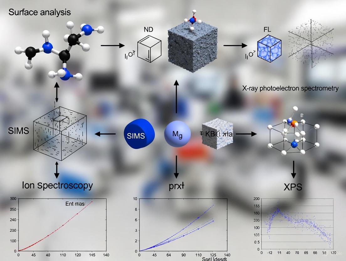
This article provides a comprehensive guide to ion spectroscopy surface analysis techniques, including XPS, AES, and SIMS, tailored for researchers and drug development professionals.
Ion Spectroscopy Surface Analysis: Techniques, Applications, and Optimization for Biomedical Research
Abstract
This article provides a comprehensive guide to ion spectroscopy surface analysis techniques, including XPS, AES, and SIMS, tailored for researchers and drug development professionals. It covers foundational principles, methodological applications in pharmaceutical research like drug and metabolite imaging, common troubleshooting and optimization strategies for data quality, and a comparative analysis of technique performance for informed method selection. The content synthesizes current research and validation protocols to support advancements in biomaterial characterization and therapeutic development.
Core Principles of Surface Analysis: Understanding XPS, AES, and SIMS
Surface analysis is a critical component in materials science, chemistry, and biological research, providing detailed information about the outermost layers of a material, which often dictate its properties and performance. This application note focuses on three principal surface analysis techniques: X-ray Photoelectron Spectroscopy (XPS), Auger Electron Spectroscopy (AES), and Secondary Ion Mass Spectrometry (SIMS). Each technique offers unique capabilities for determining elemental composition, chemical state information, and spatial distribution of components at surfaces and interfaces. The selection of an appropriate technique depends on the specific analytical requirements, including the need for spatial resolution, chemical state information, detection sensitivity, and depth profiling capabilities. Within the broader context of ion spectroscopy research, these techniques provide complementary data that can elucidate surface-mediated processes in fields ranging from semiconductor technology to drug development.
Fundamental Principles
X-ray Photoelectron Spectroscopy (XPS)
X-ray Photoelectron Spectroscopy (XPS) operates on the principle of the photoelectric effect. When a sample is irradiated with X-rays, electrons are ejected from inner atomic orbitals. The kinetic energy of these photoelectrons is measured, allowing for the determination of their binding energy according to the equation: Ek = hν - Eb - φ, where Ek is the kinetic energy of the emitted electron, hν is the energy of the X-ray photon, Eb is the binding energy of the electron, and φ is the work function of the spectrometer. Since binding energies are characteristic of specific elements and are influenced by chemical environment, XPS provides both elemental identification and chemical state information. The technique is highly surface-sensitive, with an information depth typically limited to the top 10 nanometers, as photoelectrons can only travel short distances in solids without losing energy [1].
Auger Electron Spectroscopy (AES)
Auger Electron Spectroscopy (AES) relies on the Auger process, which involves a three-step electronic relaxation mechanism. First, a high-energy electron beam (typically 3-20 keV) strikes the sample, ejecting a core-level electron and creating an excited ion. Second, an electron from a higher energy level fills the vacancy, releasing energy. Third, this energy causes the emission of a third electron, known as an Auger electron. The kinetic energy of the Auger electron is characteristic of the element from which it was emitted and is largely independent of the incident electron beam energy. AES achieves exceptional spatial resolution, with modern instruments capable of focusing the electron beam to a diameter of 5 nm or less, enabling analysis of nanoscale features [2]. The analysis depth is approximately 5 nm, similar to XPS, making it a true surface-sensitive technique [3].
Secondary Ion Mass Spectrometry (SIMS)
Secondary Ion Mass Spectrometry (SIMS) utilizes a focused primary ion beam (typically O-, O2-, Cs+, Au+, Bi3+, or C60+) to sputter and ionize atoms and molecules from the outermost surface of a sample. The ejected secondary ions are then analyzed by a mass spectrometer, which separates them according to their mass-to-charge ratio. SIMS operates in two primary modes: dynamic SIMS, which uses high primary ion currents for depth profiling and bulk analysis, and static SIMS, which uses low ion doses (below 1012 ions/cm2) to preserve molecular integrity and enable surface molecular analysis [4]. The technique offers exceptional sensitivity, capable of detecting elements and isotopes at parts-per-million to parts-per-billion levels, and can achieve spatial resolution below 100 nm with specialized instruments such as the NanoSIMS [4].
Technical Comparison
The following tables provide a comprehensive technical comparison of XPS, AES, and SIMS, highlighting their key characteristics, performance metrics, and application strengths.
Table 1: Fundamental characteristics of XPS, AES, and SIMS
| Parameter | XPS | AES | SIMS |
|---|---|---|---|
| Primary Probe | X-rays | Electron beam | Ion beam |
| Detected Signal | Photoelectrons | Auger electrons | Secondary ions |
| Information Depth | ~10 nm [1] | ~5 nm [3] | 1-2 monolayers |
| Spatial Resolution | ≥ 10 µm (conventional); ~1 µm (microprobe) | ≥ 8 nm [3] | 50-200 nm (NanoSIMS) [4]; ~200 nm (TOF-SIMS with C60+) [4] |
| Detection Limit | 0.1-1 at% | 0.1-1 at% | ppm-ppb (elements); < ppb (isotopes) |
| Chemical State Information | Yes | Limited | Limited (except with specialized MS) |
| Destructive | Non-destructive | Potentially damaging (electron beam) | Destructive (sputtering) |
Table 2: Applications, strengths, and limitations of XPS, AES, and SIMS
| Aspect | XPS | AES | SIMS |
|---|---|---|---|
| Key Strengths | Quantitative analysis, chemical state information, good for insulators | High spatial resolution, surface mapping, depth profiling | Ultra-high sensitivity, isotopic analysis, molecular information (TOF-SIMS), 3D imaging |
| Main Limitations | Lower spatial resolution, requires UHV, surface charging | Sample damage, limited chemical information, surface charging | Complex quantification, matrix effects, destructive |
| Typical Applications | Thin films, coatings, corrosion studies, catalysis, polymers [1] | Nanomaterials, failure analysis, microelectronics, grain boundary segregation [3] | Geochemistry, cell biology, pharmaceuticals, semiconductors, organic surfaces [4] |
Experimental Protocols
XPS Analysis Protocol for Battery Cathode Materials
Application: Characterization of engineered particle (Ep) battery cathodes to understand interfacial stability and degradation mechanisms [5].
Materials and Equipment:
- Thermo Fisher ESCALab QXi3 XPS instrument or equivalent [6]
- Aluminum Kα or monochromatic X-ray source
- Argon ion sputter gun for depth profiling
- Ultra-high vacuum (UHV) chamber (pressure < 1 × 10^-9 mbar)
- Electrical charge compensation system (low-energy electron flood gun)
Procedure:
- Sample Preparation: Mount the battery cathode material on a suitable holder using double-sided conductive tape or mounting clips. For powder samples, gently press onto indium foil to ensure electrical contact.
- Sample Introduction: Transfer the sample to the XPS introduction chamber and evacuate to approximately 1 × 10^-8 mbar before transferring to the analysis chamber.
- Survey Spectrum Acquisition: Collect a wide energy scan (e.g., 0-1100 eV binding energy) with pass energy of 100-150 eV to identify all elements present.
- High-Resolution Regional Scans: Acquire detailed spectra for relevant core levels (e.g., C 1s, O 1s, transition metal 2p, Li 1s) with pass energy of 20-50 eV to maximize energy resolution.
- Chemical State Analysis: Deconvolute high-resolution spectra using appropriate software to identify chemical states (e.g., different oxidation states of transition metals, various carbon species).
- Depth Profiling (if required): Perform alternating cycles of argon ion sputtering (typically 0.5-4 keV) and XPS analysis to determine composition as a function of depth.
- Data Analysis: Calculate elemental concentrations using sensitivity factors. Compare spectra from cycled and uncycled electrodes to identify degradation products.
AES Depth Profiling Protocol for Thin Film Structures
Application: Quantitative elemental and chemical state analysis from surfaces of solid materials, including depth distribution information for thin film structures [3].
Materials and Equipment:
- Physical Electronics Auger instrument or equivalent with spatial resolution ≤ 8 nm [3]
- Cylindrical Mirror Analyzer (CMA) electron analyzer
- Field emission electron gun
- Argon ion sputtering gun for depth profiling
- Charge compensation system (argon ion neutralization) for insulating samples [2]
Procedure:
- Sample Preparation: Mount the sample securely on the holder. Ensure good electrical contact for conductive samples. For insulating samples, confirm charge neutralization system is operational.
- Instrument Setup: Align the electron beam with the analysis position. Select appropriate electron beam energy (typically 3-10 keV) and current based on spatial resolution and sensitivity requirements.
- Point Analysis: Acquire AES survey spectrum from the region of interest to identify elements present.
- Elemental Mapping: Raster the focused electron beam across the sample surface to acquire spatial distribution of elements of interest.
- Depth Profiling Setup: Define the analysis area and select sputtering parameters (argon ion energy, current, and raster size).
- Sputtering and Analysis Cycle: Program the instrument to alternate between argon ion sputtering for a predetermined time and AES analysis of selected elemental peaks.
- Data Collection: Monitor peak-to-peak intensities in the derivative AES spectrum for selected elements as a function of sputtering time.
- Data Conversion: Convert sputtering time to depth using a calibrated sputter rate for the material system.
- Interpretation: Analyze the depth profile to determine layer thicknesses, interface widths, and interdiffusion between layers.
SIMS Imaging Protocol for Single Cell Analysis
Application: Mapping of elemental distributions and tracking isotopically labeled compounds in biological systems at single-cell resolution [4].
Materials and Equipment:
- NanoSIMS instrument (e.g., CAMECA NanoSIMS) or TOF-SIMS with cluster ion source
- C60+ or Bi3+ liquid metal ion gun
- Sample stubs suitable for biological specimens
- Conductive coating materials (if required)
- Standard samples for mass calibration
Procedure:
- Sample Preparation: Grow cells on appropriate substrates (e.g., silicon wafers). For metabolic studies, incubate with isotopically labeled compounds (e.g., 13C, 15N). Fix cells with appropriate fixative (e.g., glutaraldehyde). Dehydrate through graded ethanol series and critical point dry.
- Sample Mounting: Mount prepared samples on SIMS holders using conductive tape. Apply thin conductive coating if necessary to prevent charging.
- Instrument Setup: Select primary ion species (Cs+ for negative secondary ions, O- for positive secondary ions for NanoSIMS; C60+ or Bi3+ for TOF-SIMS). Optimize primary beam current and focus.
- Mass Calibration: Calibrate the mass spectrometer using known standards relevant to the masses of interest.
- Region of Interest Selection: Use optical microscopy or secondary electron imaging to locate cells of interest.
- Data Acquisition: Raster the primary ion beam over the selected area. For NanoSIMS, simultaneously detect up to 5-7 secondary ions with electron multipliers. For TOF-SIMS, pulse the primary beam and measure time-of-flight for mass separation.
- Image Generation: Construct elemental or isotopic maps from the acquired data by assigning counts to specific pixel locations.
- Quantification: For NanoSIMS, calculate isotopic ratios from regions of interest corresponding to specific cellular compartments. For TOF-SIMS, normalize counts to total ion count or matrix signals.
- Data Correlation: Correlate SIMS data with complementary techniques such as fluorescence microscopy (FISH) or electron microscopy for additional context [4].
Research Reagent Solutions
The following table outlines essential materials and reagents commonly used in surface analysis experiments across the three techniques.
Table 3: Essential research reagents and materials for surface analysis techniques
| Item | Function/Application | Technique |
|---|---|---|
| Conductive Tapes/Carbon Tapes | Sample mounting to ensure electrical and thermal contact | XPS, AES, SIMS |
| Indium Foil | Substrate for mounting powder samples | XPS, AES |
| Argon Gas (High Purity) | Sputtering source for depth profiling and charge neutralization | XPS, AES, SIMS |
| Silicon Wafers | Clean, flat substrates for mounting samples, particularly biological specimens | SIMS |
| Isotopically Labeled Compounds (13C, 15N) | Tracers for metabolic studies in biological systems | SIMS |
| Conductive Coatings (Carbon, Gold) | Applied to insulating samples to prevent charging | AES, SIMS (if charge compensation inadequate) |
| Standard Reference Materials | Quantification calibration, mass calibration, instrument performance verification | XPS, AES, SIMS |
| Charge Neutralization Flood Gun | Provides low-energy electrons to neutralize surface charge on insulating samples | XPS |
| Ultra-pure Solvents (e.g., methanol, water) | Sample cleaning prior to analysis to remove surface contaminants | XPS, AES, SIMS |
Advanced Applications and Case Studies
Combined XPS and TOF-SIMS Analysis of Battery Materials
In a study of engineered particle (Ep) battery cathodes, researchers combined XPS and TOF-SIMS to understand stabilizing effects on electrode-electrolyte interfaces. XPS provided quantitative chemical state information about the surface species, while TOF-SIMS offered high-sensitivity detection of organic and inorganic species across the interface. This combined approach revealed that Ep-coated cathodes exhibited more uniform and controlled interfaces, leading to improved battery performance and long-term stability. The TOF-SIMS data complemented XPS results by detecting trace degradation products that were below the detection limit of XPS, providing a more comprehensive understanding of degradation mechanisms [5].
NanoSIMS for Biological Systems at Single-Cell Resolution
NanoSIMS has enabled groundbreaking research in microbiology and cell biology by allowing researchers to track metabolic processes at the single-cell level. In one pioneering application, researchers used NanoSIMS to quantify N2 fixation by individual bacteria inhabiting the gills of shipworms. By introducing 15N-labeled substrates, they could measure nitrogen assimilation at unprecedented resolution. Similarly, studies on cyanobacteria used 13C and 15N tracers to track the assimilation of inorganic carbon and ammonium by individual cells, revealing significant variation in uptake rates among phylogenetically identical cells. These applications demonstrate the unique capability of SIMS to link metabolic function to phylogenetic identity in complex biological systems [4].
AES for Nanomaterials and Thin Film Characterization
AES provides critical information about surface layers and thin film structures that govern performance in various applications. With its high spatial resolution (as small as 8 nm), AES can characterize composition at nanoscale features in materials such as catalysts, semiconductors, and magnetic media. The combination of AES with ion sputtering enables depth profiling of multilayer structures, allowing researchers to determine layer thicknesses, interface quality, and interdiffusion between layers. This capability is particularly important for semiconductor devices and packaging, where nanoscale layers determine device performance and reliability [3].
XPS, AES, and SIMS represent three powerful and complementary techniques for surface analysis, each with distinct strengths and applications. XPS excels at providing quantitative elemental composition and detailed chemical state information, making it invaluable for studying surface chemistry in materials such as battery electrodes and catalysts. AES offers superior spatial resolution for mapping elemental distributions at the nanoscale, particularly useful for failure analysis and thin film characterization. SIMS provides unparalleled sensitivity for trace element and isotopic analysis, with growing applications in biological systems and materials science. The continuing development of these techniques, including improvements in spatial resolution, sensitivity, and data analysis capabilities, ensures they will remain essential tools for understanding surface and interface phenomena across numerous scientific and industrial fields.
Historical Development and Technological Evolution of Surface Analysis
Surface analysis, in analytical chemistry, is the study of the part of a solid that is in contact with a gas or a vacuum [7]. This interface dictates critical material properties and behaviors. The field has undergone a tremendous evolution since the mid-20th century, moving from classical methods that provided physical descriptions to modern spectroscopic techniques capable of providing detailed elemental and chemical state information from the outermost atomic layers [7]. This evolution has been pivotal for advancements in diverse fields, including catalysis, nanotechnology, and drug development, where understanding surface composition is essential for designing and improving materials and processes [7] [8].
This application note details the key methodologies within surface analysis, with a particular focus on Ion Scattering Spectroscopy (ISS) as a cornerstone technique for quantifying the atomic composition of the outermost surface. It provides structured protocols and data to support researchers in the effective application of these powerful analytical tools.
Key Techniques in Modern Surface Analysis
Modern surface analysis is characterized by a "beam in, beam out" mechanism, where a primary beam of photons, electrons, or ions interacts with the sample, and an emitted beam is analyzed to yield surface-specific information [7]. The sampling depth—a critical parameter defining surface sensitivity—varies significantly with the probe and its energy, being shallowest for ions (∼1 nm) and deepest for photons (∼1000 nm) [7]. During the 1970s and 1980s, four techniques emerged as particularly useful for real-world problem-solving due to their general applicability and ease of use [7].
Table 1: Major Surface Analysis Techniques and Their Characteristics
| Technique | Acronym | Probe In | Signal Out | Key Information | Sampling Depth |
|---|---|---|---|---|---|
| X-ray Photoelectron Spectroscopy | XPS/ESCA | X-rays (Photons) | Electrons | Elemental identity, chemical state, quantitative analysis [7] | ~1-10 nm [7] |
| Auger Electron Spectroscopy | AES | Electrons | Electrons | Elemental composition, surface mapping [7] | ~2 nm [7] |
| Secondary Ion Mass Spectrometry | SIMS | Ions | Ions | Elemental and molecular composition, extreme surface sensitivity, depth profiling [7] [8] | < 1 nm |
| Ion Scattering Spectroscopy | ISS/LEIS | Ions (Noble Gas) | Ions | Atomic composition of the outermost atomic layer [9] [8] | ~0.3 nm (top monolayer) [8] |
Ion Scattering Spectroscopy (ISS): Principles and Protocol
Core Principles of ISS
Ion Scattering Spectroscopy (ISS), also referred to as Low-Energy Ion Scattering (LEIS), is a surface-specific technique where a beam of noble gas ions (e.g., He⁺, Ne⁺, Ar⁺) is elastically scattered by atoms on the sample surface [9] [8]. The kinetic energy of the scattered ions, ( E_s ), is measured, and the energy loss is determined by the masses of the incident ion and the target surface atom, governed by the principles of binary elastic collision and momentum conservation [9].
The fundamental equation for ISS is: [ \frac{Es}{E0} = \left( \frac{1}{1 + M2 / M1} \right)^2 \left( \cos \theta + \sqrt{ (M2 / M1)^2 - \sin^2 \theta } \ \right)^2 ] where ( E0 ) is the primary ion energy, ( M1 ) is the mass of the incident ion, ( M_2 ) is the mass of the surface atom, and ( \theta ) is the scattering angle [9]. Because the scattered ion signal is dominated by atoms in the topmost atomic layer, ISS is uniquely sensitive to the outer surface [9] [8].
Experimental Protocol for Quantitative Surface Composition Analysis
Objective: To determine the elemental composition of the outermost atomic layer of a solid sample using ISS.
Table 2: Research Reagent Solutions and Essential Materials
| Item | Function / Specification |
|---|---|
| Noble Gas Ion Source | Generates the primary ion beam (typically He⁺, Ne⁺, or Ar⁺) with energy between 0.5 - 3 keV [9] [8]. |
| Ultra-High Vacuum (UHV) Chamber | Provides a clean environment (< 10⁻⁹ mbar) to prevent surface contamination during analysis [8]. |
| Sample Holder & Stage | Holds the sample and allows for precise positioning and, if available, heating or cooling. |
| Energy Analyzer | Measures the kinetic energy of the scattered ions (e.g., a hemispherical sector analyzer or time-of-flight system) [8]. |
| Standard Reference Sample | A pure, well-characterized material (e.g., gold foil) for instrument calibration [9]. |
| Charge Neutralization System | An electron flood gun for analyzing insulating samples to compensate for surface charging [8]. |
Step-by-Step Workflow:
Sample Preparation: Introduce the sample into the UHV chamber. For powder samples, press into a clean indium foil or mount on a suitable stub. Clean the sample surface in situ via argon ion sputtering to remove adventitious carbon and contaminants, if the analysis goal permits [8].
Instrument Calibration: a. Energy Scale Calibration: Using the standard reference sample (e.g., Au), acquire a spectrum with a known primary ion (e.g., He⁺). The scattering angle ( \theta ) is a fixed parameter of the instrument (e.g., 130°) [9]. b. Primary Energy Calibration: Measure the kinetic energy of the scattered peak from the standard (( Es )). With known ( M1 ), ( M2 ), and ( \theta ), solve the ISS equation for ( E0 ) to determine the precise primary beam energy [9]. For example, with He⁺ (( M1 = 4 )) scattered from Au (( M2 = 197 )) at ( \theta = 130^\circ ) and a measured ( Es = 877 ) eV, the calculated ( E0 ) is 937 eV [9].
Data Acquisition: Direct the calibrated primary ion beam onto the sample surface. Set the energy analyzer to scan over the appropriate kinetic energy range (from just below ( E_0 ) down to zero) and collect the spectrum of scattered ion yield versus kinetic energy.
Data Interpretation: a. Peak Identification: Identify peaks in the spectrum. Each element on the surface will produce a peak at a characteristic ( Es ) [9]. b. Elemental Assignment: For each peak at energy ( Es ), use the known ( E0 ), ( M1 ), and ( \theta ) in the ISS equation to calculate the mass of the scattering atom, ( M_2 ), thereby identifying the element [9].
Advanced Applications and Case Studies
Application in Corrosion Analysis
Background: Incoming coil steel stock conforming to a specified average roughness (Ra = 20-70 μin) showed unexpected corrosion after processing [10]. Analysis: 3D optical profilometry revealed that while both acceptable and rust-prone stock had similar Ra values, their 3D topography differed significantly. The rust-prone stock featured many deep valleys, whereas the acceptable stock was more isotropic [10]. ISS/SIMS Role: While surface topography parameters (Skewness, valley depth) identified the structural risk, compositional analysis via techniques like ISS or SIMS would be critical to determine if surface contaminants or specific alloying elements segregated in the valleys, contributing to the corrosion onset. Outcome: Bearing area curve analysis quantified the percentage of deep valleys that retained processing solutions, leading to flash rusting. This combined topographical and compositional analysis allowed for the development of a better quality control parameter than Ra alone [10].
Surface Engineering for Performance
Background: A new clutch plate design required optimization for friction and wear performance [10]. Analysis: Multiple plate designs with known performance were evaluated using 3D surface metrology parameters. ISS/SIMS Role: In such a study, ISS could be used to ensure the consistency of the outermost surface composition of the plates, while SIMS could depth profile to monitor the integrity of any surface coatings or modifications. Outcome: Statistical parameters like skewness (Ssk) and kurtosis were found to correlate strongly with wear and friction performance. These parameters were then used to control a novel manufacturing process, ensuring consistent and superior part performance [10].
Table 3: Quantitative Data from ISS Analysis of a Phosphor-Bronze Sample
| Element | Atomic Mass (M₂) | Theoretical ( Es/E0 ) | Measured ( E_s ) (eV) | Calculated ( Es/E0 ) | Identified? |
|---|---|---|---|---|---|
| Oxygen | 16 | 0.536 | ~400 eV (est. from fig.) | 0.427 (for E₀=937 eV) | Yes [9] |
| Copper | 63.5 | 0.837 | ~762 eV (est. from fig.) | 0.813 (for E₀=937 eV) | Yes [9] |
| Tin | 118.7 | 0.947 | ~877 eV (est. from fig.) | 0.936 (for E₀=937 eV) | Yes [9] |
Note: The table uses estimated values from a figure in [9] for a He⁺ beam (M₁=4) with a scattering angle of 130°. The primary beam energy E₀ is assumed to be 937 eV for calculation consistency.
Current Trends and Future Outlook
The field of surface analysis continues to evolve, driven by technological advancements and growing industrial adoption. Key trends shaping its future include:
- Democratization of Techniques: Instruments like XPS, SIMS, and AES are becoming more user-friendly, moving from tools exclusively operated by specialized experts to accessible resources for a broader range of scientists. This allows researchers to focus more on material exploration than instrumental complexity [11].
- Embracing Surface Engineering: Industries are increasingly using surface analysis as a general-purpose characterization tool to solve material challenges. This is particularly evident in the development of batteries, touch-screens, and medical devices, where surface properties are critical to performance [11].
- Automation and Data Integrity: The introduction of intelligent software, automation, and robust data analysis protocols is enhancing the reproducibility and integrity of surface analysis data, which is fundamental for scientific research and industrial quality control [11].
The precise measurement of electrons and ions forms the cornerstone of modern analytical chemistry, enabling groundbreaking research across surface science, drug development, and fundamental physics. These measurements provide critical insights into electronic structures, chemical bonding behaviors, and material properties at the atomic level. For researchers investigating surface analysis techniques, understanding these fundamental operating principles is essential for selecting appropriate methodologies and interpreting experimental data accurately. This article details the core techniques, protocols, and applications of advanced spectroscopic methods that probe the interactions of electrons and ions with matter, with particular emphasis on approaches that achieve unprecedented sensitivity for studying rare and exotic elements.
Table 1: Core Techniques for Measuring Electrons and Ions
| Technique | Measured Property | Fundamental Principle | Primary Applications |
|---|---|---|---|
| Laser Photodetachment Threshold (LPT) Spectroscopy | Electron Affinity (EA) | Neutralization of anions by collinear laser photons above a specific energy threshold [12]. | Determining electron affinities of atoms/molecules, benchmarking atomic theory [12]. |
| Multi-Reflection Time-of-Flight (MR-ToF) Analysis | Mass-to-Charge Ratio (m/z) |
Separation of ions based on flight time over extended path lengths in an electrostatic trap [12]. | High-resolution mass separation, ion storage for repeated laser probing [12]. |
| Laser-Induced Breakdown Spectroscopy (LIBS) | Elemental Composition | Analysis of atomic emission spectra from laser-generated plasma [13]. | Surface mapping, spatial element distribution in battery and material science [13]. |
| Parallel Reaction Monitoring (PRM) | Peptide/Protein Quantification | High-resolution, accurate-mass targeted analysis of selected precursor ions [14]. | Targeted proteomics, kinase activity profiling, biomarker verification [14]. |
Fundamental Operating Principles
Laser Photodetachment and Electron Affinity Determination
The electron affinity (EA) is defined as the energy released when an electron is added to a neutral atom to form a negative ion [12] [15]. It is a fundamental atomic property governed by complex electron-electron correlations and is a critical parameter for predicting chemical reactivity and bonding behavior [12]. Laser Photodetachment Threshold (LPT) spectroscopy determines this property by measuring the exact photon energy required to remove the extra electron from an anion, thereby neutralizing it [12] [15].
In conventional LPT, a beam of anions is overlapped collinearly with a laser beam. The laser frequency is tuned, and the number of resulting neutral atoms is monitored. When the photon energy (E) exceeds the electron affinity (EA), photodetachment occurs, releasing an electron with kinetic energy Ee = E - EA [12]. The threshold energy at which neutral atoms are detected corresponds directly to the EA of the element. The collinear overlap of the laser and ion beams extends the interaction time and reduces the Doppler broadening of the spectral line, leading to higher resolution [12].
Ion Trapping and Signal Amplification
A transformative advancement in this field is the integration of LPT spectroscopy with Electrostatic Ion Beam Traps, specifically Multi-Reflection Time-of-Flight (MR-ToF) devices [12] [15]. This approach, exemplified by the MIRACLS (Multi Ion Reflection Apparatus for Collinear Laser Spectroscopy) technique, confines ions between two electrostatic mirrors [12].
Within the MR-ToF device, anions oscillate back and forth through a field-free drift region, where they are repeatedly probed by a collinear spectroscopy laser. This "recycling" of ions—achieving approximately 60,000 passes in demonstrated experiments—increases the laser-ion interaction time by several orders of magnitude compared to single-pass setups [15]. Consequently, the probability of photodetachment for each anion is drastically increased. This enhanced sensitivity allows for state-of-the-art precision EA measurements while employing five orders of magnitude fewer anions than conventional techniques [12]. After neutralization, atoms maintain their momentum and exit the trap along a predictable path to a detector, ensuring high detection efficiency [12].
Experimental Protocols
Protocol: Determining Electron Affinity via MR-ToF-Enhanced LPT Spectroscopy
This protocol details the procedure for measuring electron affinity with high sensitivity using an electrostatic ion beam trap, as demonstrated for chlorine [12].
Research Reagent Solutions and Materials
Table 2: Essential Research Reagents and Materials
| Item | Function / Specification |
|---|---|
| Sample Material | Element of interest (e.g., Chlorine gas for Cl⁻ production). |
| Negative Surface Ion Source | Produces a continuous beam of negative ions (Cl⁻) [12]. |
| Paul Trap | Captures, accumulates, and cools ion bunches using helium buffer gas [12]. |
| Helium Buffer Gas | Cools ions via collisions to room temperature within the Paul trap [12]. |
| High-Voltage Pulsed Drift Tube | Adjusts the kinetic energy of ion bunches for isobaric purification [12]. |
| High-Voltage Deflector | Selects ions of a specific m/z (e.g., ³⁵Cl⁻) via time-of-flight [12]. |
| MR-ToF Device | Electrostatic trap with two mirrors for confining and cycling ions [12]. |
| Narrow-Band Continuous-Wave (cw) Laser | Spectroscopy laser for photodetachment; tunable energy around the expected EA [12]. |
| Neutral Particle Detector | High-efficiency, low-background detector for neutral atoms generated upon photodetachment [12]. |
Step-by-Step Procedure
Ion Production and Preparation:
- Generate a continuous beam of anions (e.g.,
Cl⁻) using a negative surface ion source [12]. - Guide the anion beam into a Paul trap. Capture, accumulate, and cool the ions via collisions with room-temperature helium buffer gas to reduce their kinetic energy and emittance [12].
- Extract the ions from the Paul trap in a low-emittance bunch.
- Generate a continuous beam of anions (e.g.,
Beam Purification and Energy Adjustment:
- Pass the ion bunch through a pulsed drift tube to adjust its kinetic energy to a precise value (e.g., 20-30 keV) [12].
- Direct the energy-adjusted bunch through a high-voltage deflector. Use time-of-flight selection to isolate the specific isotope of interest (e.g.,
³⁵Cl⁻) by deflecting unwanted species away from the beam axis [12].
Ion Trapping and Laser Probing:
- Inject the purified ion bunch into the MR-ToF device. Confine the ions by reflecting them between the two electrostatic mirrors for thousands of cycles [12].
- Continuously illuminate the ions in the field-free drift section of the MR-ToF with the narrow-band cw laser. The laser beam is collinear with the ion trajectory.
- Gradually tune the laser's photon energy across the theoretical electron affinity threshold of the element.
Signal Detection and Data Acquisition:
- When the photon energy exceeds the EA, photodetachment occurs. The resulting neutral atoms are no longer confined by the electrostatic mirrors and continue on a straight path, exiting the trap [12].
- Detect these neutral atoms using a high-efficiency neutral particle detector positioned downstream [12].
- Record the neutral count rate as a function of the laser photon energy.
Data Analysis and EA Determination:
- Plot the neutral count rate versus laser photon energy. The threshold energy where the signal significantly increases corresponds to the electron affinity of the atom.
- Fit the threshold curve to determine the EA with high precision. The demonstrated precision for chlorine was 3.612720(44) eV [12].
Workflow Visualization
The following diagram illustrates the core experimental workflow and ion pathways for the MR-ToF-enhanced LPT spectroscopy protocol.
Advanced Applications and Broader Context
The enhanced sensitivity of techniques like MIRACLS is pushing the boundaries of scientific inquiry. A primary application is the systematic measurement of isotope shifts and hyperfine splittings in electron affinities across isotopic chains, providing benchmarks for nuclear models [12]. Furthermore, this methodology paves the way for the first direct determination of electron affinities in superheavy elements (e.g., oganesson, Z=118), where relativistic effects are predicted to dominate and potentially颠覆 periodic table trends [12] [15].
Beyond fundamental atomic physics, these principles are vital in applied fields. In cancer research, measuring the electron affinity of rare elements like astatine and actinium is crucial for developing targeted alpha-therapy radiopharmaceuticals [15]. The technique can also be applied to molecules, providing data for theoretical calculations relevant to antimatter research and the use of radioactive molecules to probe physics beyond the Standard Model [12] [15].
The operating principles of electron and ion measurement find parallel applications in other spectroscopic methods. For instance, Laser-Induced Breakdown Spectroscopy (LIBS) uses a pulsed laser to create a micro-plasma, whose emitted light is analyzed to determine the elemental composition of a surface. This has proven highly effective for mapping lithium nucleation in anode-less solid-state batteries, distinguishing between electrolyte-derived lithium and in-situ formed metal anodes [13].
Table 3: Comparison of Electron/Ion Measurement Techniques
| Technique | Typical Sample Size/Intensity | Achievable Precision | Key Limitation |
|---|---|---|---|
| Conventional LPT | ~10¹¹ anions per second (e.g., 600 fA for Astatine) [12] | High (for abundant samples) | Inefficient use of sample; unsuitable for very rare species [12]. |
| MR-ToF-Enhanced LPT | ~10⁶ anions per second (5 orders of magnitude fewer) [12] | State-of-the-art (e.g., 3.612720(44) eV for Cl) [12] | Requires stable anion formation and specialized trap instrumentation. |
| Photodetachment Microscopy | Large ensembles | Very High [12] | Requires high beam intensity and complex electron interference pattern analysis [12]. |
| LIBS | Microscopic surface area | Semi-Quantitative (excellent for mapping) [13] | Matrix effects can influence emission spectra and quantification [13]. |
Surface analysis techniques are indispensable tools in modern scientific research, particularly in fields requiring detailed material characterization such as drug development and materials science. These techniques provide critical data on elemental composition, chemical speciation, and spatial distribution of analytes. However, each method possesses unique capabilities alongside inherent limitations. This application note provides a detailed examination of three critical aspects—elemental detection, chemical state information, and spatial resolution—within the context of ion spectroscopy and related surface analysis techniques. By framing this discussion within experimental protocols and quantitative comparisons, this document serves as a practical resource for researchers and scientists designing characterization strategies for complex biological and material systems.
Key Capabilities and Limitations: A Comparative Analysis
The table below summarizes the core capabilities and limitations of prominent surface analysis techniques, highlighting their performance across the three focus areas.
Table 1: Key Capabilities and Inherent Limitations of Surface Analysis Techniques
| Technique | Elemental Detection | Chemical State Information | Spatial Resolution | Primary Limitations |
|---|---|---|---|---|
| PIXE (Particle Induced X-ray Emission) | Quantitative mapping of metals (e.g., Fe, Mn) in biological tissues [16]. | Limited directly, often requires coupling with other techniques [16]. | ~Micrometer resolution, suitable for distinguishing brain regions like SNpc and SNpr [16]. | Requires correlation with histology for accurate anatomical location; sample preparation is critical to preserve native element distribution [16]. |
| Synchrotron XRF (X-ray Fluorescence) | Quantitative distribution of multiple elements (P, S, Cl, K, Ca, Cu, Zn) simultaneously [16]. | Can be coupled with micro-XANES to identify oxidation states (e.g., Fe(II) vs. Fe(III)) [16]. | Micro-scale resolution, capable of revealing intra-regional differences (e.g., Mn content in SNpc vs. SNpr) [16]. | Requires synchrotron radiation source; complex sample preparation using cryogenic protocols to avoid disturbing native element speciation [16]. |
| ICP-MS (Inductively Coupled Plasma Mass Spectrometry) | Exceptional sensitivity with detection limits in the low parts-per-trillion (ppt) range for most elements; high ionization efficiency for most metals (80-95%) [17]. | None; provides elemental composition only. | Essentially none for spatial mapping; typically a bulk analysis technique. | Overall inefficiency (~0.00002%); losses occur at nebulization, interface (sampler/skimmer cones), and in ion lenses [17]. |
| Imaging Mass Spectrometry (IMS) | Direct mapping of a wide variety of analytes, including lipids and metabolites, in thin tissue sections [18]. | Tandem MS (MS/MS) and ion/ion reactions provide specificity to differentiate isobaric and isomeric compounds [18]. | A key trade-off exists; higher chemical specificity (via ion/ion reactions) often forces lower spatial resolution (e.g., 125 µm) due to signal requirements [18]. | Differentiating isobaric/isomeric lipids is challenging; spatial resolution is inversely related to chemical specificity and limit of detection [18]. |
| Scanning Tunneling Microscopy (STM) | Limited elemental specificity. | Can probe electronic characteristics of surfaces [19]. | Atomic-scale resolution for conductive surfaces [19]. | Requires conductive samples; primarily for topographical and electronic mapping rather than direct chemical identification [19]. |
Quantitative Data in Analytical Performance
Understanding the numerical benchmarks for sensitivity and efficiency is crucial for technique selection. The following tables consolidate key quantitative data from the literature.
Table 2: Sensitivity and Detection Limits of Mass Spectrometry Techniques
| Technique | Typical Sensitivity | Exemplary Detection Limit | Notes |
|---|---|---|---|
| Quadrupole ICP-MS | 100-500 million counts per second per part per million (Mcps/ppm) [17]. | ~1 part per trillion (ppt) for many elements [17]. | 1 ppt corresponds to ~1.1 x 10⁹ to 4.2 x 10⁷ atoms per second introduced to the nebulizer [17]. |
| FT-ICR Imaging MS | Not explicitly quantified in results. | Enables separation of isobaric compounds [18]. | High resolving power requires long transient times [18]. |
Table 3: Component Efficiency in a Typical Quadrupole ICP-MS System
| Component / Process | Approximate Efficiency | Description of Loss |
|---|---|---|
| Nebulization & Spray Chamber | 1-2% | Majority of sample is drained to waste [17]. |
| Ionization in ICP | 40% (Hg) to >95% (most metals) | Dependent on element's first ionization potential [17]. |
| Interface (Sampler & Skimmer Cones) | ~0.04% (2% x 2%) | Intentional due to the 10⁷-fold pressure reduction from atmospheric plasma to high vacuum [17]. |
| Ion Lenses | ~50-80% | Losses from focusing and steering ions; modern "off-axis" designs improve transmission and block photons [17]. |
| Collision/Reaction Cell | Variable | Losses from space charge effects and collisional scattering [17]. |
| Quadrupole Mass Filter | ~50% | Ions filtered based on mass-to-charge ratio [17]. |
| Detector (Electron Multiplier) | ~100% (for counted ions) | One detectable pulse per ion strike [17]. |
| Overall System Efficiency | ~0.00002% | Product of individual component efficiencies [17]. |
Detailed Experimental Protocols
Protocol 1: Correlative Elemental and Chemical State Analysis in Brain Tissue
This protocol, adapted from a study on Parkinson's disease models, details the process for correlating quantitative metal mapping with chemical speciation and histology in specific brain regions [16].
1. Sample Preparation and Cryofixation
- Animal Perfusion and Brain Extraction: Anesthetize the animal and transcardially perfuse with a suitable buffer to remove blood. Rapidly extract the brain.
- Cryofixation: Immediately plunge the intact brain into a cooled cryogenic liquid (e.g., isopentane cooled with dry ice to -40°C) to preserve the native distribution and speciation of elements. Store cryofixed brains at -80°C [16].
- Cryo-Sectioning: Section the frozen brain on a cryostat (e.g., 10-20 µm thickness) and thaw-mount onto appropriate substrates (e.g., indium tin oxide (ITO)-coated slides for SXRF) [16].
2. Correlative Immunohistochemistry (on Adjacent Sections)
- Purpose: To unambiguously identify anatomical regions of interest (e.g., Substantia Nigra pars compacta (SNpc) and pars reticulata (SNpr)) for subsequent chemical analysis.
- Procedure: On a separate, adjacent cryo-section, perform standard tyrosine hydroxylase (TH) immunohistochemical staining. This provides a high-resolution anatomical map.
- Correlation: Superimpose the optical image of the stained section with the elemental maps from the analyzed section to define the regions for quantitative analysis and speciation [16].
3. Quantitative Elemental Mapping via PIXE/SXRF
- Data Acquisition: Irradiate the unstained cryo-section with a focused micro-beam of protons (PIXE) or synchrotron X-rays (SXRF). Collect the emitted X-rays to generate quantitative distribution maps for elements like Fe, Mn, P, S, K, Ca, Cu, and Zn [16].
- Spatial Registration: Use the correlated histology image to assign the elemental content to specific brain structures (SNpc, SNpr, VTA).
4. Chemical Speciation via micro-XANES
- Site Selection: Based on the correlated maps, select specific micron-scale spots within the SNpr and SNpc for speciation analysis.
- Data Collection: Acquire X-ray absorption spectra at the Fe K-edge across a range of energies around the absorption edge.
- Data Analysis: Compare the experimental XANES spectra (edge position, pre-edge features) with spectra from reference compounds (e.g., ferritin) to identify the dominant oxidation state and chemical environment of iron [16].
Protocol 2: Resolving Isobaric Lipids via Ion/Ion Reaction Imaging MS
This protocol describes a tandem MS workflow to differentiate isobaric lipids in tissue sections, addressing a key challenge in imaging mass spectrometry [18].
1. Tissue Preparation and Matrix Application
- Cryo-Sectioning: Fresh-frozen mouse brain tissue is sectioned at 10 µm thickness using a research cryostat and thaw-mounted onto ITO-coated slides [18].
- Matrix Deposition: Coat the tissue section with a matrix (e.g., 2’,5’-Dihydroxyacetophenone - DHA) using a sublimation apparatus (e.g., 3 minutes at 180°C) to ensure homogeneous crystallization [18].
2. High-Resolution Full Scan (MS1) Imaging
- Acquisition: Acquire full scan mass spectra using a MALDI-FT-ICR instrument or similar high-resolution mass spectrometer.
- Parameters: Use a high spatial resolution (e.g., 25 µm pixel spacing) to capture the detailed spatial distribution of all detectable lipids. This dataset will serve as the high-spatial-resolution input for later fusion [18].
3. Low-Resolution Ion/Ion Reaction (MS2) Imaging
- Acquisition: Acquire MS/MS images on the same tissue section using a targeted approach.
- Ion/Ion Reaction: For each pixel, isolate lipid anion precursors, mutually store them with TMODA dications from an ESI source to form Schiff base products, and perform CID to generate diagnostic fragment ions [18].
- Parameters: Use a lower spatial resolution (e.g., 125 µm pixel spacing) to compensate for the lower signal intensity resulting from the multi-step reaction and fragmentation process [18].
4. Computational Image Fusion
- Co-registration: Spatially align the high-resolution MS1 and low-resolution MS2 imaging datasets.
- Dimensionality Reduction: Apply Non-Negative Matrix Factorization (NMF) to the full MS1 dataset to reduce its complexity while retaining spatial features.
- Model Training: Use machine learning models (e.g., Linear Regression, 2-D Convolutional Neural Networks) to learn the relationship between the NMF components of the MS1 data and the specific ion/ion reaction product ion image from the MS2 data.
- Prediction: Apply the trained model to the high-resolution MS1 data to generate a predicted image of the ion/ion reaction product with a 5-fold enhanced spatial resolution (e.g., predicted at 25 µm) [18].
The Scientist's Toolkit: Essential Research Reagents and Materials
Table 4: Key Reagents and Materials for Featured Surface Analysis Experiments
| Item | Function / Application | Specific Example / Note |
|---|---|---|
| Cryogenic Fluids | Rapid cryofixation of biological tissues to preserve native elemental distribution and speciation [16]. | Isopentane cooled with dry ice (-40°C to -80°C) [16]. |
| Primary Antibodies for IHC | Histological staining for anatomical correlation in correlative elemental imaging [16]. | Anti-Tyrosine Hydroxylase (TH) antibody for identifying dopaminergic regions in brain tissue [16]. |
| TMODA Reagent | Gas-phase derivatization reagent for charge inversion ion/ion reactions; selectively targets phosphatidylserine lipids [18]. | Synthesized from N,N,N′,N′-Tetramethyl-1,6-hexanediamine and 6-Bromohexanal [18]. |
| MALDI Matrix | Assists in the desorption and ionization of analytes from tissue surfaces for mass spectrometry analysis. | 2’,5’-Dihydroxyacetophenone (DHA) for lipid analysis [18]. |
| Indium Tin Oxide (ITO) Coated Slides | Conductive substrate required for mounting tissue sections in imaging mass spectrometry and micro-XRF to prevent charging [18]. | -- |
| Reference Standards | Critical for calibrating quantitative analyses and for identifying chemical species via fingerprinting in XANES. | Pure ferritin for matching Fe speciation in brain tissue [16]. |
Current Trends and Publication Growth in Surface Analysis Techniques
Surface analysis is a methodical examination of a material's outermost layers to ascertain its composition, structure, and properties at atomic and molecular levels [20]. These techniques are foundational for understanding surface characteristics that dictate chemical activity, adhesion, wetness, electrical properties, corrosion-resistance, and biocompatibility [20]. The field has evolved significantly since the 1960s with the introduction of commercial instrumentation, and today encompasses a range of sophisticated techniques, the most widespread being X-ray photoelectron spectroscopy (XPS), Auger electron spectroscopy (AES), and secondary ion mass spectrometry (SIMS) [21]. The growing demand for precise surface characterization in sectors like semiconductors, healthcare, and advanced materials is propelling both technological innovation and market expansion, with the global surface analysis market projected to reach between USD 9.19 billion and USD 9.38 billion by 2032 [19] [22].
This application note frames the current landscape and experimental protocols within the context of advanced ion spectroscopy and surface analysis techniques research. It is designed to equip researchers, scientists, and drug development professionals with actionable data and methodologies to navigate this dynamic field.
Quantitative Market Growth Analysis
The surface analysis market demonstrates robust growth, driven by technological advancements and increasing demand across industrial sectors. Table 1 summarizes the quantitative market projections from key industry reports, which show some variation based on methodology and forecast periods [19] [22] [23].
Table 1: Surface Analysis Market Size and Growth Projections
| Source | Base Year Value | Projected Year Value | CAGR | Forecast Period |
|---|---|---|---|---|
| Coherent Market Insights [19] | USD 6.45 Bn (2025) | USD 9.19 Bn (2032) | 5.18% | 2025-2032 |
| The Business Research Company [22] | USD 6.61 Bn (2025) | USD 9.38 Bn (2029) | 9.1% | 2024-2029 |
| Prophecy Market Insights [24] | USD 6.1 Bn (2025) | USD 10.7 Bn (2035) | 6.3% | 2025-2035 |
| SERPVision [23] | USD 5.77 Bn (2025) | USD 14.68 Bn (2033) | 16.84% | 2026-2033 |
This growth is fueled by several macro and microeconomic factors. Key drivers include the expansion of the semiconductor and electronics industry, stringent quality control regulations in aerospace and medical devices, rising R&D investments in healthcare and nanotechnology, and government programs supporting metrology and environmental laws [19] [22] [24]. A notable restraint is the high cost of advanced equipment and a global shortage of skilled professionals to operate these sophisticated instruments [24].
Technique Adoption and Publication Growth
An analysis of publication rates reveals clear trends in the adoption and scientific application of different surface analysis techniques. As illustrated in Figure 1, the number of publications utilizing XPS (including related terms like ESCA, PES, HAXPES, and NAP-XPS) has seen a rapid and continuous increase over the past two decades. In contrast, publication rates for AES and SIMS have remained relatively constant during the same period [21]. This trend underscores XPS's position as the most commonly used technique, owing to its relatively simple spectra, ease of quantification, excellent chemical state information, and lower instrument cost compared to AES and SIMS [21].
Advanced Techniques and Methodologies
Dominant Surface Analysis Techniques
The surface analysis landscape is characterized by several powerful techniques, each with unique strengths and applications, particularly in ion spectroscopy research.
X-ray Photoelectron Spectroscopy (XPS): Also known as Electron Spectroscopy for Chemical Analysis (ESCA), XPS operates by irradiating a sample with X-rays and analyzing the kinetic energy of emitted photoelectrons [21] [25]. It provides quantitative data on atomic composition and chemical bonding states of the surface [25] [20]. Its key advantage is the ability to analyze various materials, both organic and inorganic, making it the most widespread technique [21] [20]. XPS cannot directly detect hydrogen or helium [21].
Time-of-Flight Secondary Ion Mass Spectrometry (TOF-SIMS): This technique uses a pulsed primary ion beam to desorb and ionize species from the sample surface. The secondary ions are accelerated into a time-of-flight mass analyzer, separating ions based on their mass-to-charge ratio [20]. TOF-SIMS offers extremely high surface sensitivity, can detect all elements including isotopes, and provides molecular mass information for organic compounds [21] [20]. Its spectra can be complex due to large molecular fragments.
Auger Electron Spectroscopy (AES): AES employs a focused electron beam to excite the sample, resulting in the emission of Auger electrons that are analyzed for their kinetic energy [21] [25]. It provides elemental and some chemical state information with very high spatial resolution, making it particularly suitable for analyzing micro-level foreign substances and for failure analysis in semiconductors and metals [25] [20].
Emerging and Specialized Methodologies
Recent years have seen the development and commercialization of several advanced XPS methodologies that address previous limitations and open new research avenues.
Hard X-ray Photoelectron Spectroscopy (HAXPES): Traditionally conducted at synchrotron facilities, HAXPES is now available in laboratories using silver, chromium, or gallium X-ray sources instead of standard aluminum or magnesium sources [21]. The higher energy X-rays enable deeper analysis (up to several tens of nanometers), reduce the effects of surface contamination, and allow access to higher binding energy core levels, facilitating the study of buried interfaces [21] [25].
Near-Ambient Pressure XPS (NAP-XPS): A significant advancement beyond traditional ultra-high vacuum requirements, NAP-XPS allows for the chemical analysis of surfaces in reactive environments or under modest gas pressures [21]. This capability is crucial for in-situ studies of catalytic reactions, corrosion processes, and the interaction of microorganisms with surfaces [21].
XPS Depth Profiling: This technique combines controlled material removal (typically with an ion beam) with sequential XPS analysis to construct a high-resolution composition profile from the surface into the bulk [25]. The recent development of gas cluster ion sources has significantly improved the depth profiling of soft materials like organic polymers and biomaterials, which were previously damaged by monatomic ion beams [25].
Experimental Protocols for Surface Analysis
Protocol 1: XPS Analysis of Thin Film Coatings
This protocol details the procedure for characterizing the composition and chemical state of a thin film coating on a metallic substrate, relevant for biomedical implant materials.
Step 1: Sample Preparation
- Cut the sample to a maximum size of 10mm x 10mm to fit the sample holder.
- For electrically insulating materials, ensure the use of a charge compensation system [25].
- Avoid any contact with the analysis area. Use powder-free gloves and clean tweezers.
- If the sample is prone to degradation, consider rapid transfer to the introduction chamber.
Step 2: Instrument Setup
- Mount the sample on the holder using double-sided conductive tape or clamping.
- Insert the sample into the introduction chamber and pump down to ultra-high vacuum (UHV), typically ≤ 10⁻⁸ mbar [20].
- Select an Al Kα X-ray source (1486.6 eV) for standard analysis. For HAXPES, switch to a Ag Kα or Cr Kα source [21].
- Set the pass energy to 20-50 eV for high-resolution scans (high sensitivity) and 100-200 eV for survey scans (wide energy range).
Step 3: Data Acquisition
- Acquire a survey spectrum from 0-1100 eV binding energy to identify all elements present.
- Collect high-resolution spectra for each identified element, ensuring sufficient signal-to-noise ratio.
- For non-destructive depth information, perform Angle-Resolved XPS (ARXPS) by acquiring data at emission angles from 0° (normal) to 60° [25].
- Use a step size of 0.1 eV and acquisition time of 50-100 ms/step for high-resolution spectra.
Step 4: Data Processing and Peak Fitting
- Calibrate the energy scale using the C 1s peak for adventitious carbon at 284.8 eV.
- Subtract a Shirley or Tougaard background from the high-resolution spectra.
- For peak fitting, use appropriate line shapes (asymmetric for metals, symmetric for oxides/polymers) and apply correct constraints for spin-orbit doublets (e.g., fixed area ratio and separation for the Pt 4f doublet) [21].
- Report the atomic concentrations calculated from the peak areas using instrument-specific sensitivity factors.
Protocol 2: TOF-SIMS Depth Profiling for Organic Interfaces
This protocol is optimized for investigating the vertical distribution of organic molecules in a multilayer drug-eluting film.
Step 1: Sample Preparation and Handling
- Prepare samples as described in Protocol 1, Step 1.
- Due to the extreme surface sensitivity of TOF-SIMS, ensure samples are not exposed to volatile organic compounds.
- For non-conductive samples, a thin metal coating (e.g., ~10 nm Au) may be necessary, unless using a charge compensation system designed for SIMS.
Step 2: Instrument Configuration
- Select a bismuth (Biₙᵐ⁺) liquid metal ion gun as the primary analysis beam for high yield of molecular ions.
- Use a cesium (Cs⁺) or argon (Arₙ⁺) gas cluster ion source for sputtering to minimize damage to the organic matrix [25].
- Optimize the primary ion beam current and focus for the desired spatial resolution.
- Set the time-of-flight analyzer for high mass resolution (M/ΔM > 10,000).
Step 3: Depth Profiling Acquisition
- Set the analysis area and ensure it is smaller than the sputter crater to avoid crater edge effects.
- Alternate between short pulses of the analysis beam (Biₙᵐ⁺) and continuous etching with the sputter beam (Cs⁺ or Arₙ⁺).
- Adjust the sputter beam energy and current to achieve a suitable erosion rate. Calibrate the depth scale post-analysis using a profilometer.
- Acquire positive and negative ion spectra at each depth increment.
Step 4: Data Analysis and Interpretation
- Reconstruct 2D chemical images from the ion data for specific depths.
- Extract intensity profiles for characteristic molecular ions and fragments as a function of sputter time/depth.
- Use multivariate analysis techniques (PCA, MCR) to deconvolute complex spectral data from overlapping layers.
- Correlate the SIMS depth profile with XPS data from Protocol 1 for comprehensive chemical and molecular information.
The Scientist's Toolkit: Essential Research Reagents and Materials
Successful surface analysis requires careful selection of reagents and materials. Table 2 lists key solutions and items crucial for preparing and analyzing surfaces, especially in a pharmaceutical or biomaterials context.
Table 2: Essential Research Reagents and Materials for Surface Analysis
| Item/Reagent | Function/Application | Critical Notes |
|---|---|---|
| High-Purity Solvents (e.g., HPLC-grade Water, Ethanol, Toluene) | Sample cleaning and residue removal prior to analysis. | Prevents introduction of contaminants from solvents that could adsorb onto the surface. |
| Conductive Adhesive Tapes (Carbon, Copper) | Mounting powder or irregularly shaped samples onto holders. | Ensures good electrical and thermal contact between sample and holder, critical for non-conductive samples. |
| Certified Reference Materials | Instrument calibration and quantification. | Required for validating analytical results; includes pure metal foils (Au, Ag, Cu) for energy scale calibration. |
| Charge Compensation Source (Low-energy Electron Flood Gun) | Neutralizing surface charge on insulating samples. | Essential for analyzing polymers, ceramics, and pharmaceutical powders to prevent peak shifting and broadening in XPS [25]. |
| Sputter Ion Sources (Argon, Cesium, Gas Clusters) | Surface cleaning and depth profiling. | Monatomic ions (Ar⁺) for inorganic materials; gas clusters (Arₙ⁺) for organic and soft materials to reduce damage [25]. |
| Standardized Substrata (e.g., Silicon Wafers) | Model surfaces for method development and calibration. | Provides a well-defined, atomically flat, and reproducible surface for validating analytical protocols [26]. |
| Cryogenic Preparation Stage | Sample preparation and transfer for volatile or biological samples. | Preserves the native state of samples containing liquids or sensitive biomolecules by freezing. |
A critical consideration, particularly for biological samples, is that cell preparation protocols—including centrifugation, washing, desiccation (air-drying or freeze-drying), and contact with hydrocarbons—can severely modify the physicochemical properties of cell surfaces [26]. Therefore, preparation methods must be critically investigated for each microorganism to ensure results reflect the in-situ cell surface as closely as possible.
The field of surface analysis is characterized by consistent growth in both market value and scientific publication output, with XPS maintaining its dominant position. The integration of artificial intelligence and machine learning for data interpretation and automation is a key trend enhancing precision and efficiency [19] [24]. Furthermore, the development of more accessible specialized techniques like HAXPES and NAP-XPS is expanding the boundaries of what can be studied, allowing researchers to probe buried interfaces and operate in reactive environments [21].
For researchers in drug development and material science, this translates to an increasingly powerful toolkit for solving complex problems related to surface composition and reactivity. However, challenges remain, including the high cost of instrumentation, the need for skilled operators, and pervasive issues with data interpretation, as evidenced by incorrect peak fitting in a significant portion of XPS publications [21] [24]. Addressing these challenges through improved training, robust data analysis software, and the development of more cost-effective instruments will be crucial for the future advancement and accessibility of these indispensable analytical techniques.
Practical Applications in Drug Development and Material Characterization
Mass Spectrometric Imaging (MSI) for Drug and Metabolite Distribution in Tissues
Mass Spectrometric Imaging (MSI) has emerged as a transformative analytical technique in pharmacology and toxicology, enabling the direct detection and spatial mapping of active pharmaceutical ingredients (APIs), their metabolites, and endogenous biomarkers directly from biological tissue sections [27]. Unlike traditional analytical methods that require tissue homogenization, MSI preserves the spatial context of molecular distributions, providing crucial insights into drug distribution, metabolism, and target engagement at the site of action [27]. This capability is particularly valuable for understanding tissue pharmacokinetics, which often offers a more accurate representation of drug activity than plasma measurements alone [27].
The fundamental strength of MSI lies in its label-free, multiplexed capability to simultaneously detect parent compounds, known and potential metabolites, and endogenous biomarkers in a single experiment without the need for radioactive labeling or fluorescent tags [27]. When integrated with traditional histology techniques such as haematoxylin and eosin (H&E) staining and immunohistochemistry (IHC), MSI creates a comprehensive picture that combines analytical and spatial information [27]. This integration provides researchers with a powerful tool to optimize dosing strategies, mitigate off-target effects, and identify reliable biomarkers for early toxicity detection [27].
Application Notes: Key MSI Approaches
Quantitative MSI (qMSI) for Neurotransmitters
Overview and Principle: Precise quantitation of endogenous compounds in heterogeneous tissue samples presents significant challenges due to matrix effects and variations in ionization efficiency [28]. Advanced MALDI-MSI protocols employing a standard addition approach have been developed to address these limitations, enabling accurate spatial quantification of neurotransmitters and their metabolites in rodent brain tissue [28]. These methods involve homogeneous spraying of standard solutions onto tissue sections to minimize matrix effects associated with spatially heterogeneous samples [28].
Experimental Workflow: The quantitative workflow incorporates two distinct methods. Method A utilizes spraying of deuterated analogues of neurotransmitters across all tissue sections for normalization, with calibration standards applied quantitatively to consecutive tissue sections [28]. Method B employs two stable isotope-labeled compounds: one for calibration and the other for normalization [28]. Both methods demonstrated strong linearity between signal intensities and analyte concentrations across brain tissue sections, with values comparable to those obtained using high-performance liquid chromatography-electrochemical detection [28].
Key Advantages: The standard addition approach significantly enhances quantitation accuracy by accounting for tissue-specific matrix effects, providing a robust method for spatial quantification of neurotransmitters in complex brain tissue environments [28]. This capability is particularly valuable for studying neuropharmacology and the distribution of neuroactive compounds.
Tissue-Expansion Mass Spectrometry Imaging (TEMI)
Technology Principle: TEMI represents a recent innovation that addresses the longstanding challenge of low spatial resolution in conventional MSI [29]. This method involves chemically anchoring proteins into a hydrogel synthesized in situ, followed by controlled swelling of the tissue-hydrogel material to achieve physical expansion of the tissue sample [29]. By eliminating the proteolysis, detergent, and high-temperature treatments used in conventional expansion methods, TEMI better retains biomolecules including lipids, metabolites, peptides, and N-glycans through interactions with anchored, native-state proteins [29].
Performance Characteristics: TEMI achieves single-cell spatial resolution without sacrificing voxel throughput, enabling the profiling of hundreds of biomolecules simultaneously [29]. Through optimized protocols involving multiple rounds of gel embedding without denaturation, TEMI achieves expansion factors of approximately 2.5–3.5-fold linearly, resulting in effective lateral resolutions of ~20 μm down to ~2.9 μm [29]. This enhanced resolution enables visualization of individual Purkinje cells in mouse cerebellum and reveals previously unknown spatial heterogeneity in lipid distributions across cerebellar layers [29].
Application Potential: The significantly improved spatial resolution offered by TEMI facilitates uncovering metabolic heterogeneity in tumors and detailed mapping of biomolecule distributions across various mammalian tissues [29]. This advancement opens new possibilities for studying drug distribution at cellular resolution and investigating subcellular compartmentalization of pharmaceuticals and their metabolites.
MSI for Toxicological Investigations
Integration in Drug Safety Assessment: MSI plays an increasingly important role in toxicological investigations by providing spatial context for drug-induced injury [27]. The technique helps establish mechanistic hypotheses of toxicity, guiding critical go/no-go decisions in drug development by balancing therapeutic benefits against potential risks [27]. MSI integrates seamlessly with histopathology, clinical chemistry, pharmacodynamics, and drug metabolism data to provide a comprehensive understanding of drug safety profiles [27].
Strategic Implementation: Incorporating MSI into toxicological studies requires careful planning and collaboration across multidisciplinary teams including toxicology, histopathology, drug metabolism, bioanalysis, and quality assurance [27]. The initial step involves determining the MSI limit of detection (LOD) for the parent drug and its metabolites [27]. Prospective studies offer greater flexibility for optimizing necropsy timing and flash-freezing of tissues for MSI analysis with input from study toxicologists and histopathologists [27].
Case Example: A recent study demonstrated MSI's ability to distinguish biliary toxicants by revealing distinct pharmacokinetics within hepatocytes and bile duct cells, informing the development of safer alternatives [27]. This application highlights how spatial distribution data can provide crucial insights into organ-specific toxicity mechanisms that might be missed using traditional analytical approaches.
Experimental Protocols
Protocol 1: Quantitative MALDI-MSI with Standard Addition
This protocol describes a robust method for absolute quantification of drugs and metabolites in tissue sections using a standard addition approach to account for matrix effects [28].
Materials and Equipment
- Biological Samples: Fresh-frozen tissue specimens (e.g., rodent brain, liver)
- Chemical Standards: Deuterated or stable isotope-labeled analogues of target analytes, calibration standards (e.g., dopamine, norepinephrine, 3-methoxytyramine)
- Matrix Compounds: FMP-10, 1,5-diaminonaphthalene (DAN), 9-aminoacridine (9-AA)
- Solvents: HPLC-grade methanol, acetonitrile, deionized water, 0.1 N HCl/50% MeOH (1/9 v/v)
- Equipment: Cryostat, robotic sprayer (e.g., TM-sprayer, HTX-Technologies), MALDI-FTICR-MS instrument (e.g., Solarix XR 7T-2Ω)
Step-by-Step Procedure
Tissue Preparation
- Surgically remove tissue and snap-freeze in liquid nitrogen or over dry ice.
- Store at -80°C in appropriate containers to maintain spatial integrity.
- Cut sagittal tissue sections at 12 μm thickness using a cryostat.
- Mount sections centrally on ITO-coated slides with sufficient distance between sections to avoid cross-contamination.
Standard Solution Preparation
- Prepare stock solutions of neurotransmitters and corresponding SIL analogues in 0.1 N HCl/50% MeOH (1/9 v/v).
- Sonicate for 20 minutes to ensure complete dissolution.
- Dilute calibration standards and SIL compounds for normalization in 50% methanol.
Standard Application via Robotic Sprayer
- Apply SIL internal standards (e.g., DA-d4, 3-MT-d3, NE-d6) over sagittal mouse brain tissue sections in six passes to achieve a concentration of 7.2 pmol/mg tissue.
- For Method A: Spray different concentrations of calibration standards in four passes over intended brain tissue sections, while covering other tissues with a coverslip.
- For Method B: Prepare samples similarly to Method A, but use different stable isotope-labeled compounds as calibration standards.
- Use the following spraying parameters: nozzle temperature 90°C, solvent flow rate 70 μL/min, nozzle velocity 1100 mm/min, nitrogen gas pressure 6 psi, track spacing 2.0 mm.
- Calculate the density of deposited standard using: ( w = n \times C \times \frac{F}{V \times d} ), where ( n ) is number of passes, ( C ) is solution concentration, ( F ) is flow rate, ( V ) is sprayer head velocity, and ( d ) is track spacing.
Matrix Deposition
- Vacuum-dry samples for 10 minutes after standard application.
- Apply derivatizing MALDI matrix FMP-10 (4.4 mM in 70% acetonitrile) using 20 passes with horizontal lines.
- Use spraying parameters: solvent flow rate 80 μL/min, nozzle temperature 90°C, nozzle velocity 1100 mm/min, nitrogen gas pressure 6 psi, track spacing 2 mm.
MALDI-MSI Acquisition
- Perform imaging using a MALDI-FTICR-MS instrument equipped with a Smartbeam II 2 kHz Nd:YAG laser.
- Acquire data in positive ion mode using quadrature phase detection (QPD) mode over a mass range of m/z 150-1500.
- Record spectra by summing signals from 100 laser shots per pixel.
- Use a matrix-derived peak at m/z 555.2231 as a lock mass for internal m/z calibration.
- Utilize red phosphorus for external calibration.
Data Analysis and Quantification
- Convert acquired data to imzML format using flexImaging, then to msIQuant format for quantitative analysis.
- Extract signal intensity values of neurotransmitters and metabolites from specific regions of interest.
- Plot data against amounts of added analytes and perform least-squares analysis.
- Determine endogenous concentration of each analyte from the x-intercept of the trend line.
Table 1: Quantitative Performance of MALDI-MSI for Neurotransmitters
| Analyte | Linear Range | R² Value | Tissue Region | Validation Method |
|---|---|---|---|---|
| Dopamine (DA) | Not specified | >0.99 | Striatum | HPLC-ECD |
| Norepinephrine (NE) | Not specified | >0.99 | Striatum | HPLC-ECD |
| 3-Methoxytyramine (3-MT) | Not specified | >0.99 | Striatum | HPLC-ECD |
| 5-HT | Not specified | >0.99 | Striatum | HPLC-ECD |
| 5-HIAA | Not specified | >0.99 | Striatum | HPLC-ECD |
Protocol 2: Tissue-Expansion MSI (TEMI)
This protocol describes the TEMI method for achieving single-cell resolution in mass spectrometry imaging through physical expansion of tissue samples [29].
Materials and Equipment
- Biological Samples: Fresh mammalian tissues (e.g., mouse cerebellar tissue)
- Hydrogel Components: High-monomer, high-toughness gel components
- Solutions: 30% sucrose solution, 1× PBS, deionized water
- Matrix Compounds: 1,5-diaminonaphthalene (DAN), 2,5-dihydroxybenzoic acid
- Equipment: Cryostat, MALDI-MS instrument with high spatial resolution capability
Step-by-Step Procedure
Tissue Preparation and Hydrogel Embedding
- Chemically anchor proteins into a hydrogel synthesized in situ using a high-monomer, high-toughness gel formulation.
- Avoid proteolysis, detergent, or high-temperature treatments to maximize retention of biomolecules.
- Perform a second round of gel embedding to create an interpenetrating gel network with sufficient toughness for isotropic expansion.
Controlled Expansion
- Expand embedded tissue slices approximately 2.5–3.5-fold linearly in 1× PBS.
- Avoid full expansion in deionized water to prevent cracks or macroscale gel deformation.
- Lay expanded gels flat before freezing without macroscopic tissue distortion.
Cryosectioning
- Use 30% sucrose for optimal cryosectioning of expanded tissue-gel material.
- Cut sections at 30 μm thickness, as thinner sections become increasingly difficult to obtain intact.
- Ensure even surface without uneven hydrogel layers that could interfere with MALDI laser penetrance.
Matrix Application and MALDI-MSI Acquisition
- Apply appropriate MALDI matrix (e.g., DAN for lipids in positive and negative ionization modes).
- Perform imaging with 50-μm raster laser scan for ~2.5-fold expanded tissue, resulting in effective lateral resolution of ~20 μm.
- For higher resolution, image 3.5-fold expanded tissue sections with 10-μm raster laser scan, achieving ~2.9-μm effective scanning resolution.
Data Analysis
- Identify lipid molecules specifically enriched in different tissue structures and functional layers.
- Generate spatial distribution maps revealing biomolecular heterogeneity.
- Correlate molecular distributions with tissue morphology and cellular features.
Table 2: TEMI Performance Characteristics for Different Biomolecule Classes
| Biomolecule Class | Spatial Resolution | Key Findings | Tissue Application |
|---|---|---|---|
| Lipids | ~2.9 μm effective | Revealed PC(32:0), PC(38:1), PC(38:6) enrichment in distinct cerebellar layers | Mouse cerebellum |
| Metabolites | ~20 μm effective | Identified 187 metabolite features with distinctive spatial organization | Mouse cerebellum |
| Peptides/Proteins | ~2.9 μm effective | Detected 57 features including myelin basic protein and histone H2B groups | Mouse cerebellum |
| N-glycans | ~2.9 μm effective | Spatial distribution mapping of N-linked glycans | Mouse cerebellum |
The Scientist's Toolkit: Essential Research Reagents and Materials
Table 3: Essential Research Reagents and Materials for MSI Experiments
| Item | Function/Application | Examples/Specifications |
|---|---|---|
| MALDI Matrices | Facilitates desorption/ionization of analytes | CHCA, DHB (peptides, metabolites); DAN, 9-AA (lipids); FMP-10 (derivatizing matrix) [28] [30] |
| Internal Standards | Normalization and quantification | Deuterated or stable isotope-labeled analogues of target analytes (e.g., DA-d4, 3-MT-d3, NE-d6) [28] |
| Tissue Preservation | Maintain tissue integrity and molecular preservation | Liquid nitrogen, dry ice for snap-freezing; -80°C storage [30] |
| Sectioning Materials | Tissue mounting and sectioning | ITO-coated slides, cryostat, gelatin-embedding materials [28] [30] |
| Spraying Equipment | Uniform application of standards and matrix | Robotic sprayer (e.g., TM-sprayer) with controlled parameters (nozzle temperature 90°C, flow rate 70-80 μL/min) [28] |
| Hydrogel Components | Tissue expansion for enhanced resolution | High-monomer, high-toughness gel formulations for TEMI [29] |
| Solvent Systems | Standard preparation and matrix application | HPLC-grade methanol, acetonitrile, 0.1 N HCl/50% MeOH, 50% MeOH [28] |
Mass Spectrometric Imaging has evolved into a powerful analytical platform that provides unique spatial context for drug distribution and metabolism studies. The continued advancement of quantitative methods like standard addition approaches and innovative technologies such as TEMI addresses previous limitations in accuracy and resolution [28] [29]. These technical improvements, combined with strategic integration into toxicological investigations and therapeutic development workflows, position MSI as an indispensable tool in modern drug development [27]. As MSI continues to evolve alongside complementary spatial omics technologies, its role in understanding drug dynamics within tissues at increasingly higher resolutions will further expand, ultimately contributing to the development of safer and more effective therapeutics.
Techniques for Biomaterial and Implant Surface Characterization
The comprehensive characterization of biomaterial and implant surfaces is a critical prerequisite for understanding their biological performance, particularly their biocompatibility and osseointegration potential. Surface properties, including topography, chemistry, and energy, directly influence protein adsorption, cellular response, and long-term integration with host tissue. This Application Note provides a detailed overview of key surface characterization techniques, with a specific focus on ion spectroscopy methods, and standardizes the experimental protocols required to generate reproducible, high-quality data for research and development in the field of biomaterials.
The biological response to an implant is intrinsically governed by its surface properties at the nanoscale to microscale. Surface characteristics such as roughness, chemical composition, crystallinity, and wettability are now understood to be primary factors determining the success or failure of a biomedical device [31]. These properties directly modulate protein adsorption, which is the initial event upon implantation, and subsequently dictate cell behaviours such as adhesion, proliferation, and differentiation [31]. Consequently, a meticulous analysis of these surface qualities is not merely a material science exercise but a fundamental step in predicting and enhancing in vivo performance. This document situates itself within a broader research thesis exploring the efficacy of ion spectroscopy techniques, which offer unparalleled surface sensitivity, for solving complex challenges in biomaterial analysis.
Comparative Analysis of Surface Characterization Techniques
A suite of analytical techniques is employed to probe the various aspects of implant surfaces. The selection of a technique depends on the specific information required—elemental composition, chemical state, or topographic structure—and the necessary depth of analysis and spatial resolution. [32]
Table 1: Key Techniques for Biomaterial and Implant Surface Analysis
| Technique | Acronym | Primary Information | Information Depth | Detection Sensitivity | Lateral Resolution |
|---|---|---|---|---|---|
| Ion Scattering Spectroscopy [9] | ISS | Elemental composition of the topmost atomic layer | 1 monolayer | Varies with element and primary ion | ~100 µm |
| Auger Electron Spectroscopy [33] [32] | AES | Elemental composition, chemical mapping | 1-15 monolayers | ~0.1 - 1 at% | < 10 nm |
| X-ray Photoelectron Spectroscopy [33] [32] | XPS | Elemental & chemical state composition | 1-15 monolayers | ~0.1 - 1 at% | 10 µm |
| Secondary Ion Mass Spectrometry [33] [32] | SIMS | Elemental & molecular composition, isotopic, trace analysis | 1-15 monolayers | < 10-6 of a monolayer (ppb) | < 1 µm |
| Scanning Electron Microscopy [34] [35] | SEM | Surface morphology, topography | - | - | < 1 nm |
Table 2: Categorization of Implant Surface Roughness (Sa Values) [34]
| Roughness Category | Sa Value (µm) | Example Implant Systems |
|---|---|---|
| Smooth Surface | 0.0 - 0.4 | "Machined" experimental implants |
| Minimally Rough Surface | 0.5 - 1.0 | Brånemark System (Nobel Biocare) |
| Moderately Rough Surface | 1.0 - 2.0 | Straumann SLA, Nobel Biocare TiUnite, BIOMET 3i Osseotite |
| Rough Surface | > 2.0 | Titanium Plasma-Sprayed (TPS) implants, Hydroxyapatite (HA)-coated implants |
Detailed Experimental Protocols
Protocol 1: Ion Scattering Spectroscopy (ISS) for Topmost Surface Composition
1. Principle: ISS is a technique in which a beam of primary ions is elastically scattered by atoms at the very surface of a sample. The kinetic energy of the scattered ions is measured, and the energy loss upon collision is determined by the masses of the incident ion and the target surface atom, allowing for elemental identification of the topmost atomic layer [9].
2. Materials and Equipment:
- ISS Spectrometer (e.g., Thermo Scientific ESCALAB Xi+)
- Primary ion source (He+, Ne+, or Ar+ gas)
- High-purity sample substrates (e.g., gold for calibration)
- Sample holder and introduction system
3. Procedure: Step 1: Sample Preparation. Clean the biomaterial surface thoroughly using appropriate solvents and/or plasma cleaning to remove adventitious carbon and other contaminants. Even minor contamination can significantly alter the ISS spectrum [9]. Step 2: Energy Calibration (if E₀ is unknown). - Mount a standard sample of known composition, such as pure gold. - Acquire a scattering spectrum from the standard. A strong peak from gold will be observed. - Measure the kinetic energy (ES) of the scattered ion peak from gold. - Using the known masses (M1 for He+ = 4; M2 for Au = 197) and the instrument's fixed scattering angle (θ, e.g., 130°), calculate the primary beam energy (E₀) using the fundamental ISS equation [9]: ES/E₀ = {[M₁/(M₁ + M₂)] * cos θ + [M₂² - M₁² sin² θ]^{1/2}/(M₁ + M₂)]}² Step 3: Data Acquisition. - Mount the test sample and introduce it into the ultra-high vacuum (UHV) analysis chamber. - Set the primary ion beam energy (E₀, now known from calibration, typically ~1 keV for He+) and initiate the beam. - Measure the kinetic energy distribution of the ions scattered from the sample surface. Step 4: Data Analysis. - Identify the peaks in the scattered ion energy spectrum. - For each peak at energy ES, calculate the ratio E = ES/E₀. - Calculate the atomic mass of the scattering surface atom (M2) using the derived equation [9]: M₂ = M₁ * [ (1 + E - 2C√E) / (1 - E) ] where C = cos θ.
4. Data Interpretation: Each peak in the final ISS spectrum corresponds to a different element present in the outermost layer of the biomaterial surface. The technique is particularly sensitive to heavy elements, and mass resolution decreases with increasing atomic mass when using light primary ions like He+ [9].
Protocol 2: Scanning Electron Microscopy (SEM) for Surface Morphology
1. Principle: SEM produces high-resolution images of a sample surface by scanning it with a focused beam of electrons. The interactions between the electrons and the atoms in the sample generate various signals, including secondary electrons, which reveal information about the surface topography [34] [35].
2. Materials and Equipment:
- Scanning Electron Microscope
- Sample stubs and conductive tape
- Sputter coater (for non-conductive samples)
3. Procedure: Step 1: Sample Preparation. For biomaterials that are non-conductive (e.g., polymers, ceramics), it is necessary to render the surface conductive by coating it with an ultra-thin layer of a conductive material like gold or carbon using a sputter coater. This prevents charging and improves image quality [35]. Step 2: Sample Loading. Securely mount the sample onto a stub using conductive tape to ensure electrical contact. Step 3: Microscope Setup. Place the stub in the microscope chamber and evacuate to high vacuum. Select an appropriate accelerating voltage (e.g., 5-15 kV) and beam current. Step 4: Imaging. Navigate to the region of interest at low magnification. Progressively increase magnification to focus on specific surface features. Adjust contrast and brightness to optimize the image. Capture micrographs of representative areas.
4. Data Interpretation: SEM micrographs provide a qualitative and semi-quantitative assessment of surface morphology. They can reveal the presence of pores, cracks, surface roughness, and the uniformity of surface treatments at high resolution [34] [35]. When equipped with Energy Dispersive X-ray Spectroscopy (EDS), SEM can also provide elemental composition of the analyzed area [35].
Protocol 3: Water Contact Angle for Surface Energy and Wettability
1. Principle: The contact angle of a water droplet on a solid surface is a direct measure of the surface's wettability and is related to its surface free energy. A low contact angle indicates hydrophilicity (high wettability), while a high contact angle indicates hydrophobicity [35].
2. Materials and Equipment:
- Goniometer
- High-purity deionized water
- Microliter syringe
- Flat, clean test surface
3. Procedure: Step 1: Sample Preparation. Clean the biomaterial surface thoroughly to remove any contaminants that could affect the measurement. Ensure the surface is dry. Step 2: Droplet Deposition. Using the syringe, carefully dispense a small, consistent volume of water (e.g., 2-5 µL) onto the sample surface. Step 3: Image Capture. Immediately capture a high-contrast image of the static water droplet profile. Step 4: Angle Measurement. Use the goniometer's software to manually or automatically fit the droplet profile and calculate the contact angle on both the left and right sides of the droplet. Average multiple measurements from different locations on the sample.
4. Data Interpretation: Surfaces with contact angles less than 90° are generally considered hydrophilic, which is often desirable for biomaterials as it promotes protein adsorption and cell adhesion. Contact angles greater than 90° indicate hydrophobicity [35]. The measured angle provides critical insight into the surface energy, which influences biological interactions [31].
Workflow and Data Relationship Visualization
The following diagram illustrates the logical sequence for selecting and applying surface characterization techniques to answer specific biomaterial research questions.
Surface Analysis Technique Selection Workflow
The Scientist's Toolkit: Essential Research Reagent Solutions
Table 3: Key Materials and Reagents for Surface Characterization Experiments
| Item / Reagent | Function / Application |
|---|---|
| High-Purity Noble Gases (He, Ne, Ar) | Source for primary ions in Ion Scattering Spectroscopy (ISS). Their inert nature avoids surface contamination [9]. |
| Gold Calibration Standard | Standard reference sample for calibrating the primary beam energy in ISS and for assessing instrument performance [9]. |
| Conductive Coatings (Gold, Carbon) | Applied via sputter coater to non-conductive biomaterials to prevent charging during SEM analysis, ensuring clear imaging [35]. |
| High-Purity Solvents | Used for rigorous cleaning of sample surfaces to remove organic contaminants prior to any surface analysis (e.g., ISS, XPS, Contact Angle) [9]. |
| Plasma Cleaner | Utilized for ultra-cleaning and surface activation of samples immediately before analysis, removing monolayers of contamination [35]. |
| Goniometer & High-Purity Water | Core system for measuring water contact angle to determine surface wettability and free energy [35]. |
Thin Film and Interface Analysis in Drug Delivery Systems
The efficacy and safety of modern drug delivery systems (DDS) are profoundly influenced by their surface characteristics and interfacial properties. Thin films and nanocarriers function within biological environments where their surface composition, chemistry, and structure dictate critical interactions with proteins, cells, and tissues [36]. The biological response to engineered biomaterials is almost entirely mediated by this interface, controlling processes such as protein adsorption, cell attachment, and the self-assembly of tissues [36]. For nanomedicines, even minor variations in physicochemical surface characteristics can significantly impact biological performance, making accurate and detailed surface characterization essential during development and clinical application to ensure both safety and reproducibility [37].
Surface analysis faces unique challenges in the context of biological materials and drug delivery applications. The surface region represents only a minute portion of the entire material, requiring specialized techniques that can selectively probe this interface without interference from the bulk material [36]. Furthermore, biological interfaces are complex and often fragile, necessitating careful sample preparation and, where possible, characterization under conditions that mimic their native aqueous environment, as transfer to ultra-high vacuum (UHV) can alter surface structure and composition [36]. This application note provides a structured framework of protocols and analytical techniques for comprehensive thin film and interface analysis, contextualized within the broader research scope of ion spectroscopy and surface analysis.
Key Surface Analysis Techniques and Their Applications
A multi-technique approach is essential for complete surface characterization, as no single method can provide all necessary information about composition, structure, and functionality [36]. The following techniques are particularly relevant for analyzing drug delivery systems.
Table 1: Major Surface Analysis Techniques for Drug Delivery Systems
| Technique | Primary Information Obtained | Sampling Depth | Key Strengths | Key Limitations |
|---|---|---|---|---|
| X-ray Photoelectron Spectroscopy (XPS) [36] [37] | Elemental composition, chemical state quantification | ~2-10 nm | Detects all elements except H and He; semi-quantitative; good for surface contamination assessment | Requires UHV; can damage sensitive surfaces; surface restructuring possible |
| Time-of-Flight Secondary Ion Mass Spectrometry (ToF-SIMS) [36] [37] | Molecular structure, chemical mapping, extreme surface sensitivity | < 2 nm | Very high sensitivity (ppm-ppb); detailed molecular information; high-resolution imaging | UHV required; complex data interpretation; less quantitative than XPS |
| Atomic Force Microscopy (AFM) [37] | Topography, surface morphology, nanomechanical properties | Atomic layer to surface | Operates in liquid/air; provides 3D topography; no UHV needed | Limited chemical information; slow scan speed; potential tip artifacts |
| Zeta (ζ) Potential Measurement [37] | Surface charge, colloidal stability | Shear plane (few nm) | Predicts suspension stability; simple and rapid measurement | Highly sensitive to dispersion medium (pH, ionic strength) |
The selection of an appropriate technique combination depends on specific analysis objectives. XPS is generally recommended for initial surface analysis to determine elemental composition and identify major contaminants [36]. ToF-SIMS provides complementary molecular information with higher sensitivity and spatial resolution, while AFM reveals nanoscale topography and mechanical properties. Zeta potential measurements are crucial for predicting the stability of nanoparticulate systems in various biological media [37].
Experimental Protocols for Surface Characterization
Protocol: Surface Analysis of Polymeric Thin Films Using XPS and ToF-SIMS
Objective: To determine the elemental and molecular surface composition of a polymeric drug delivery thin film, identifying potential contaminants and verifying surface modification.
Materials:
- Polymeric thin film samples (e.g., PLGA, chitosan-based)
- Solvent-cleaned tweezers (e.g., stainless steel)
- Indium tin oxide (ITO) or silicon wafer substrates
- Argon gas source (for XPS depth profiling)
- Conductive double-sided tape (for ToF-SIMS)
Procedure:
- Sample Preparation:
- Handle samples exclusively with solvent-cleaned tweezers, contacting only the edges or non-analysis areas [36].
- Mount samples on appropriate holders using conductive double-sided tape for ToF-SIMS or standard XPS stubs.
- Avoid any exposure to potential contaminants such as hydrocarbons, poly(dimethyl siloxane) (PDMS), salts, and skin oils [36].
- If analysis requires, store and transport samples in pre-cleaned tissue culture polystyrene dishes.
XPS Analysis:
- Insert samples into the XPS instrument load lock, minimizing air exposure time.
- Pump down to ultra-high vacuum (typically < 1 × 10⁻⁸ mbar).
- Acquire a survey spectrum over a binding energy range of 0-1200 eV to identify all detectable elements [36].
- Collect high-resolution spectra for key elements (e.g., C 1s, O 1s, N 1s) to determine chemical states.
- Use a low-energy electron flood gun for charge compensation if analyzing insulating samples.
- Process data to calculate atomic concentrations and deconvolute high-resolution peaks to identify functional groups.
ToF-SIMS Analysis:
- Transfer prepared samples to the ToF-SIMS instrument under minimal atmospheric exposure.
- Pump down to analysis vacuum (typically < 5 × 10⁻⁹ mbar).
- For static SIMS, use a primary ion beam (e.g., Bi₃⁺ or C₆₀⁺) with a total ion dose below the static limit (10¹² ions/cm²) to preserve molecular information.
- Collect positive and negative ion spectra from multiple areas (e.g., 500 × 500 µm) to assess homogeneity.
- For imaging, use the primary ion beam in raster mode to acquire chemical maps of specific molecular fragments over a defined area (typically 100 × 100 µm to 500 × 500 µm).
- Process data to identify characteristic peaks, generate chemical maps, and perform multivariate analysis (e.g., PCA) for complex datasets.
Data Interpretation:
- Correlate XPS atomic concentrations with expected surface composition.
- Identify unexpected elements (e.g., Si from PDMS contamination) via XPS survey scans [36].
- Use ToF-SIMS fragment peaks to confirm the presence of specific polymer additives, drug molecules, or surface modifiers.
- Combine XPS and ToF-SIMS data to build a comprehensive model of the surface chemistry.
Protocol: Nanoparticle Size and Surface Charge Characterization
Objective: To determine the hydrodynamic size, size distribution, and surface charge (zeta potential) of nanoparticle-based drug delivery systems.
Materials:
- Nanoparticle suspension (e.g., liposomes, polymeric NPs)
- Appropriate dispersion medium (deionized water, phosphate-buffered saline, or biologically relevant fluid)
- Disposable folded capillary zeta cells
- Disposable sizing cuvettes
Procedure:
- Sample Preparation:
- Dilute the nanoparticle suspension to an appropriate concentration for the measurement technique (e.g., for DLS, a concentration that yields a count rate of 100-500 kcps is typically suitable).
- Filter the dispersion medium through a 0.2 µm filter to remove particulate contaminants.
- For zeta potential measurements, ensure the ionic strength and pH of the dispersion medium are controlled and documented, as these strongly influence the results [37].
Dynamic Light Scattering (DLS) Measurement:
- Transfer the diluted nanoparticle suspension into a clean, disposable sizing cuvette.
- Equilibrate the sample in the instrument at the desired temperature (typically 25°C) for 2 minutes.
- Set the measurement angle and duration according to instrument specifications.
- Perform a minimum of three measurements to calculate the average hydrodynamic diameter and polydispersity index (PDI).
Zeta Potential Measurement:
- Load the sample into a disposable folded capillary zeta cell, ensuring no air bubbles are trapped.
- Insert the cell into the instrument and allow temperature equilibration.
- Set the voltage and measurement parameters.
- Conduct a minimum of three measurements, each consisting of multiple runs, to determine the average zeta potential and electrophoretic mobility.
Data Interpretation:
- Report the Z-average hydrodynamic diameter and PDI from DLS. A PDI < 0.1 indicates a highly monodisperse sample, while >0.3 suggests broad size distribution [37].
- Interpret zeta potential values: ±0-10 mV (high aggregation tendency), ±10-20 mV (relative stability), ±30-60 mV (good stability) [37].
- Note that DLS results can be biased by a small population of large particles or aggregates; complementary techniques like NTA or DCS provide higher resolution for polydisperse samples [37].
Analytical Workflows and Logical Relationships
The following diagram illustrates the decision-making workflow for selecting and applying surface analysis techniques to solve common challenges in drug delivery system development.
Diagram 1: Surface Analysis Technique Selection Workflow for Drug Delivery Systems
The Scientist's Toolkit: Research Reagent Solutions
Table 2: Essential Materials for Surface Analysis of Drug Delivery Systems
| Item | Function/Application | Technical Considerations |
|---|---|---|
| ITO-coated Glass Slides [36] | Provides conductive substrate for analysis of insulating thin films by XPS and ToF-SIMS | Pre-cleaning with solvents and plasma treatment is essential to remove surface contaminants. |
| Solvent-Cleaned Tweezers [36] | Handling samples without contaminating analysis area | Use stainless steel tweezers, cleaned with HPLC-grade solvents and only contact sample edges. |
| Tissue Culture Polystyrene Dishes [36] | Sample storage and shipping containers | Low in surface contaminants; test for cleanliness before use to avoid plasticizer transfer. |
| Phosphate Buffered Saline (PBS) [37] | Dispersion medium for zeta potential measurements | Filter through 0.2 µm filter; control pH and ionic strength as they dramatically affect results. |
| Conductive Double-Sided Tape | Mounting powder samples or thin films for XPS/ToF-SIMS | Ensure tape is free of silicone (PDMS) contaminants which are easily detected by ToF-SIMS. |
| Reference Nanospheres [37] | Instrument calibration for DLS and NTA | Use monodisperse polystyrene or silica standards of known size (e.g., 100 nm) for validation. |
Advanced Applications and Future Directions
The integration of surface analysis with artificial intelligence (AI) and advanced manufacturing is shaping the future of drug delivery system design. AI is revolutionizing formulation optimization and critical parameter prediction, with comprehensive guidelines such as the "Rule of Five" (Ro5) emerging to standardize AI applications. This includes criteria like formulation datasets containing at least 500 entries, coverage of a minimum of 10 drugs, and the use of appropriate molecular representations [38]. Furthermore, intelligent deep learning models are being developed for targeted cancer therapy, creating bio-cyber interfaces that enable precise control of drug concentration at diseased sites through molecular communication [39].
Advanced formulation strategies are increasingly focusing on overcoming the biological barriers that limit clinical translation of nanomedicines. Research is shifting from nanoparticle design alone to integrated formulation platforms that address stability, administration route, and bioavailability challenges [40]. This includes the development of sterile injectables, hydrogels for topical delivery, microspheres for oral delivery, dry powder inhalers, and polymer implants for controlled release [40]. Surface analysis techniques provide critical quality control for these advanced systems, ensuring consistent performance and facilitating their path through regulatory approval.
Near-Ambient Pressure X-ray Photoelectron Spectroscopy (NAP-XPS) represents a significant evolution in surface analysis techniques, enabling researchers to investigate samples in the presence of gases rather than requiring ultra-high vacuum (UHV) conditions. Conventional XPS operates under UHV (P ~ 10⁻⁸ mbar), which restricts analysis to solid samples or liquids with very low vapor pressure, limiting the study of real-world interfacial chemical processes [41] [42]. NAP-XPS systems overcome this limitation by employing differentially pumped electrostatic lens systems and specialized analyzer designs, allowing operation at pressures of up to 20-30 mbar while maintaining detector functionality [43] [41]. This capability opens new possibilities for studying surfaces under environmentally relevant conditions, including catalytic reactions, electrochemical interfaces, and biological systems in their native hydrated states [44].
The fundamental principle of NAP-XPS remains identical to conventional XPS: a material is irradiated with X-rays (typically monochromatic Al Kα at 1486.7 eV), and the kinetic energy of emitted photoelectrons is measured to determine elemental composition, empirical formula, and chemical state of elements within the top 0-10 nm of a material [41] [42]. However, when measuring XPS in a gas atmosphere, the emitted photoelectrons undergo scattering through collisions with surrounding gas molecules before entering the hemispherical electron analyzer, which must be accounted for in quantitative analysis [44] [42]. The technical advancements in NAP-XPS have created opportunities for investigating surface chemistry in fields ranging from materials science to life sciences, particularly for samples that undergo structural or chemical changes under vacuum conditions.
Technical Specifications and Methodological Considerations
Laboratory-based NAP-XPS instruments, such as the EnviroESCA (SPECS Surface Nano Analysis GmbH) and similar custom systems, form the technological foundation for contemporary NAP-XPS research. These systems typically incorporate several key components: a PHOIBOS 150 Hemispherical Energy Analyzer coupled with a differentially pumped electrostatic pre-lens system, a monochromatized Al Kα X-ray source of high intensity, and often a "reaction cell" or "chamber-in-chamber" design that enables in situ XPS studies at pressures ranging from 10⁻¹⁰ mbar up to 20 mbar [44] [41]. This pressure range bridges the gap between traditional UHV-XPS and true ambient conditions. Additionally, these systems often include preparation chambers, load-lock systems, sputter guns, LEED optics, flood guns for charge compensation, and specialized sample holders designed for various sample types [41]. The temperature control capabilities are typically extensive, ranging from cryogenic conditions (-200K) to elevated temperatures (1000K), enabling studies of thermal processes and phase transitions [41].
Quantitative Analysis in Gas Environments
A critical consideration for NAP-XPS is characterizing electron attenuation in different gas environments to enable accurate quantitative analysis. The presence of gas molecules between the sample and analyzer causes inelastic scattering of photoelectrons, reducing signal intensity in an energy-dependent manner. Systematic characterization of electron attenuation has been performed for nitrogen, argon, and water vapor – common gases in NAP-XPS experiments [44]. This characterization is essential for correcting spectral intensities and performing reliable quantitative analysis. The pressure-dependent attenuation follows an exponential decay model, with the degree of attenuation varying with photoelectron kinetic energy and the specific gas composition. For experiments involving water vapor, which is particularly relevant for biological and electrochemical studies, the attenuation characteristics must be carefully calibrated to distinguish genuine surface composition changes from artifacts introduced by the gas environment [44].
Table 1: Comparison of XPS Modalities for Surface Analysis
| Parameter | Conventional XPS (UHV) | Cryo-XPS | NAP-XPS |
|---|---|---|---|
| Pressure Range | 10⁻⁸ to 10⁻¹⁰ mbar | UHV conditions (10⁻⁹ mbar range) | Up to 20-30 mbar |
| Sample State | Freeze-dried or low vapor pressure solids | Frozen hydrated specimens | Hydrated samples in controlled atmospheres |
| Temperature Range | Room temperature to ~1000K | Liquid nitrogen temperature (~77K) | -200K to 1000K |
| Information Depth | <10 nm with Al Kα excitation | <10 nm with Al Kα excitation | <10 nm (with gas-dependent attenuation) |
| Advantages | High signal-to-noise; established protocols | Preserves hydrated structure; reduces beam damage | Realistic environmental conditions; dynamic studies |
| Limitations | Requires vacuum-compatible samples | Complex sample preparation; water sublimation | Signal attenuation in gas phase; more complex quantification |
Application to Corrosion Studies in Perovskite Materials
Experimental Protocol for Activated Corrosion Studies
The application of NAP-XPS to corrosion studies is exemplified by recent investigations into lead mixed-halide perovskites, materials with significant promise for optoelectronic applications but limited by environmental instability [45]. The following protocol outlines the methodology for studying activated corrosion processes:
Sample Preparation: Triple cation perovskite thin films with composition (FA₀.₇₉MA₀.₁₆Cs₀.₀₅)Pb(I₀.₈₇Br₀.₁₃)₃ are prepared on appropriate substrates using standardized deposition techniques. Film quality is verified through X-ray diffraction, photoluminescence, and time-resolved photoluminescence characterization [45].
NAP-XPS Configuration: The spectrometer is configured with a monochromatized Al Kα X-ray source (1486.7 eV) with an illumination spot size of 0.25 × 0.25 mm². The analyzer nozzle is positioned close to the sample surface (approximately 1 mm) to minimize photoelectron path length through the gas environment [45].
Control Experiment (UHV Baseline):
- Establish UHV conditions (<10⁻⁸ mbar) in the analysis chamber
- Acquire core-level spectra (Pb 4f, I 3d, Br 3d, O 1s, C 1s) to establish baseline stoichiometry
- Monitor samples for 60-90 minutes under UHV to verify stability under X-ray illumination [45]
Corrosion Experiment (O₂ Exposure):
- Introduce dry O₂ gas to achieve a pressure of 2 mbar in the analysis chamber
- Simultaneously expose samples to white light illumination and X-rays
- Acquire time-series spectra of core levels with 5-10 minute intervals
- Continue monitoring for 2-4 hours to track degradation kinetics [45]
Recovery Phase:
- Return to UHV conditions while continuing XPS monitoring
- Track compositional changes and potential recovery of initial stoichiometry
- Continue measurement for 60-120 minutes to assess self-healing behavior [45]
Data Analysis:
- Quantify elemental composition using integrated peak areas with appropriate sensitivity factors
- Analyze chemical shifts to identify oxidation states and reaction products
- Calculate rate coefficients from time-dependent concentration changes [45]
Key Findings and Quantitative Analysis
Dynamic NAP-XPS studies of perovskite corrosion have revealed detailed mechanisms of photoactivated degradation in the presence of oxygen. When perovskite films are exposed to both O₂ and light, an electron transfer process occurs, leading to iodide oxidation and partial reduction of lead centers [45]. This activated corrosion manifests as a transformation of corner-sharing PbX₆⁴⁻ octahedra to weakly coordinated Pb sites (PbWC), with approximately half of the Pb centers in the illuminated near-surface region being converted during the process [45]. The rate coefficient for this transformation has been quantified at approximately 3 (±0.3) × 10⁻⁴ atomic percent/s [45]. Simultaneously, hole capture by I⁻ produces I₃⁻, accompanied by increased near-surface bromide concentrations, suggesting anion vacancy formation and ion demixing processes [45].
Table 2: Quantitative Analysis of Perovskite Corrosion via NAP-XPS
| Parameter | Value | Experimental Conditions | Significance |
|---|---|---|---|
| Pb Reduction Rate | 3 (±0.3) × 10⁻⁴ atomic %/s | 2 mbar dry O₂, white light + X-ray illumination | Quantifies cathodic reaction rate in corrosion process |
| Fraction of Pb Converted to PbWC | ~50% of near-surface Pb | 2 mbar dry O₂, extended exposure | Indicates extent of structural degradation in illuminated area |
| I₃⁻ Formation | Concentration increases with time | Correlated with PbWC formation | Demonstrates anodic reaction (iodide oxidation) |
| Near-surface Br⁻ Concentration | Increases during corrosion | Accompanying I⁻ depletion | Suggests anion vacancies and ion demixing |
| Recovery Rate under UHV | Slow return to initial Pb/I ratio | After O₂ removal, diffusion-limited | Evidence of self-healing capability in perovskites |
The corrosion process is quasi-reversible; when the O₂/light catalyst is removed, the initial perovskite stoichiometry slowly recovers, attributed to mobile halide species diffusing from regions beneath the XPS sampling depth [45]. This self-healing behavior has important implications for the operational stability of perovskite-based devices. Furthermore, studies comparing stoichiometric, FAI-rich, and PbI₂-rich formulations demonstrate that degradation rates are highly dependent on initial composition and defect concentrations, with nonstoichiometric films showing altered reaction kinetics [45].
Diagram 1: Reaction Pathways in Perovskite Corrosion and Recovery. This workflow illustrates the photoactivated corrosion mechanism observed in lead mixed-halide perovskites under O₂ atmosphere and the subsequent recovery process under UHV conditions.
Application to Biological Studies
Sample Preparation and Analysis Protocol for Bacterial Cells
NAP-XPS enables direct analysis of biological samples, including bacterial cells, in hydrated states without requiring extensive dehydration that may alter surface chemistry or collapse delicate structures. The following protocol details methodology for bacterial surface analysis:
Bacterial Culture and Preparation:
- Grow reference strain bacterial cultures (e.g., Pseudomonas fluorescens DSM 50090 or Escherichia coli) under standardized conditions to desired growth phase [43]
- Harvest cells through gentle centrifugation (e.g., 3000-5000 × g for 5-10 minutes)
- Wash cells in appropriate buffer solution (e.g., phosphate-buffered saline) to remove media components
- Concentrate cells to create a dense suspension or pellet for analysis [43]
Sample Loading:
- Apply bacterial suspension to appropriate sample holder
- For comparative studies, prepare parallel samples for NAP-XPS, cryo-XPS, and conventional UHV-XPS
- Load sample into NAP-XPS instrument using load-lock system to maintain main chamber environment [43]
NAP-XPS Analysis in Hydrating Conditions:
- Introduce water vapor into analysis chamber to maintain hydrated state (typical pressures: 5-15 mbar)
- Maintain sample temperature appropriate for biological integrity (typically 20-30°C)
- Acquire survey spectra and high-resolution core-level spectra (C 1s, N 1s, O 1s, P 2p)
- Use appropriate pass energy and step sizes for optimal resolution and signal-to-noise ratio [43] [44]
Comparative Analysis:
Data Processing:
- Apply charge correction referencing to adventitious carbon (C-C/C-H component at 284.8 eV) or known biological reference peaks
- Perform peak fitting of high-resolution spectra using appropriate line shapes and constraints
- Calculate elemental composition from integrated peak areas with sensitivity factors
- Compare spectral profiles between different measurement conditions [43]
Key Findings in Bacterial Surface Chemistry
Comparative studies of bacterial surfaces using NAP-XPS and cryo-XPS have demonstrated generally good agreement between the two techniques, validating both approaches for biological surface analysis [43] [44]. The carbon 1s spectra of bacterial surfaces typically display characteristic components representing carbon in different functional environments: C-C/C-H (284.8 eV) from lipid-like compounds, C-O/C-N (286.1-286.5 eV) from polysaccharides and peptide components, and C=O/O-C-O (287.8-288.2 eV) from amide and carboxyl functionalities [43]. Nitrogen 1s spectra are dominated by the peptide nitrogen of amide functionalities (399.9-400.2 eV), providing specific information about protein content at the cell surface [43].
Table 3: Bacterial Surface Analysis by NAP-XPS and Cryo-XPS
| Analysis Parameter | NAP-XPS Results | Cryo-XPS Results | Interpretation |
|---|---|---|---|
| C 1s Spectral Profile | Three main components: C-C/C-H, C-O/C-N, C=O/O-C-O | Similar three-component structure | Comparable chemical states at surface |
| N 1s Signal | Dominated by amide nitrogen (~400.0 eV) | Similar amide dominance | Consistent protein detection at surface |
| O/C Atomic Ratio | ~0.20-0.30 (species-dependent) | ~0.20-0.30 (species-dependent) | Agreement in surface composition |
| N/C Atomic Ratio | ~0.05-0.08 (species-dependent) | ~0.05-0.08 (species-dependent) | Agreement in surface protein content |
| Adventitious Carbon | Lower levels due to hydrated state | Minimal contamination | Advantage over freeze-dried samples |
For Gram-negative bacteria like Pseudomonas fluorescens, the information depth of XPS (<10 nm with Al Kα excitation) primarily probes the outer membrane and possibly the outermost portion of the thin peptidoglycan layer in the periplasmic space [43]. The asymmetric outer membrane, composed of lipopolysaccharides, lipids, and embedded proteins, thus dominates the spectral signatures. The presence of a hydrated surface in NAP-XPS measurements, combined with the elimination of freeze-drying, has been shown to reduce adventitious carbon contamination – a common challenge in biological XPS analysis [43]. Studies comparing spectra acquired in air versus water vapor atmospheres have demonstrated measurable differences in C 1s and N 1s spectra, emphasizing the importance of carefully controlled measurement conditions that reflect the biological environment of interest [44].
Diagram 2: Bacterial Surface Analysis Workflow. This diagram outlines the parallel pathways for preparing and analyzing bacterial samples using different XPS modalities, culminating in comparative surface chemistry data.
The Scientist's Toolkit: Essential Research Reagents and Materials
Table 4: Essential Research Reagents and Materials for NAP-XPS Studies
| Item | Function/Application | Specifications/Notes |
|---|---|---|
| Reference Bacterial Strains | Standardized biological samples | Pseudomonas fluorescens DSM 50090 recommended as reference material for biological studies [43] |
| Perovskite Precursor Solutions | Model system for corrosion studies | Triple cation formulations: (FA₀.₇₉MA₀.₁₆Cs₀.₀₅)Pb(I₀.₈₇Br₀.₁₃)₃ for corrosion mechanism studies [45] |
| High-Purity Gases | Controlled atmosphere for NAP experiments | Dry O₂ for corrosion studies; Water vapor for hydrated biological samples; N₂/Ar for inert environments [44] [45] |
| Specialized Sample Holders | Sample presentation for NAP environments | Electrically conductive holders; Temperature-controlled stages; Electrochemical cells for operando studies [46] [41] |
| NAP-XPS Instrumentation | Core analytical system | EnviroESCA or similar with differential pumping; PHOIBOS NAP 150 analyzer; Monochromatic Al Kα source [44] [41] |
| Electrochemical Flow Cell | Operando electrocatalysis studies | 3D printed cell with membrane-electrode-graphene assembly; Compatible with aqueous reactions [46] |
NAP-XPS has established itself as a powerful surface analysis technique that bridges the gap between conventional UHV-XPS and real-world sample environments. In corrosion science, it enables precise quantification of degradation mechanisms and rates under controlled gas atmospheres, as demonstrated in perovskite stability studies where reaction kinetics and self-healing behavior can be monitored in real time [45]. In biological research, NAP-XPS preserves hydrated states of delicate samples, providing more physiologically relevant surface chemistry information while minimizing artifacts associated with freeze-drying [43] [44]. The continued development of NAP-XPS methodology, including specialized sample environments and operando capabilities, promises to expand applications across materials science and life sciences, offering unprecedented insights into surface processes under realistic environmental conditions.
Application Note: Surface and Subsurface Analysis of Pharmaceutical Tablets using ToF-SIMS
Time-of-Flight Secondary Ion Mass Spectrometry (ToF-SIMS) is a powerful surface analysis technique characterized by its high sensitivity, high mass resolution, and capability for both imaging and depth profiling. This makes it exceptionally suitable for investigating the distribution of active pharmaceutical ingredients (APIs) and excipients within solid dosage forms, a critical factor influencing drug release kinetics, stability, and overall therapeutic performance [47] [48] [49]. Unlike conventional techniques like dissolution testing, which only provide bulk concentrations, ToF-SIMS can reveal the heterogeneous distribution of components at the molecular level without the need for sample pretreatment, thereby preserving spatial context [47] [49]. This application note details the protocol for analyzing pharmaceutical tablets, using specific studies on paracetamol and dexamethasone (DEX) as case studies.
Experimental Protocol for ToF-SIMS Tablet Analysis
1. Sample Preparation:
- Obtain commercial tablets directly from packaging. For the referenced studies, 4-mg DEX tablets and 0.5-mg DEX tablets were used, alongside paracetamol tablets [47] [49].
- Remove tablets from their blister packs by carefully cutting the foil along the edge to ensure no direct contact with the tablet surface.
- Mount the entire tablet onto a standard ToF-SIMS sample holder using double-sided, silicon-free tape. Avoid any contact with the analysis surface [49].
- For reference standards, a pure API standard (e.g., DEX reference standard powder) can be pressed into a pellet using a hydraulic press (e.g., Atlas 25 T hydraulic press) to form a 5-mm diameter tablet approximately 2-mm thick [49].
2. ToF-SIMS Measurement Conditions:
- Instrument: Utilize a ToF-SIMS instrument such as the IONTOF M6 [49].
- Primary Ion Source: Employ a liquid metal ion gun (LMIG) generating a pulsed, high-energy (e.g., 30-keV)
Bi3+primary ion beam [47] [49]. - Primary Ion Current: Use a low target current, typically 0.6 pA, to operate in static SIMS mode, which minimizes surface damage [48] [49].
- Charge Compensation: Activate an electron flood gun to neutralize charging effects on the insulating tablet surface [49].
- Spectral Acquisition: Collect both MS1 (survey) and MS/MS (tandem) spectra to identify characteristic signals and assign fragmentation mechanisms for the API [47].
- Imaging Mode: Operate in the delayed-extraction ToF analyzer mode for high mass and lateral resolution 2D imaging. Acquire images with a sufficient number of pixels (e.g., 256 x 256) to resolve API distribution at the micron scale [47] [49].
3. Depth Profiling for 3D Analysis:
- Sputter Source: Use a Gas Cluster Ion Beam (GCIB), such as a
10-keV Ar1000+source, for sputtering [47] [49]. - Sputtering Area: Target a relatively large sputter area (e.g., 2 x 2 mm) to ensure a uniform crater [49].
- Process: Alternate between GCIB sputtering for material removal and the
Bi3+analysis beam for data acquisition. This enables the visualization of the three-dimensional distribution of species within the tablet [47].
4. Data Analysis:
- Multivariate Statistical Analysis (MVSA): Apply techniques like principal component analysis (PCA) to complex mass spectra to differentiate signals originating from the API and those from the complex tablet matrix (excipients like lactose, corn starch, SiO₂, Mg stearate) and assign unique signals to paracetamol [47].
- Spectral Interpretation: Identify characteristic secondary ions and fragment patterns for the API. For example, the study on paracetamol reassigned some previously reported signals and identified unique fragments [47].
- Image Processing: Reconstruct 2D and 3D chemical images from the spectral data to visualize the spatial distribution of the API and excipients on the surface and in the subsurface regions [47] [49].
Table 1: Key Experimental Parameters for ToF-SIMS Analysis of Pharmaceutical Tablets
| Parameter | Setting/Description | Purpose/Rationale |
|---|---|---|
| Primary Ion Beam | 30 keV Bi3+ [49] |
High sensitivity for molecular ion emission |
| Sputter Ion Beam | 10 keV Ar1000+ (GCIB) [49] |
Low-damage, molecular-level depth profiling |
| Operation Mode | Static SIMS [48] | Preserves molecular information of the top surface layers |
| Mass Analyzer | Time-of-Flight (ToF) [48] | High mass resolution and parallel detection of all masses |
| Data Acquisition | MS & MS/MS spectra, 2D/3D imaging [47] | Comprehensive elemental and molecular-specific information |
The following table summarizes key findings from the cited ToF-SIMS studies on paracetamol and dexamethasone tablets.
Table 2: Summary of Quantitative Findings from ToF-SIMS Pharmaceutical Studies
| Tablet Formulation | API Distribution Finding | Spatial Scale | Technique Highlight |
|---|---|---|---|
| Paracetamol Tablets | Non-homogeneous distribution of paracetamol [47] | Micron scale [47] | ToF-SIMS 2D imaging without sample pretreatment [47] |
| 4-mg DEX Tablets | Continuous and extended DEX distribution into the tablet matrix [49] | Surface and subsurface (3D) [49] | GCIB sputtering for 3D chemical imaging [49] |
| 0.5-mg DEX Tablets | DEX localized in distinct, isolated domains [49] | Surface and subsurface (3D) [49] | GCIB sputtering for 3D chemical imaging [49] |
The Scientist's Toolkit: Research Reagent Solutions
Table 3: Essential Materials and Reagents for ToF-SIMS Tablet Analysis
| Item | Function / Role in Analysis |
|---|---|
| Pharmaceutical Tablets | The sample of interest, used without pretreatment (e.g., cutting or dissolution) to preserve surface and subsurface chemical integrity [47] [49]. |
| API Reference Standard | A pure powder of the active ingredient (e.g., Dexamethasone USP standard). Pressed into a pellet to obtain reference spectra for definitive identification and fragmentation pattern assignment [49]. |
| Double-Sided Si-Free Tape | Used to mount tablets onto the sample holder. Silicon-free to avoid introducing interfering signals from the tape in the mass spectra [49]. |
| Gas Cluster Ion Beam (GCIB) | A sputter source using large gas clusters (e.g., Ar1000+). Enables low-damage, molecular-level depth profiling to construct 3D chemical images of the tablet matrix [47] [49]. |
Application Note: Surface Composition Analysis of AuPd Alloys using ISS and XPS
The surface composition of alloys often differs significantly from their bulk composition, a phenomenon critical to understanding material properties like catalytic activity, corrosion resistance, and adhesion. This case study revisits classic research on gold-palladium (AuPd) alloy systems, demonstrating how the complementary use of Ion Scattering Spectroscopy (ISS) and X-ray Photoelectron Spectroscopy (XPS) reveals such surface segregation. While XPS probes the top several nanometers of a surface, ISS is uniquely sensitive to the outermost atomic layer, making this combination powerful for determining surface enrichment [50].
Experimental Protocol for Alloy Surface Analysis
1. Sample Preparation:
- Materials: Prepare targets of pure Au, pure Pd, and a series of AuPd alloys with varying bulk compositions (e.g., 8 different alloys) [50].
- Surface Cleaning: Clean all target surfaces in the analysis chamber via sputtering with an
Ar+ion beam to remove ambient contaminants and adventitious carbon [50].
2. XPS Measurement Conditions:
- Instrument: Use a standard XPS instrument with an Al Kα X-ray excitation source [50].
- Charge Compensation: Employ an electron neutralizer, especially for insulating surfaces, to mitigate charging effects [49].
- Spectra Acquisition:
- Acquire both survey spectra (e.g., at 160 eV pass energy) for general elemental composition and high-resolution spectra (e.g., at 40 eV pass energy) for chemical state information [49].
- For AuPd alloys, high-resolution spectra would typically be collected for the Au 4f and Pd 3d core levels.
- Data Processing:
3. ISS Measurement Conditions:
- Primary Ions: Typically use noble gas ions like
He+orNe+[50]. - Data Interpretation: The energy of the scattered ions is measured. The mass of the surface atom from which scattering occurs is determined by the kinetic energy loss of the primary ion, providing elemental identification of the outermost layer [50].
4. Data Correlation:
- Directly compare the surface composition as determined by ISS (extremely surface-sensitive) with the composition determined by XPS (more bulk-information, ~10 nm) for the same sample set [50].
Table 4: Key Experimental Parameters for Alloy Surface Analysis
| Parameter | XPS Setting/Description | ISS Setting/Description |
|---|---|---|
| Probe Beam | Al Kα X-rays [50] | He+ or Ne+ ion beam [50] |
| Analysis Depth | ~5-10 nm (several atomic layers) [49] | Topmost atomic layer (~0.3 nm) [50] |
| Information Obtained | Elemental composition, chemical bonding states [49] [50] | Elemental composition of the outermost surface [50] |
| Sample Preparation | Sputter cleaning with Ar+ ions [50] |
Sputter cleaning with Ar+ ions [50] |
The referenced study on AuPd alloys provided a clear comparison of the two techniques [50].
Table 5: Summary of Findings from AuPd Alloy Surface Analysis
| Analysis Technique | Reported Surface Composition | Interpretation |
|---|---|---|
| Ion Scattering Spectroscopy (ISS) | Gold (Au) enrichment at the surface after Ar+ sputtering [50] |
The outermost atomic layer of the alloy is richer in Au than the bulk composition. |
| X-ray Photoelectron Spectroscopy (XPS) | No significant difference between surface and bulk compositions [50] | The average composition within the XPS information depth (top ~10 nm) matches the known bulk composition. |
The Scientist's Toolkit: Research Reagent Solutions
Table 6: Essential Materials for Alloy Surface Analysis
| Item | Function / Role in Analysis |
|---|---|
| Alloy Samples | Well-characterized samples of pure metals and their alloys with known bulk composition, essential as a reference for identifying surface segregation [50]. |
| High-Purity Sputter Gas | High-purity Argon gas used to generate the Ar+ ion beam for in-situ surface cleaning to remove oxides and contaminants prior to analysis [50]. |
| Reference Materials | Pure elemental standards (e.g., pure Au and Pd foil) for calibrating relative sensitivity factors in both XPS and ISS quantification [50]. |
Optimizing Data Quality: Overcoming Common Challenges in Surface Analysis
Addressing Matrix Effects and Signal Suppression in Complex Samples
Matrix effects represent a significant challenge in modern bioanalysis, particularly when using sensitive techniques like liquid chromatography-tandem mass spectrometry (LC-MS/MS). A matrix effect is defined as the combined influence of all components of a sample other than the analyte on the measurement of the quantity, according to the International Union of Pure and Applied Chemistry (IUPAC) [51]. In practical terms, this manifests as an alteration in the ionization efficiency of the target analyte due to co-eluted compounds from the sample matrix, resulting in either a loss (ion suppression) or an increase (ion enhancement) in signal response [52]. These effects directly impact key analytical figures of merit including detection capability, precision, and accuracy, potentially leading to false negatives or false positives in severe cases [53].
The mechanisms of ion suppression differ between ionization techniques. In electrospray ionization (ESI), competition for limited charge or space on the surface of evaporating droplets occurs between the analyte and co-eluting matrix components [53]. This competition is influenced by characteristics such as surface activity and basicity, with biological matrices often containing abundant endogenous compounds that readily cause suppression [53]. In atmospheric-pressure chemical ionization (APCI), ionization occurs in the gas phase after nebulization, typically resulting in less pronounced matrix effects, though suppression can still occur through mechanisms such as efficiency reduction in charge transfer from the corona discharge needle [53]. The complexity of these effects is compounded by their variability across different matrix lots and sample types, necessitating systematic assessment protocols during method validation [52].
Experimental Protocols for Matrix Effect Assessment
Comprehensive Validation Experiment Design
A robust approach for evaluating matrix effects, recovery, and process efficiency integrates three complementary strategies within a single experiment based on pre- and post-extraction spiking methods [52]. This design efficiently addresses regulatory guidelines while providing comprehensive method understanding, which is particularly valuable for challenging scenarios such as analyses with limited sample volume or endogenous analytes [52].
Materials and Reagents:
- Matrix Lots: A minimum of 6 independent lots of the biological matrix (e.g., human cerebrospinal fluid, plasma, urine)
- Standard Solutions: Analyte standard solutions at appropriate concentrations
- Internal Standard (IS) Solution: Stable isotope-labeled IS is strongly recommended
- Solvents: LC-MS grade methanol, acetonitrile, water, and appropriate mobile phase additives
- Sample Preparation Supplies: Appropriate extraction plates, tubes, and equipment
Experimental Sets Preparation (adapted from Matuszewski et al. approach) [52]:
- Set 1 (Neat Solution): Prepare analyte and IS in neat mobile phase solution in triplicate. This set represents the maximum signal response without matrix.
- Set 2 (Post-extraction Spiked): Spike analyte and IS into extracted blank matrix from different lots. This set evaluates the combined matrix effect and recovery.
- Set 3 (Pre-extraction Spiked): Spike analyte into blank matrix before extraction, then add IS after extraction. This set determines process efficiency.
The experiment should be conducted at least two concentration levels (e.g., low and high quality control levels) with appropriate replicates to assess precision [52].
Data Analysis and Calculation
The following parameters should be calculated for each matrix lot and concentration level:
- Matrix Factor (MF):
MF = Peak area in post-extraction spiked sample / Peak area in neat solution - IS-normalized MF:
MF_{IS-norm} = MF_{analyte} / MF_{IS} - Reccovery (RE):
RE = Peak area in pre-extraction spiked sample / Peak area in post-extraction spiked sample - Process Efficiency (PE):
PE = Peak area in pre-extraction spiked sample / Peak area in neat solutionORPE = ME × RE
Acceptance criteria typically include a coefficient of variation (CV) for the IS-normalized MF of less than 15% across different matrix lots, demonstrating that the IS adequately compensates for matrix effects [52].
Table 1: Key Parameters for Assessing Matrix Effects in Method Validation
| Parameter | Calculation Formula | Acceptance Criteria | Information Provided |
|---|---|---|---|
| Matrix Factor (MF) | Peak areapost-spike / Peak areaneat | CV < 15% | Absolute ionization effect |
| IS-normalized MF | MFanalyte / MFIS | CV < 15% | Effectiveness of IS compensation |
| Recovery (RE) | Peak areapre-spike / Peak areapost-spike | Based on method requirements | Extraction efficiency |
| Process Efficiency (PE) | (Peak areapre-spike / Peak areaneat) × 100 | Based on method requirements | Overall method efficiency |
Visualization of Matrix Effect Assessment Workflow
The following workflow diagram illustrates the comprehensive strategy for assessing matrix effects, recovery, and process efficiency:
Matrix Effect Assessment Workflow: This diagram illustrates the comprehensive experimental strategy for evaluating matrix effects, recovery, and process efficiency during bioanalytical method validation, incorporating multiple sample sets and key parameter calculations.
Advanced Matrix Matching Strategy
For techniques beyond LC-MS/MS, advanced chemometric approaches can address matrix effects. The Multivariate Curve Resolution-Alternating Least Squares (MCR-ALS) method provides a robust framework for matrix matching in multivariate calibration [51]. This approach systematically selects calibration subsets that optimally match unknown samples in both spectral characteristics and concentration domains, significantly improving prediction accuracy in complex matrices [51].
MCR-ALS Matrix Matching Protocol:
- Data Collection: Acquire spectral data (e.g., NIR, NMR) for calibration sets with varying matrix compositions.
- MCR-ALS Decomposition: Apply MCR-ALS to decompose the data matrix (D) into concentration (C) and spectral (S) profiles using the bilinear model:
D = CS^T + E - Spectral Matching: Assess similarity between unknown samples and calibration sets using:
- Net Analyte Signal (NAS) projections
- Euclidean distance calculations
- Concentration Matching: Evaluate alignment of predicted concentration ranges between unknown samples and calibration sets.
- Optimal Subset Selection: Identify calibration subsets that minimize both spectral and concentration mismatches.
This approach has been successfully validated using simulated datasets, NIR spectra of corn, and NMR spectra of alcohol mixtures, demonstrating substantially improved prediction performance by effectively reducing errors caused by spectral shifts, intensity fluctuations, and concentration mismatches [51].
Research Reagent Solutions for Matrix Effect Mitigation
Table 2: Essential Research Reagents and Materials for Addressing Matrix Effects
| Product Category | Specific Examples | Function in Matrix Effect Management |
|---|---|---|
| Enhanced Matrix Removal Cartridges | Captiva EMR-PFAS Food Cartridge (Agilent), Captiva EMR-Lipid HF [54] | Selective removal of phospholipids, fats, and other matrix interferents through size exclusion and hydrophobic interactions |
| Dual-bed SPE Cartridges | Restek Resprep PFAS SPE, InertSep WAX FF/GCB (GL Sciences) [54] | Comprehensive cleanup through multiple retention mechanisms (weak anion exchange + graphitized carbon black) |
| QuEChERS Kits | InertSep QuEChERS Kit (GL Sciences), Restek extraction salt packets [54] | Efficient extraction with minimized matrix interference for pesticides, veterinary drugs, and contaminants in food |
| Automated Sample Preparation | Samplify System (Sielc Technologies), Alltesta Mini-Autosampler [54] | Improved reproducibility, reduced cross-contamination, and capabilities for in-vial extraction and precise reagent quenching |
Quantitative Comparison of Matrix Effect Assessment Guidelines
Table 3: International Guideline Recommendations for Matrix Effect Assessment
| Guideline | Matrix Lots | Concentration Levels | Assessment Protocol | Key Acceptance Criteria |
|---|---|---|---|---|
| EMA 2011 | 6 | 2 | Post-extraction spiked matrix vs neat solution; IS-normalized MF | CV < 15% for MF; Fewer lots acceptable for rare matrices |
| ICH M10 2022 | 6 | 2 | Matrix effect precision and accuracy; Recovery in separate experiments | Accuracy < 15% of nominal; Precision < 15% for each matrix lot |
| CLSI C62A 2022 | 5 | 7 | Post-extraction spiked matrix vs neat solution; Absolute %ME and IS-norm %ME | CV < 15% for peak areas; Evaluation based on TEa limits |
| CLSI C50A 2007 | 5 | Not specified | Pre- and post-extraction spiked matrix and neat solvent (Sets 1, 2, 3) | Refers to Matuszewski et al. as best practice |
Matrix effects and signal suppression present significant challenges in the analysis of complex samples, particularly in regulated bioanalysis and method validation. A systematic approach incorporating multiple assessment strategies provides comprehensive understanding of these phenomena and their impact on method performance. The integrated protocol combining post-extraction and pre-extraction spiking experiments enables simultaneous evaluation of matrix effects, recovery, and process efficiency while facilitating compliance with various regulatory guidelines. Implementation of advanced mitigation strategies, including selective sample clean-up technologies, automated preparation systems, and sophisticated chemometric approaches like MCR-ALS matrix matching, significantly enhances method robustness. As analytical techniques continue to evolve toward greater sensitivity and application to increasingly complex samples, rigorous assessment and control of matrix effects remains fundamental to generating reliable, high-quality data in both research and regulatory contexts.
Best Practices for Peak Fitting and Data Interpretation in XPS
X-ray Photoelectron Spectroscopy (XPS) has become the most widely used method of surface analysis, providing essential information about the elemental composition and chemical states of material surfaces [55]. In the broader context of ion spectroscopy surface analysis techniques, peak-fitting—the process of decomposing complex spectral envelopes into their individual chemical components—stands as a critical, yet often misapplied, step in data interpretation [56] [55]. The reliability of scientific conclusions drawn from XPS data depends heavily on correct peak-fitting procedures, especially as the technique expands into biomedical, nanotechnology, and catalysis research [57] [58].
This guide outlines established best practices for peak-fitting monochromatic XPS spectra, providing researchers with a structured framework to enhance the reproducibility and accuracy of their surface analysis [56]. The protocols detailed herein are particularly vital for drug development professionals and materials scientists who rely on precise surface characterization for applications ranging from implant biocompatibility to biosensor optimization [57].
Fundamental Principles of XPS Peak-Fitting
Critical Spectral Parameters
Successful peak-fitting requires simultaneous optimization of multiple spectral parameters based on experimentally derived facts from pure metals and chemical compounds [56].
Table 1: Key Spectral Parameters for XPS Peak-Fitting
| Parameter | Typical Range for Pure Elements | Typical Range for Compounds | Special Cases |
|---|---|---|---|
| FWHM | 0.3 - 1.0 eV | 0.9 - 1.9 eV | Re(4f7): 0.32 eV (smallest recorded) |
| Peak-shape (G:L ratio) | 0-500 eV: 80:20>700 eV: 50:50 | 70:30 to 90:20Most common: 80:20 | Asymmetric tails for some pure metals (Doniach-Sunjic) |
| Spin-Orbit Area Ratios | Theory-defined (e.g., Si 2p3:2p1 = 2.0:1) | Empirical constraints based on known chemical states | Scofield cross-sections provide alternative ratios (e.g., 1.96 for Si 2p) |
| Chemical Shift Differences | 0.05 eV (metal alloys) | 1.0-1.2 eV per oxidation state | Up to 4.0 eV (S vs SO4) |
Background Subtraction Considerations
The choice of background shape significantly impacts the relative peak areas produced by peak-fitting [56]. The iterated Shirley background is most commonly used, though Tougaard, Smart, and Linear backgrounds may be appropriate for specific applications [56]. Endpoint selection, particularly at the high binding energy end, has a substantial effect on results, with best practices recommending averaging 3-10 data points at both starting and ending endpoints to ensure the background resides within the background noise [56].
Experimental Protocols for Peak-Fitting
Systematic Peak-Fitting Workflow
A structured approach to peak-fitting ensures consistent, reproducible results. The following workflow, developed from years of practical experience with XPS instrumentation, provides a reliable methodology [56]:
Data Collection Protocol for Chemical State Spectra
Proper data collection is prerequisite to successful peak-fitting. The following table summarizes recommended settings for acquiring high-quality chemical state spectra [56]:
Table 2: Data Collection Parameters for Chemical State Analysis
| Parameter | Recommended Setting | Purpose |
|---|---|---|
| Pass Energy | 10-50 eV | Optimizes energy resolution while maintaining adequate signal intensity |
| Spectral Window | Appropriate to element of interest | Ensures complete capture of peak and background regions |
| Step Size | 0.05-0.1 eV | Provides sufficient data points for accurate peak definition |
| Dwell Time | 50-200 ms | Balances signal-to-noise with acquisition time |
| Number of Scans | 5-20 | Improves signal-to-noise through signal averaging |
Quality Assessment Metrics
Evaluating peak-fit quality requires both quantitative metrics and visual inspection [56]:
- Chi-Squared (χ²): Values between 1-2 indicate a very good fit; 2-4 is acceptable but not fully optimized; values >5 suggest missing components in the fit [56].
- Residual Plot: Should appear as a relatively flat uniform line with only statistical noise; systematic deviations indicate poor fit requiring additional peaks or constraint adjustments [56].
- Physical Meaning: The final fit must make chemical sense, with binding energies and component ratios consistent with the sample composition and expected chemistry.
Advanced Applications in Biomedical Research
XPS peak-fitting plays a crucial role in biomedical surface characterization, where precise chemical state information is essential for understanding material-biological interactions [57].
Table 3: XPS Applications in Biomedical Surface Analysis
| Application Area | Key XPS Information | Peak-Fitting Considerations |
|---|---|---|
| Implant & Medical Device Analysis | Surface composition, coatings, corrosion products | Multiple chemical states of metals (Ti, Co, Cr) and their oxides |
| Surface Functionalization | Verification of functional groups (amines, carboxylates) | C 1s peak fitting for hydrocarbon, ether, ester components |
| Protein Adsorption Studies | Composition of adsorbed protein layers | N 1s analysis for amine/amide contributions; elemental ratios |
| Biosensor Development | Surface chemistry of sensing interfaces | Constrained peak area ratios based on expected chemistry |
| Drug Delivery Systems | Chemical analysis of coatings and carriers | C 1s and O 1s curve fitting with empirical constraints |
For biomaterial studies, the C 1s peak fitting requires special attention due to the presence of multiple carbon functional groups. In polyethylene terephthalate (PET), for example, the C 1s envelope requires three peaks constrained to a 3:1:1 area ratio for hydrocarbon, ether, and ester components respectively [56]. Contamination peaks may need inclusion if adventitious carbon is present.
The Scientist's Toolkit: Essential Research Reagents & Materials
Table 4: Essential Materials for XPS Analysis
| Item | Function | Application Notes |
|---|---|---|
| Monochromatic X-ray Source | Excitation of photoelectrons | Al Kα (1486.7 eV) most common; FWHM limited by X-ray energy spread (0.16-0.25 eV) [56] |
| Charge Neutralization System | Compensation of surface charging on insulators | Electron flood gun essential for analyzing biomedical materials (polymers, ceramics) [59] |
| Ion Etching Source | Depth profiling through surface erosion | Monatomic (Ar+) for inorganic materials; gas cluster sources for organic/biomaterials [59] [60] |
| Reference Standards | Energy scale calibration | Pure Au, Ag, Cu for instrument verification; well-characterized oxides for chemical state validation [55] |
| XPS Database Software | Peak identification and chemical state assignment | Contains reference spectra for pure elements and common compounds; essential for initial peak assignment [56] |
Common Pitfalls and Challenges
Even experienced analysts encounter challenges in XPS peak-fitting. Awareness of these common issues improves interpretation accuracy:
- Over-fitting: Using too many components without physical justification, often resulting in physically meaningless FWHM values outside typical ranges [56] [55].
- Inadequate Constraints: Failure to apply appropriate constraints for FWHM, peak-shape ratios, and spin-orbit splitting based on established databases [56].
- Ignoring Peak Asymmetry: Using symmetric peak-shapes for pure metals that require Doniach-Sunjic asymmetric tails [56].
- Charge Referencing Errors: Incorrect charge compensation leading to systematic binding energy shifts and misassignment of chemical states [56] [55].
- Artifacts from Ion Etching: During depth profiling, ion-induced damage (preferential sputtering, atomic mixing) can alter surface chemistry and complicate interpretation [60].
For complex spectra, particularly in the valence band region or with overlapping core levels, advanced peak-shapes such as Voigt, Gelius, or Doniach-Sunjic functions may provide better fits than standard Gaussian-Lorentzian sums [56]. Valence band analysis has emerged as a powerful approach for phase composition identification, especially for nanoscale thin films [58].
Proper peak-fitting and data interpretation in XPS requires meticulous attention to fundamental parameters, systematic application of constraints, and rigorous quality assessment. By adhering to the protocols outlined in this guide—particularly regarding FWHM limitations, peak-shape selection, and appropriate constraint application—researchers can generate more reliable, reproducible surface analysis data. This is especially critical in biomedical applications where surface chemistry directly influences material performance and biological responses. As XPS continues to evolve with improved instrumentation and data analysis capabilities, these foundational peak-fitting principles remain essential for extracting meaningful chemical information from complex spectral data.
Electrospray Ionization (ESI) and Matrix-Assisted Laser Desorption/Ionization (MALDI) represent the two primary soft ionization techniques that have revolutionized mass spectrometry (MS) for biomolecular analysis. The performance of these ionization sources is highly dependent on the optimization of numerous instrument parameters and sample preparation conditions. Within the broader context of ion spectroscopy surface analysis techniques research, understanding and systematically optimizing these variables is crucial for achieving accurate, reproducible, and sensitive results, particularly in fields like drug development where characterizing noncovalent protein-ligand interactions or polymer properties is routine. This application note provides detailed protocols and optimization strategies for both ESI and MALDI, framed within a systematic, data-driven approach essential for modern scientific research.
Fundamental Principles and Comparative Analysis
ESI and MALDI, while both being soft ionization techniques, operate on fundamentally different principles, leading to distinct advantages and applications. ESI involves the generation of charged droplets from a liquid sample under the influence of a high-voltage electric field, followed by solvent evaporation and ion release into the gas phase. A key characteristic of ESI is its tendency to produce multiply charged ions, which extends the effective mass range of mass analyzers and is particularly beneficial for the analysis of large biomolecules like proteins [61] [62]. MALDI, in contrast, involves co-crystallizing the analyte with a light-absorbing matrix. A pulsed laser is then used to desorb and ionize the analyte, predominantly generating singly charged ions. This results in simpler spectra and makes MALDI especially well-suited for the analysis of complex mixtures, such as synthetic polymer distributions and microbial identification [61] [63].
The following table summarizes the core characteristics and optimal application spaces for each technique:
Table 1: Fundamental Comparison of ESI and MALDI Techniques
| Characteristic | Electrospray Ionization (ESI) | Matrix-Assisted Laser Desorption/Ionization (MALDI) |
|---|---|---|
| Ion Charge State | Predominantly multiply charged ions [61] | Predominantly singly charged ions [61] |
| Sample State | Liquid solution [61] | Solid, co-crystallized with matrix [61] |
| Typical Speed | Relatively slower analysis [61] | Rapid analysis [61] |
| Throughput | Lower capacity for high-throughput screening [61] | High capacity, suitable for large sample numbers [61] |
| MS/MS Capability | Strong, excellent for structural elucidation [61] | Weaker compared to ESI [61] |
| Ideal for | Protein-ligand binding studies, LC-MS coupling, quantification [64] [65] | Synthetic polymer characterization, microbial ID, imaging (MALDI-MSI) [63] [66] |
Optimization of Electrospray Ionization (ESI)
Systematic Parameter Optimization Using Design of Experiments (DoE)
Optimizing an ESI source via the univariate "one-variable-at-a-time" (OVAT) approach is inefficient and fails to account for interactions between parameters. The Design of Experiments (DoE) methodology is a superior, multivariate statistical approach that allows for the effective evaluation of multiple factors and their interactions with a minimal number of experimental runs [64] [67]. A common workflow begins with screening designs, such as a Two-Level Fractional Factorial Design (FFD), to identify the most influential parameters. This is followed by response surface methodology (RSM), using designs like Central Composite Design (CCD) or Box-Behnken Design (BBD), to model the response and locate the true optimum conditions [67].
The following workflow diagram illustrates this systematic approach for ESI optimization:
Key ESI Parameters and Optimal Ranges
The critical parameters for ESI source optimization can be grouped into several categories. The optimal values depend on the specific instrument and analyte, but general ranges and their effects are provided below.
Table 2: Key ESI Source Parameters for Optimization [64] [65] [67]
| Parameter Category | Specific Parameter | Typical Range/Consideration | Effect on Response |
|---|---|---|---|
| Voltages | Capillary Voltage | 2000 - 4000 V [67] | Affects droplet charging and initial ion formation. |
| Capillary Exit Voltage | Tunable [64] | Influences ion transfer into the vacuum stages. | |
| Skimmer 1 & 2 Voltages | Tunable [64] | Controls ion focusing and declustering. | |
| Gas & Nebulization | Nebulizer Gas Pressure | 10 - 50 psi [67] | Aids in droplet formation and stability. |
| Drying Gas Flow Rate | 4 - 12 L/min [67] | Promotes solvent evaporation from charged droplets. | |
| Drying Gas Temperature | 200 - 340 °C [67] | Enhances desolvation; too high can denature labile complexes. | |
| Sample Introduction | Sample Flow Rate | Instrument-specific [64] | Lower flows often improve ionization efficiency. |
| Solution Conditions | Mobile Phase pH | e.g., pH 2.8 vs. 8.2 [65] | Critical for analytes that gain/lose protons (acids/bases). |
| Buffer/Additive Choice | e.g., Ammonium acetate/formate [64] [65] | Must be volatile; can affect analyte protonation/adduct formation. |
Application Note: ESI-MS for Protein-Ligand Binding Constant (KD) Determination
Objective: To determine the equilibrium dissociation constant (KD) for a protein-ligand complex (e.g., Plasmodium vivax guanylate kinase with GMP/GDP) under native conditions, preserving noncovalent interactions [64].
Protocol:
- Sample Preparation:
- ESI Source Optimization (DoE):
- Goal: Maximize the relative abundance ratio of protein-ligand complex to free protein (PL/P) while minimizing in-source dissociation [64].
- Design: Use an inscribed central composite design (CCI) focusing on key parameters from Table 2 (e.g., capillary voltage, drying gas T and flow, nebulizer pressure) [64].
- Execution: Acquire mass spectra for each experimental run in the design.
- Analysis: Use RSM to build a model and predict optimal parameter settings for the KD titration experiment.
- Data Acquisition & KD Calculation:
- Acquire mass spectra for each titration point under the optimized source conditions.
- Calculate the relative ion abundance of the bound complex for each charge state: (∑ I(PL)n+/n) / [ (∑ I(P)n+/n) + (∑ I(PL)n+/n) ] [64].
- Plot the fraction bound against the total ligand concentration.
- Fit the data to a nonlinear least-squares curve using the equation for a 1:1 binding model to obtain the KD value [64].
Optimization of Matrix-Assisted Laser Desorption/Ionization (MALDI)
A Strategic Workflow for MALDI Success
Successful MALDI analysis is highly dependent on proper sample preparation. A systematic strategy is essential, especially for challenging analytes like synthetic polymers or oligonucleotides. The following workflow outlines the key decision points:
Critical MALDI Parameters: Matrices, Additives, and Instrumentation
The choice of matrix is arguably the most critical factor in MALDI. The principle of "like dissolves like" applies; the matrix's relative polarity should match that of the analyte [63]. For instance, 2,5-dihydroxybenzoic acid (DHB) is suitable for more polar polymers like PEG, while trans-indoleacrylic acid (IAA) or dithranol are better for less polar polymers like polystyrene (PS) and PMMA [69] [63].
Table 3: Common MALDI Matrices and Their Applications [70] [63]
| Matrix (Common Name) | Chemical Name | Typical Analyte Applications | Notes |
|---|---|---|---|
| DHB | 2,5-Dihydroxybenzoic Acid | Peptides, Proteins, PEG, PPO [63] | "Sweet spot" formation; universal for many applications. |
| CHCA | α-Cyano-4-hydroxycinnamic acid | Peptides, Proteins, PTMEG [63] | High sensitivity for peptides; fine microcrystals. |
| SA | Sinapic Acid | Peptides, Proteins, PMMA [63] | Good for higher MW proteins. |
| DCTB | trans-2-[3-(4-tert-Butylphenyl)-2-methyl-2-propenylidene]malononitrile | Synthetic Polymers (universal) [63] | Does not require salt addition; good for low-polarity polymers. |
| 3-HPA | 3-Hydroxypicolinic Acid | Oligonucleotides [70] | Often used with additives for oligonucleotide analysis. |
| ATT | 6-Aza-2-thiothymine | Oligonucleotides [70] | Can be used to form ionic matrices with organic bases. |
Additives and Ionic Matrices: The use of additives is common to improve signal quality. Diammonium hydrogen citrate (DAC) or EDTA can suppress alkali metal adducts in oligonucleotide analysis [70]. For synthetic polymers without a readily ionizable group, cationization agents like sodium or potassium trifluoroacetate are essential to promote the formation of [M+Na]+ or [M+K]+ ions [63]. A modern advancement is the use of Ionic Matrices (IMs), prepared by mixing a conventional matrix (e.g., ATT, 3-HPA) with an organic base (e.g., 1-methylimidazole, pyridine) in equimolar amounts. IMs produce more homogeneous sample spots, leading to improved reproducibility and sensitivity [70].
Instrument Parameters: Key instrumental parameters that require optimization include:
- Laser Energy: Must be set above the threshold for ionization but below the level causing excessive fragmentation. It is a primary parameter for optimizing signal-to-noise (S/N) [69].
- Detector Voltage: Significantly influences S/N, particularly for high-mass analytes [69].
- Analysis Mode: Reflector mode offers higher mass resolution for analytes typically below 40 kDa, while linear mode is used for higher molecular weight species [63].
- Delay Time: An extraction delay time can also be a critical parameter for optimizing S/N [69].
Application Note: MALDI-TOF MS Analysis of Synthetic Polymers
Objective: To obtain high-quality mass spectra for determining the molecular weight distribution and end-group analysis of a synthetic polymer (e.g., Polystyrene).
Protocol:
- Analyte Assessment:
- Determine the polymer's solubility, polarity, and functional groups. Uncharged, non-polar polymers like PS will require a cationization agent [63].
- Matrix and Salt Selection:
- Select a matrix of matching polarity. For PS, DCTB is an excellent choice [63].
- Prepare a cationization agent, e.g., Sodium Trifluoroacetate (NaTFA) solution in a suitable solvent (e.g., THF, acetone).
- Sample Preparation (Dried Droplet Method):
- Prepare stock solutions: Matrix (e.g., 20-40 mg/mL in THF), Polymer (1-10 mg/mL in THF), Salt (e.g., 10 mg/mL NaTFA in THF).
- Mix in a vial with a typical volume ratio of Polymer : Matrix : Salt = 1 : 10 : 1 [63].
- Vortex thoroughly and spot 0.5-1 µL of the mixture onto the MALDI target plate.
- Allow to dry under ambient conditions to form homogeneous crystals.
- Data Acquisition:
- Load the target into the instrument.
- Begin with a medium laser energy and adjust upwards until a strong signal is observed with minimal chemical noise.
- Acquire data in the appropriate mode (e.g., reflector mode for PS standards below 40 kDa).
- Collect spectra from multiple spots across the sample deposit to account for heterogeneity.
The Scientist's Toolkit: Essential Research Reagents and Materials
Table 4: Essential Reagents and Materials for ESI and MALDI Studies
| Item | Function/Application | Example(s) |
|---|---|---|
| Volatile Buffers | To maintain solution-phase conditions compatible with ESI and to preserve native structures. | Ammonium acetate, Ammonium formate [64] [65] [68] |
| Common MALDI Matrices | To absorb laser energy and facilitate soft desorption/ionization of the analyte. | DHB, CHCA, SA, DCTB, 3-HPA, ATT [70] [63] |
| Cationization Agents | To promote ionization of neutral synthetic polymers by forming [M+Cation]+ adducts. | Sodium/Potassium Trifluoroacetate [63] |
| Ionic Matrix Components | To create homogeneous ionic matrices that improve spot reproducibility and signal. | 1-Methylimidazole, Pyridine, Butylamine [70] |
| Adduct Suppressors | To reduce heterogeneous metal adduction, particularly in oligonucleotide analysis. | Diammonium Hydrogen Citrate (DAC), EDTA [70] |
| Desalting Columns | To remove non-volatile salts and impurities from protein samples prior to native ESI-MS. | Zeba Spin Desalting Columns, NAP-5 Columns [64] [68] |
Troubleshooting and Advanced Techniques
Common ESI Issues:
- Low Signal Intensity: Optimize source parameters via DoE. Check for improper solvent pH (should ionize analyte) and ensure nebulizer gas is not blocked.
- Source-Induced Dissociation: Lower capillary exit and skimmer voltages, reduce drying gas temperature to preserve noncovalent complexes [64].
- Irreproducible Results: Use an internal standard. For MALDI, ensure homogeneous sample-matrix crystallization; consider using ionic matrices [70].
Advanced and Hybrid Techniques:
- IR-MALDESI (Infrared Matrix-Assisted Laser Desorption Electrospray Ionization): A hybrid technique combining benefits of both ESI and MALDI. It allows for rapid, automated analysis under native conditions and has been demonstrated for high-throughput determination of protein-ligand binding constants (KD and Bmax) in less than 13 seconds per sample [68].
- Laserspray Ionization (LSI): A technique that allows for the production of ESI-like multiply charged ions directly from a solid MALDI-type sample preparation, enabling the analysis of high molecular weight compounds on mass spectrometers with limited m/z range [62].
Sample Preparation Techniques to Minimize Contamination and Artifacts
In ion spectroscopy surface analysis, the quality of sample preparation directly determines the validity and accuracy of analytical results. Inadequate preparation is a leading cause of analytical errors, accounting for up to 60% of problematic data in spectroscopic applications [71]. For sensitive techniques like Time-of-Flight Secondary Ion Mass Spectrometry (ToF-SIMS), even minimal contamination or improper handling can generate significant artifacts that compromise data interpretation and lead to incorrect scientific conclusions.
Artifacts in high-resolution analysis may originate from multiple sources, including the ion source itself, which can release unintended charged particles and energetic neutral atoms that interfere with analysis [72]. The selection of appropriate preparation protocols is therefore not merely a preliminary step but a critical determinant of analytical success, particularly for researchers and drug development professionals working with sophisticated nanomedicines and advanced materials.
This application note provides detailed protocols and methodologies designed to minimize contamination and artifacts specifically for ion spectroscopy surface analysis, with a focus on practical implementation within a research and development context.
Understanding potential contamination sources enables researchers to implement targeted prevention strategies. The table below summarizes major artifact categories and their effects on ion spectroscopy analysis.
Table 1: Common Contamination Sources and Their Effects in Ion Spectroscopy
| Contamination Source | Typical Manifestation | Impact on Analysis | Primary Prevention Strategies |
|---|---|---|---|
| Substrate Incompatibility | Charge accumulation on non-conductive surfaces [73] | Signal distortion, mass resolution degradation, imaging artifacts | Carbon coating, metallic grids, conductive substrates |
| Ion Source Artifacts | Charged micro-droplets, molecular ions from liquid metal ion sources [72] | Secondary ion interference, reduced signal-to-noise ratio | Source maintenance, beam parameter optimization |
| Sample Handling Residues | Alkali metal contamination from skin contact, polymer transfer from tools [74] | False positive identification, spectral interference | Cleanroom gloves, high-purity tweezers, minimal handling protocols |
| Preparation-Induced Morphology | Rough surfaces, uneven particle distribution [71] | Inhomogeneous sputtering, topographic artifacts, quantification errors | Precision milling, controlled grinding, pelletizing |
| Environmental Volatiles | Adsorption of airborne siloxanes, organic acids [75] | Surface contamination, interference with trace analysis | Controlled atmosphere preparation, rapid transfer to UHV |
| Cryo-Preparation Artifacts | Incomplete vitrification, ice crystal formation [76] | Loss of native structure, delocalization of analytes | Optimized plunge-freezing protocols, cryo-protectants |
Protocols for Specific Analytical Scenarios
Cryogenic ToF-SIMS Preparation for PEGylated Liposomal Nanomedicines
Principle: Preserving the native state of temperature-sensitive pharmaceutical nanoparticles during transfer and analysis under ultra-high vacuum (UHV) conditions.
Materials:
- High-purity phosphate buffered saline (PBS)
- Liquid nitrogen or slush nitrogen
- Cryo-preparation chamber with transfer system
- Conductive substrates (silicon wafers, ITO-coated slides)
- Specimen carriers compatible with cryo-transfer
Procedure:
- Liposome Formulation and Purification: Prepare PEGylated liposomes via controlled nanoprecipitation using a microfluidic system. Composition: POPC:Chol:DSPE-PEG2k with variable PEG-lipid molar concentrations (e.g., 3.0, 5.8, 8.5, 15.5 mol%) [76].
- Dialysis: Perform solvent exchange via dialysis to remove ethanol residues using PBS or MilliQ water.
- Quality Control: Verify liposome size distribution and total lipid concentration using Dynamic Light Scattering (DLS) and High-Performance Liquid Chromatography (HPLC) before UHV analysis.
- Cryo-Fixation: Apply 2-5 μL of liposome suspension to a pre-cleaned conductive substrate. Immediately plunge-freeze into slush nitrogen (-210°C) to achieve vitrification.
- Cryo-Transfer: Transfer the frozen-hydrated sample under continuous liquid nitrogen cooling to the ToF-SIMS precooled stage (-170°C to -150°C).
- SIMS Analysis: Acquire data using optimized primary ion beam parameters (typically Bi₃⁺ or Ar-GCIB clusters) with charge compensation enabled.
Critical Considerations:
- Maintain uninterrupted temperature control below -150°C throughout transfer and analysis
- Optimize beam dose to minimize sample degradation during analysis
- Include control samples with known PEG densities for calibration
Figure 1: Cryo-ToF-SIMS workflow for liposome surface analysis.
Multimodal Ion Beam Imaging Sample Preparation
Principle: Enabling sequential analysis using Secondary Ion Mass Spectrometry (SIMS) and X-ray elemental mapping on the same tissue section by addressing substrate compatibility challenges.
Materials:
- Thin polymer films (1.4 μm PET, 4 μm PEN membrane slides)
- High-purity carbon coating source
- Conductive metallic grids (aluminum, 2mm grid pitch)
- Cryostat maintained at -20°C
- Argon gas supply for sample drying
Procedure:
- Tissue Sectioning: Cut fresh or frozen tissue (porcine skin, rat liver) to 12-18 μm thickness using a cryostat at -20°C [73].
- Substrate Mounting: Thaw-mount sections onto specially prepared substrates:
- Option A: Carbon-coated PET membranes (10-20 nm thickness)
- Option B: PEN membranes with metallic grid overlay
- Control: Standard conductive substrates (Si wafers, ITO slides)
- Drying: Lightly dry mounted sections under argon gas flow to prevent contamination.
- Storage: Vacuum pack samples and store at -80°C until analysis.
- SIMS Analysis: First perform SIMS imaging using either:
- Bismuth cluster source (25 keV Bi₃⁺, high current bunched mode)
- Water cluster GCIB source for enhanced molecular ion yields
- Elemental Mapping: Transfer the same sample to MeV ion beam analysis system for PIXE/RBS without intermediate treatment.
Critical Considerations:
- Carbon coating thickness must balance conductivity requirements with X-ray transparency
- Metallic grids must provide sufficient grounding points without obscuring regions of interest
- Sequential analysis must account for previous beam exposure effects
Table 2: Research Reagent Solutions for Multimodal Imaging
| Material/Reagent | Function | Application Specifics |
|---|---|---|
| Polyethylene Naphthalate (PEN) Membranes | Low-background substrate for X-ray spectrometry | 4 μm thickness; compatible with both SIMS and PIXE [73] |
| Carbon Coating Source | Applied conductivity for charge compensation | 10-20 nm thickness; Edwards E306 Auto Vacuum Coater recommended [73] |
| Aluminum Metallic Grids | Charge dissipation during SIMS analysis | 2mm grid pitch; positioned over polymer substrates [73] |
| Cryostat | Tissue sectioning at preserved molecular state | Leica CM 1850 or equivalent; -20°C operating temperature [73] |
| Argon Gas | Non-reactive drying atmosphere | Prevents oxidation and contamination during sample preparation [73] |
Surface Analysis for Contamination Identification on Semiconductor Devices
Principle: Sequential application of complementary techniques to identify and characterize unknown contaminants while preserving evidence for root cause analysis.
Materials:
- Scanning Electron Microscope with EDS capability
- Raman spectrometer with multiple laser wavelengths
- High-purity solvents (isopropanol, acetone)
- Low-particulate tweezers and handling tools
- Conductive adhesive tabs
Procedure:
- Initial Inspection: Perform visual examination under optical microscope to locate particles or defects of interest.
- SEM/EDS Analysis:
- Mount device on SEM stub using conductive adhesive
- Acquire secondary electron images at multiple magnifications
- Perform EDS elemental mapping at particle location and surrounding area
- Raman Analysis:
- Transfer same sample to Raman spectrometer without cleaning
- Align laser spot precisely on previously characterized particle
- Acquire spectra using multiple excitation wavelengths (532 nm, 785 nm)
- Compare with spectral libraries for compound identification
- Data Correlation: Overlay elemental and molecular information to identify contaminant composition and potential sources.
Critical Considerations:
- Maintain consistent coordinate systems between instruments
- Minimize sample exposure to ambient atmosphere between analyses
- Use low-vacuum SEM mode for non-conductive contaminants if needed
Figure 2: Contaminant identification workflow for semiconductor devices.
Quality Assurance and Validation
Analytical Blank Implementation
Incorporating analytical blanks is critical for identifying and controlling background contamination in trace analysis [77]. Prepare method blanks that undergo identical preparation procedures as actual samples but without the analyte of interest. For ion spectroscopy analysis, this includes:
- Substrate blanks: Analysis of clean substrates to identify inherent contaminants
- Preparation blanks: Samples taken through all preparation steps except sample application
- Instrument blanks: Analysis without sample introduction to identify system background
Contamination Monitoring and Control
Establish regular monitoring protocols for critical preparation areas:
- Cleanroom certification: Regular particulate counting and surface contamination checks
- Solvent purity verification: LC-MS analysis of high-purity solvents before use
- Tool contamination assessment: Swab tests followed by extraction and analysis of extracts
Document all contamination incidents and their resolution to build an institutional knowledge base for continuous improvement of preparation protocols.
Proper sample preparation is a foundational element of reliable ion spectroscopy surface analysis. The protocols detailed in this application note provide structured methodologies for minimizing contamination and artifacts across diverse analytical scenarios, from pharmaceutical nanomedicines to semiconductor devices. Implementation of these techniques, coupled with rigorous quality assurance practices, enables researchers to obtain data that accurately represents sample properties rather than preparation artifacts.
As ion spectroscopy techniques continue to evolve toward higher sensitivity and spatial resolution, the importance of optimized sample preparation will only increase. Future developments will likely focus on integrated preparation systems that minimize environmental exposure and enable direct correlation between multiple analytical modalities without sample transfer.
Software Limitations and Strategies for Reliable Automated Analysis
The adoption of automated software is transforming surface analysis techniques such as Ion Scattering Spectroscopy (ISS), X-ray Photoelectron Spectroscopy (XPS), and Secondary Ion Mass Spectrometry (SIMS). Automation delivers enhanced throughput, improved reproducibility, and reduced operator intervention, enabling more ambitious research programs in drug development and materials science [21] [78]. However, these benefits are contingent upon a clear understanding of inherent software limitations and the implementation of robust validation protocols. Uncritical reliance on automated outputs, particularly in data processing stages like peak identification and fitting, can introduce significant errors, compromising the integrity of scientific conclusions [21]. This application note details common software limitations encountered in ion spectroscopy and provides structured protocols and strategies to ensure reliable, reproducible automated analysis.
Common Software Limitations and Challenges
Automated analysis software, while powerful, is not infallible. Recognizing its limitations is the first step toward developing effective mitigation strategies. The primary challenges are cataloged in Table 1 and elaborated in the following sections.
Table 1: Common Software Limitations in Automated Ion Spectroscopy Analysis
| Challenge Category | Specific Limitation | Potential Impact on Data Integrity |
|---|---|---|
| Data Processing | Unreliable automated peak identification [21] | Misidentification of elemental or chemical species |
| Incorrect peak fitting, especially for asymmetric metal peaks [21] | Inaccurate quantification of chemical states | |
| Failure to check relative peak intensities for the same element [21] | Overlooked spectral inconsistencies | |
| Instrument Control & Automation | Integration challenges with specialized analytical equipment [78] | Inefficient workflows and potential for manual error |
| Difficulty automating complex, multi-step analytical workflows [78] | Limited throughput and reproducibility | |
| Data Reporting | Automated composition reports generating errors without user input [21] | Propagation of incorrect data into publications and decisions |
Data Processing and Peak Fitting Inaccuracies
Peak fitting is a fundamental yet error-prone area. Studies indicate that in approximately 40% of published papers utilizing XPS, peak fitting is performed incorrectly [21]. A prevalent issue is the use of symmetrical line shapes for metal peaks that are inherently asymmetrical, leading to the addition of unnecessary, spurious components to achieve a fit. Furthermore, software may not properly apply constraints for doublets, such as fixed peak separations and known intensity ratios, or may apply them incorrectly—for instance, by forcing the full-width at half-maximum (FWHM) of doublet peaks to be identical when they are not (e.g., the Ti 2p1/2 FWHM is typically ~20% larger than that of the Ti 2p3/sub> peak) [21].
Workflow Automation and Integration Hurdles
A significant bottleneck in modern laboratories is integrating cutting-edge, specialized analytical equipment—such as ion mobility mass spectrometers or ISS instruments—into seamless automated workflows [78]. Much of this equipment is designed for human operation, requiring manual control and intervention. Automating these processes often necessitates custom software solutions to coordinate instrument control, resource allocation, and data processing across multiple systems, which can be a complex and technically demanding task [78].
Strategies and Protocols for Reliable Automated Analysis
To overcome these limitations, a systematic approach emphasizing validation, robust workflow design, and critical human oversight is essential. The following protocols provide a framework for achieving reliable results.
Protocol for Validating Automated Peak Identification and Fitting
This protocol ensures the accuracy of automated spectral data processing.
- Step 1: Initial Automated Processing. Run the automated peak identification and fitting routines provided by the instrument software.
- Step 2: Manual Verification of Peak Identification. Correlate identified peaks with known binding energies (XPS) or scattering energies (ISS) for expected elements. Manually check for the presence of confirming peaks (e.g., auger lines for XPS) that the software may have missed or misassigned [21].
- Step 3: Critical Assessment of Peak Fit.
- Line Shape: Verify that the correct line shape (e.g., asymmetric for pure metals, symmetric for oxides/polymers) is used for each component [21].
- Constraints: Apply appropriate physical constraints during fitting, including fixed peak separations for doublets and correct relative intensities. Ensure constraints reflect physical reality (e.g., do not force identical FWHM for spin-orbit doublets if unjustified) [21].
- Background Subtraction: Validate that the chosen background subtraction method (e.g., Shirley, Tougaard, linear) is appropriate for the sample and analysis type.
- Step 4: Documentation. Record all fitting parameters, including line shapes, full-width at half-maximum (FWHM) values, and constraints, in the experimental metadata to ensure reproducibility.
Protocol for Implementing a High-Throughput Automated Workflow
Inspired by platforms like AutonoMS, this protocol outlines the steps for automating an ion spectroscopy analysis sequence [78].
- Step 1: Experimental Definition. Create a machine-readable experimental definition file (e.g., in XML or CSV format) that specifies sample locations, analytical methods, sequence order, and required calibrations.
- Step 2: Workflow Orchestration. Utilize a workflow manager (e.g., Prefect, Nextflow) to orchestrate the sequence. The workflow should execute tasks modularly: loading the method, starting the instrument run, monitoring pressure and system state, and triggering data processing upon acquisition completion [78].
- Step 3: Automated Data Processing and Calibration. The workflow should automatically execute post-acquisition tasks. For ion mobility spectrometry, this includes demultiplexing and collision cross section (CCS) calibration. A key step is using a standard tune mixture, analyzed within the same sequence, to calculate CCS correction coefficients that are applied to all subsequent sample data [78].
- Step 4: Automated Analysis and Reporting. The final step involves automated peak detection and quantification using a defined method, generating structured results (e.g., via Skyline) for researcher review [78].
General Best Practices for Mitigating Software Limitations
- Prioritize Reproducibility: The ultimate goal is a fully documented analysis that can be reproduced by other researchers. This requires detailed reporting of all software settings and processing parameters [21].
- Maintain Human Oversight: Automation should augment, not replace, expert judgment. A scientist's critical evaluation remains irreplaceable for interpreting complex data and validating automated outputs.
- Leverage Open-Source and Custom Tools: When commercial software is lacking, consider or develop custom scripts to bridge functionality gaps, ensuring these tools are also rigorously validated.
The Scientist's Toolkit: Research Reagent Solutions
Table 2: Essential Research Reagents and Materials for Ion Spectroscopy
| Item | Function / Application |
|---|---|
| Noble Gas Ion Sources (He, Ne, Ar) | Primary ions for Ion Scattering Spectroscopy (ISS); He provides the widest mass range for elemental detection [9]. |
| Standard Reference Samples (e.g., Gold, Silicon Dioxide) | Essential for daily instrumental energy scale calibration and resolution checks for XPS and ISS [21] [9]. |
| Agilent ESI Tuning Mix / Other CCS Calibrants | Standard with known ion mobility values (Collision Cross Section, CCS) for calibrating ion mobility spectrometers within automated workflows [78]. |
| Ultra-High Purity Sputtering Gases (Ar, Xe) | Used for in-situ sample cleaning and depth profiling to expose fresh, uncontaminated surfaces for analysis [21]. |
Workflow Visualization
The following diagram illustrates the automated analysis workflow integrating the protocols and strategies discussed in this document.
Automated Analysis with Expert Oversight
Automated software is an indispensable tool in ion spectroscopy surface analysis, but its reliability is not absolute. The path to trustworthy results lies in a balanced approach that leverages automation for efficiency while rigorously enforcing validation protocols and maintaining critical human oversight. By understanding common pitfalls in data processing, implementing structured experimental workflows, and meticulously documenting all procedures, researchers can harness the full power of automation to accelerate drug development and materials research without compromising data quality. The strategies outlined herein provide a foundation for developing a culture of rigorous and reproducible automated analysis.
Method Validation and Comparative Analysis: Selecting the Right Technique
In the field of ion spectroscopy and surface analysis, the accurate determination of detection limits is paramount for validating analytical methods, ensuring data reliability, and pushing the boundaries of what is measurably possible. Detection limits define the frontiers of an analytical technique's capability, indicating the smallest amount of an analyte that can be reliably detected or quantified. For researchers working with sophisticated surface analysis techniques such as Time-of-Flight Secondary Ion Mass Spectrometry (ToF-SIMS), X-ray Photoelectron Spectroscopy (XPS), and other spectroscopic methods, a thorough understanding of these parameters is not merely academic—it directly impacts experimental design, data interpretation, and methodological credibility [79] [80]. This application note provides a comprehensive framework for defining, calculating, and applying key detection limits—LLD, LOD, LOQ, and ILD—within the context of modern spectroscopic research, with a special focus on surface analysis applications driving innovation in material science, semiconductor technology, and drug development [19].
Defining the Detection Limit Framework
Core Terminology and Concepts
The following detection limit parameters form the essential vocabulary for characterizing analytical method performance in spectroscopic measurements.
Limit of Detection (LOD): The lowest concentration of an analyte that can be reliably distinguished from the background noise with a specified level of confidence. According to IUPAC guidelines, the LOD typically corresponds to a signal-to-noise ratio of 3:1, representing a concentration that produces a signal three times the standard deviation of blank measurements [81] [82]. The LOD confirms the presence of an analyte but does not guarantee accurate quantification.
Limit of Quantification (LOQ): The lowest concentration at which an analyte can not only be reliably detected but also quantified with acceptable accuracy and precision. The LOQ is typically set at a signal-to-noise ratio of 10:1 or 10 times the standard deviation of blank measurements, ensuring the measurement falls within an acceptable range of uncertainty [83] [82].
Lower Limit of Detection (LLD): A term often used interchangeably with LOD, though it specifically denotes the smallest amount of analyte detectable with 95% confidence, equivalent to two standard errors (2σB) of the measured background beneath the analyte's peak [80].
Instrumental Limit of Detection (ILD): The minimum net peak intensity of an analyte detectable by the instrument itself in a given analytical context with a 99.95% confidence level. For a given analyte in a specific sample, the ILD depends solely on the measuring instrument's capabilities and is independent of sample preparation variations [80].
Table 1: Comparative Overview of Key Detection Limit Parameters
| Parameter | Definition | Typical Calculation | Statistical Confidence | Primary Application |
|---|---|---|---|---|
| LLD | Lowest amount detectable with 95% confidence | 2σB (background standard deviation) | 95% | General spectroscopy [80] |
| LOD | Lowest concentration reliably distinguished from blank | 3.3σ/S or 3σB | 99.7% (for k=3.3) | Method validation [83] [81] |
| LOQ | Lowest concentration quantifiable with acceptable precision | 10σ/S or 10σB | 99.95% (for k=10) | Quantitative analysis [83] [82] |
| ILD | Minimum signal detectable by instrument | Specific to instrument capabilities | 99.95% | Instrument specification [80] |
The Distinction Between Detection and Quantification
The fundamental relationship between LOD and LOQ warrants particular emphasis in research settings. While the LOD represents the threshold at which an analyte's presence can be confirmed, the LOQ establishes the minimum level for meaningful numerical analysis. A clinical analogy effectively illustrates this distinction: an assay with a LOD of 10 mg/dL for blood glucose can indicate when glucose is present below 10 mg/dL, but only at or above its LOQ of 50 mg/dL can it accurately quantify the concentration for clinical decision-making [82]. In surface analysis, this distinction determines whether a technique can merely detect an element or compound on a surface or reliably measure its abundance for comparative studies.
Experimental Protocols for Determination
Protocol 1: Detection Limit Determination via Blank Measurement
This approach is recommended when the analytical method exhibits measurable background noise and blank samples are readily available.
- Scope: Quantitative assays with background noise; Identification assays; Visual assays [83]
- Special Equipment: Matrix-matched blank standards; High-precision analytical balance
- Reagents: High-purity solvents; Ultrapure water; Matrix-matched blank solutions
Procedure:
- Prepare a minimum of 10 replicate blank samples using the identical matrix as test samples but containing no analyte [83].
- Process all blanks through the complete analytical procedure under standardized conditions.
- Measure the response for each blank sample using the calibrated spectroscopic instrument.
- Calculate the mean (( \bar{x}_B )) and standard deviation (σB) of the blank measurements.
- Compute the detection parameters using the following relationships:
Validation Notes:
- For non-Gaussian distributions, non-parametric statistical methods should be employed [84].
- The sample size should be selected based on delta, power, and alpha considerations, with a minimum of 10 determinations recommended [83].
Protocol 2: Detection Limit Determination via Signal-to-Noise Ratio
This method is particularly suited to techniques where background noise is measurable and consistent, such as chromatography and spectroscopy.
- Scope: LC-MS; HPLC; Infrared spectroscopy; ICP-MS [85] [86]
- Special Equipment: Signal-to-noise calculation software; Stable source for blank measurement
- Reagents: High-purity analyte standards; Matrix-matched solutions
Procedure:
- Prepare 5-7 concentrations of the analyte in the range of the anticipated detection limits, with 6 or more determinations for each concentration [83].
- For each concentration, measure the signal (S) from the analyte and the noise (N) from a blank control.
- Calculate the signal-to-noise ratio (S/N) for each concentration.
- Establish the LOD at S/N = 2-3:1 and LOQ at S/N = 10:1 [83] [82].
- Use nonlinear modeling with log concentration and 4PL logistics to determine exact LOD and LOQ values if needed.
Validation Notes:
- The exact S/N ratio requirements may vary by regulatory body, with NIDA recommending 3:1 for detection [81].
- For complex samples, a S/N threshold of 5:1 to 10:1 is recommended for reliable quantification [81].
Protocol 3: Detection Limit Determination via Calibration Curve
This approach is recommended for methods without significant background noise where a calibration curve can be established with high precision.
- Scope: Quantitative assays without background noise; Calibration curve methods [83]
- Special Equipment: High-precision pipettes; Volumetric flasks; Calibration standards
- Reagents: Certified reference materials; High-purity analyte stocks
Procedure:
- Prepare a series of standard solutions at 5 or more concentrations in the range of the anticipated LOD/LOQ [83].
- Analyze each concentration with a minimum of 6 replicates [83].
- Record the instrumental response for each measurement.
- Generate a calibration curve and determine the slope (S) of the curve.
- Calculate σ as the standard deviation of the response at low concentrations or the root mean squared error (RMSE) from the regression line.
- Compute:
Validation Notes:
- Samples must be taken in the range of the LOD and LOQ, not extrapolated into the range [83].
- The estimate of σ may be the root mean squared error (RMSE) or standard deviation of the residuals from the regression line [83].
Advanced Protocol: Binary Classification Approach for LOD
For cutting-edge research requiring maximum precision, particularly in spectroscopic imaging, LOD determination can be framed as a binary classification problem.
- Scope: FT-IR imaging; Discrete frequency IR imaging; ToF-SIMS; Advanced spectroscopic techniques [84]
- Special Equipment: Advanced statistical software; High-purity reference standards
- Reagents: Certified reference materials; Matrix-matched blanks
Procedure:
- Prepare uniform samples at 8 or more concentrations over a wide range, including blank controls [84].
- Acquire spectral data using standardized instrumental parameters.
- Frame the detection problem as hypothesis testing: H0 (blank) vs. H1 (analyte present) [84].
- Employ one of three spectral analysis approaches:
- Single Wavenumber Absorbance (SWA)
- Discrete Wavenumber Average Absorbance (DWA)
- Spectral Distance Analysis (SDA) [84]
- Calculate the separation parameter ( k = (\bar{x}D - \bar{x}B)/σ ) [84].
- Determine LOD based on specified Type I (α) and Type II (β) error tolerances.
Validation Notes:
- This approach explicitly accounts for both Type I (false positive) and Type II (false negative) errors [84].
- For Type I error of 0.5%, use k=3.3; for Type I error of 1.3%, use k=3.0 [84].
Critical Considerations in Surface Analysis Research
Matrix Effects and Method Validation
In surface analysis techniques such as XRF and ToF-SIMS, the sample matrix profoundly influences detection capabilities. Research on Ag-Cu alloys demonstrates that detection limits vary significantly with matrix composition, emphasizing the necessity for matrix-matched standards and validation [80]. Method validation must include accuracy estimation, calibration, and detection limit determination to ensure reliability and precision [80]. The "cube" nature of analytical methods—representing the nonlinear response from no signal through detection, quantitation, linear range, and finally saturation—must be considered when defining limits [83].
Instrument-Specific Considerations
Different spectroscopic techniques present unique considerations for detection limit determination:
- ToF-SIMS: Offers high surface sensitivity, mass resolution, and capabilities for chemical imaging and depth profiling, but requires careful calibration for quantitative detection limits [79].
- XRF Spectroscopy: Detection limits are significantly influenced by the sample matrix, requiring matrix-matched standards for accurate determination [80].
- ICP-MS: Provides exceptional detection limits (typically 0.1-10 ppt) but requires high dilution factors and is susceptible to matrix effects [85] [81].
- IR Spectroscopic Imaging: Detection limits can be optimized through advanced statistical approaches and careful selection of spectral analysis methods [84].
Table 2: Typical Detection Limit Ranges for Selected Analytical Techniques
| Technique | Typical LOD Range | Matrix Considerations | Key Applications in Surface Analysis |
|---|---|---|---|
| ToF-SIMS | Variable by element/ molecule | High surface sensitivity requires clean surfaces | Surface composition, chemical imaging, depth profiling [79] |
| XRF | Few ppm for elements > iron | Strong matrix effects require matched standards | Alloy composition, elemental analysis [80] |
| ICP-MS | 0.1-10 ppt | Requires high dilution (500-1000x) | Trace element analysis, impurity detection [81] |
| FT-IR Imaging | ~0.1 mg/mL for proteins | Sample uniformity critical for imaging | Chemical imaging, protein distribution [84] |
| ICP-OES | 1-50 ppb | Moderate matrix effects | Elemental analysis, concentration measurement [81] |
The Scientist's Toolkit: Essential Research Reagents and Materials
Table 3: Essential Research Reagents and Materials for Detection Limit Studies
| Item | Function | Application Notes |
|---|---|---|
| Certified Reference Materials | Calibration and accuracy verification | Required for method validation; should match sample matrix [80] |
| High-Purity Blank Matrix | Blank measurement and background characterization | Must be identical to sample matrix without analytes [83] |
| Matrix-Matched Standards | Calibration curve establishment | Minimizes matrix effects; essential for accurate LOD/LOQ [80] |
| Stable Isotope-Labeled Analytes | Internal standards for complex samples | 13C, 15N labeled compounds correct for matrix effects [81] |
| Ultrapure Solvents | Sample preparation and dilution | Minimize background contamination; essential for trace analysis [85] |
Emerging Trends and Future Perspectives
The field of detection limit analysis is evolving rapidly, with several trends shaping future research:
- AI and Machine Learning Integration: Instrument manufacturers are increasingly incorporating AI-enabled data analysis tools that enhance detection capabilities through improved signal processing and pattern recognition [19].
- Advanced Mass Spectrometry: Detection limits in mass spectrometry have improved at a rate approaching Moore's Law, with some industry data showing a million-fold improvement over 30 years [86].
- In Situ and Operando Analysis: Emerging capabilities in real-time surface analysis with high spatial resolution are pushing detection limits in dynamic systems [79].
- Standardization Initiatives: Programs such as NIST-integrated testbeds and reference wafers are improving cross-laboratory comparability for surface measurements [19].
For researchers pursuing ion spectroscopy surface analysis, these protocols and considerations provide a rigorous framework for determining and validating detection limits. The appropriate selection of methodology—whether based on blank measurements, signal-to-noise ratios, calibration curves, or advanced statistical approaches—must be guided by the specific analytical technique, matrix composition, and research objectives. As detection capabilities continue to advance through technological innovation, the fundamental principles outlined in this application note will remain essential for ensuring the validity and reliability of surface analysis research.
Within the framework of ion spectroscopy and surface analysis techniques research, selecting the appropriate analytical method is paramount for obtaining accurate and meaningful data. This application note provides a detailed comparative analysis of three predominant surface analysis techniques: X-ray Photoelectron Spectroscopy (XPS), Auger Electron Spectroscopy (AES), and Secondary Ion Mass Spectrometry (SIMS). Each technique possesses unique strengths, weaknesses, and optimal application domains, driven by their underlying physical principles. This document aims to guide researchers, scientists, and drug development professionals in making informed methodological choices by providing a structured comparison, detailed experimental protocols, and clear visualization of workflows. The content is structured to support experimental design in academic and industrial research settings, particularly where surface composition, chemical state, and elemental distribution are critical parameters.
Theoretical Background & Core Principles
The fundamental differences between XPS, AES, and SIMS originate from their distinct physical mechanisms for probing surface properties.
X-ray Photoelectron Spectroscopy (XPS): This technique operates on the photoelectric effect. A sample is irradiated with X-rays (e.g., Al Kα, Mg Kα), causing the emission of core-level photoelectrons. The kinetic energy ((Ek)) of these electrons is measured, and their binding energy ((Eb)) is calculated using the equation (Eb = h\nu - Ek - \phi), where (h\nu) is the incident X-ray energy and (\phi) is the spectrometer work function [87] [88]. The binding energy provides an elemental "fingerprint" that is sensitive to the chemical state of the atom, resulting in chemical shifts that allow for the identification of oxidation states and functional groups [87]. Notably, the XPS process can also generate Auger electrons as a secondary effect during the de-excitation of the ionized atom [89].
Auger Electron Spectroscopy (AES): AES also relies on the initial creation of a core-hole electron vacancy. However, this is typically achieved using a focused, high-energy electron beam as the primary excitation source. The de-excitation process involves an electron from a higher energy level filling the core hole, with the released energy causing the emission of a second electron, known as the Auger electron [89]. The kinetic energy of the Auger electron, which is characteristic of the element and independent of the incident beam energy, is the primary measured quantity. While AES excels at high-resolution elemental mapping and depth profiling, its chemical state sensitivity is generally inferior to that of XPS [89] [90].
Secondary Ion Mass Spectrometry (SIMS): SIMS is based on a sputtering process. A focused primary ion beam (e.g., Cs⁺, O₂⁺, Ar⁺) bombards the sample surface, causing the ejection (sputtering) of neutral atoms, as well as positive and negative secondary ions [91]. These secondary ions are then analyzed based on their mass-to-charge ratio (m/z) using a mass spectrometer. SIMS is distinguished by its extremely high sensitivity (detection limits down to ppm or ppb levels) and its ability to detect hydrogen and isotopes [91]. The process can be tuned to be relatively destructive (in dynamic SIMS depth profiling) or minimally destructive (in static SIMS for molecular surface analysis).
A critical distinction lies in the excitation source and the emitted particle being analyzed. XPS uses X-rays and measures photoelectrons, while AES uses an electron beam and measures Auger electrons. Despite both techniques sometimes involving Auger electrons, the mechanisms differ: in XPS, the core hole is created by a photon, whereas in AES, it is created by an electron beam [89]. This fundamental difference means that even with the same excitation energy, the resulting spectra are not identical.
Table 1: Core Physical Principles of XPS, AES, and SIMS
| Technique | Primary Excitation Source | Primary Detected Particle | Key Measured Quantity |
|---|---|---|---|
| XPS | X-ray Photon (e.g., Al Kα) | Photoelectron | Binding Energy ((E_b)) |
| AES | High-Energy Electron Beam | Auger Electron | Kinetic Energy ((E_k)) |
| SIMS | Primary Ion Beam (e.g., Cs⁺, O₂⁺) | Secondary Ion | Mass-to-Charge Ratio (m/z) |
Comparative Strengths and Weaknesses
A side-by-side comparison of the key performance metrics for XPS, AES, and SIMS reveals their complementary nature. The following table summarizes their analytical capabilities, which guide technique selection for specific research problems.
Table 2: Comparative Analysis of Key Performance Metrics for XPS, AES, and SIMS
| Parameter | XPS | AES | SIMS |
|---|---|---|---|
| Chemical State Information | Excellent (Primary strength, direct via chemical shift) [87] | Moderate (Limited chemical state sensitivity) [89] [90] | Limited (For elemental ions; more for molecular clusters in ToF-SIMS) |
| Detection Limits | ~0.1 - 1 at% (Major/minor constituents) [87] | ~0.1 - 1 at% | Excellent (ppm to ppb for many elements) [91] |
| Spatial Resolution | ~10s of µm (Lab-based); sub-µm (with modern probes) | Excellent (< 10 nm possible) [89] | Good (Sub-µm for ToF-SIMS) [91] [92] |
| Depth Profiling | Good (With ion sputtering) | Excellent (High-speed, high-resolution with electron beam) | Excellent (Inherent to the technique; dynamic SIMS for ultra-high depth resolution) [91] |
| Quantitative Analysis | Excellent (Relatively straightforward with sensitivity factors) | Good (Requires standards, can be affected by topography) | Semi-Quantitative (Heavily matrix-dependent, requires standards) [91] |
| Damage to Sample | Low (X-ray radiation can damage sensitive organics) | Moderate (Local heating and decomposition from electron beam) | High (Inherently destructive due to sputtering) [91] |
| Isobaric Interference | Not applicable (Separates by energy) | Not applicable (Separates by energy) | Yes (Requires high mass resolution) |
| Sample Type | Solids (conducting, semiconducting, insulating) [87] | Primarily Conducting Solids (Charge buildup on insulators) | All Solids (Conducting and insulating) |
| Information Depth | ~1-10 nm (Shallow) | ~2-10 nm (Varies with electron energy) | < 1 nm (Static SIMS) to continuous (Dynamic SIMS) |
Key Application Domains
Based on their respective strengths, the primary application domains for each technique are:
XPS is the "gold standard" for determining surface chemical composition and oxidation states [87]. It is indispensable for studying catalyst surfaces, corrosion layers, functionalized polymers, and the chemical composition of thin films. Its ability to provide a quantitative overview of the top few nanometers of a surface makes it a versatile tool across materials science, chemistry, and increasingly in bio-interface studies.
AES excels in high-spatial resolution elemental mapping and high-speed depth profiling of microelectronic devices, metallurgical inclusions, and grain boundary segregation [89]. Its strength lies in solving problems where micron or sub-micron lateral distribution of elements is critical.
SIMS is unparalleled for ultra-trace level impurity detection, isotope analysis, and 3D elemental/molecular mapping [91] [92]. It is a cornerstone technique in semiconductor manufacturing for dopant profiling, in geology for isotope ratio analysis, and in life sciences for mapping drugs and metabolites in tissues (using ToF-SIMS imaging).
Experimental Protocols
This section outlines detailed, step-by-step protocols for conducting analyses using XPS, AES, and SIMS, emphasizing critical steps for generating high-quality, reproducible data.
Protocol 1: XPS for Chemical State Analysis of a Transition Metal Oxide
Objective: To identify the chemical states of copper in a mixed Cu/Cu₂O/CuO sample.
Materials & Reagents:
- Sample: Copper foil with native and thermally grown oxides.
- Mounting: XPS sample stub (conductive, e.g., stainless steel).
- Conductive Adhesive: Carbon tape or silver paste (for charge stabilization).
- Charge Compensation: Low-energy electron flood gun (for insulating oxides).
Procedure:
- Sample Preparation: Clean the copper foil with a suitable solvent (e.g., isopropanol) in an ultrasonic bath to remove organic contaminants. Dry under a stream of inert gas (N₂). Mount the sample securely on the stub using conductive carbon tape.
- Load into UHV: Transfer the mounted sample into the ultra-high vacuum (UHV) introduction chamber of the XPS instrument. Pump down to a base pressure typically better than 5 × 10⁻⁹ mbar.
- Data Collection - Survey Scan: Position the sample in analysis position. Acquire a wide energy range survey scan (e.g., 0-1200 eV binding energy) with a pass energy of 100-150 eV to identify all elements present.
- Data Collection - High-Resolution Scans: Acquire high-resolution spectra of the Cu 2p, O 1s, and C 1s (for adventitious carbon reference) regions. Use a lower pass energy (e.g., 20-50 eV) for better energy resolution.
- Charge Referencing: Calibrate the binding energy scale by referencing the C 1s peak from adventitious carbon to 284.8 eV. If using a flood gun, optimize its settings on a standard sample to achieve peak narrowing without shifting.
- Data Analysis - Peak Fitting:
- Background Subtraction: Apply a Shirley or Tougaard background to the high-resolution Cu 2p spectrum.
- Identify Fingerprints: Note the presence of strong satellite peaks at ~941-945 eV, which are characteristic of Cu²⁺ (CuO) [87] [88].
- Curve Fitting: Using NLLSF (Non-Linear Least Squares Fitting) in the analysis software, fit the Cu 2p₃/₂ peak with components for:
- Metallic Cu (Cu⁰): Position ~932.6 eV, asymmetric line shape.
- Cu⁺ (in Cu₂O): Position ~932.5 eV, no satellite peaks, symmetric line shape.
- Cu²⁺ (in CuO): Position ~933.7 eV, accompanied by broad satellite peaks at higher BE.
- Constraints: Maintain physically meaningful full-width-at-half-maximum (FWHM) values (e.g., 0.5-2.5 eV) and consistent Gaussian-Lorentzian mixing ratios (L/G Mix) for all components [87] [93].
- Quantification: Use the integrated peak areas and relative sensitivity factors (RSFs) provided by the instrument manufacturer to calculate the atomic percentage of each chemical state.
Protocol 2: AES Depth Profiling of a Thin Film Coating
Objective: To determine the elemental composition as a function of depth for a 100 nm TiN coating on a steel substrate.
Materials & Reagents:
- Sample: TiN-coated steel coupon.
- Mounting: AES-specific sample holder.
- Conductive Adhesive: Silver paint or epoxy.
Procedure:
- Sample Preparation & Loading: Cut a small piece of the coated coupon. Ensure the surface is clean and free of fingerprints. Mount the sample to the holder using conductive silver paint, ensuring a good electrical path to minimize charging. Load into the UHV system of the AES instrument.
- Initial Surface Analysis: Using a focused electron beam (e.g., 10 keV, 10 nA), acquire a survey Auger spectrum from the surface to confirm the presence of Ti, N, O, and C.
- Sputter Source Setup: Select an appropriate sputter ion gun (e.g., Ar⁺) with an energy of 1-5 keV. Raster the ion beam over a large, uniform area (e.g., 2x2 mm²) to ensure a flat-bottomed crater.
- Depth Profiling Cycle:
- Sputtering: Sputter the sample surface for a fixed time interval (e.g., 5-30 seconds, depending on desired depth resolution and sputter rate).
- Analysis: Move the electron beam to the center of the sputtered crater. Acquire high-resolution multiplex spectra for the key elements (Ti, N, O, C, Fe) or monitor specific Auger peaks in a continuous manner.
- Iteration: Repeat the sputter-analysis cycle until the substrate (Fe) signal stabilizes, indicating complete penetration of the coating.
- Data Processing:
- Convert the acquired peak-to-peak intensities (in the differential AES spectrum) or peak areas to atomic concentrations using RSFs.
- Convert sputter time to depth by measuring the final crater depth with a profilometer and assuming a constant sputter rate.
- Analysis: Plot the atomic concentration of each element as a function of depth to visualize the interface sharpness, contamination layers, and coating stoichiometry.
Protocol 3: ToF-SIMS Imaging of a Pharmaceutical-Loaded Polymer
Objective: To map the distribution of an active pharmaceutical ingredient (API) within a polymer matrix cross-section.
Materials & Reagents:
- Sample: Polymer film containing API, cryo-fractured or microtomed to expose a cross-section.
- Mounting: Indium foil or a silicon wafer.
- Cryo-Preparation (if needed): Liquid N₂ for freezing.
Procedure:
- Sample Preparation: For a cross-sectional analysis, carefully mount the polymer film on a substrate (e.g., Si wafer) and prepare a clean cross-section using cryo-ultramicrotomy. This preserves the spatial distribution of the API. Mount the prepared sample on a SIMS holder, ideally using indium foil for optimal electrical and thermal contact.
- Load into UHV: Transfer the sample into the UHV chamber of the ToF-SIMS instrument. For sensitive organic samples, consider using a cold stage to limit molecular degradation under the ion beam.
- Static SIMS Conditions: Ensure the primary ion dose is kept below the "static SIMS limit" (typically < 10¹² ions/cm²) to preserve the molecular integrity of the surface for analysis.
- Data Acquisition - High-Resolution Imaging:
- Select a primary ion source suitable for high spatial resolution and good secondary ion yield (e.g., Biₙ⁺ cluster source).
- Define the analysis area (e.g., 100x100 µm²).
- Acquire mass spectra at every pixel (raster point) across the defined area. The Time-of-Flight mass analyzer will simultaneously collect all secondary ions with high mass resolution.
- Data Analysis - Image Generation:
- In the analysis software, select the specific molecular ion peak(s) representative of the API (e.g., [M+H]⁺) and the polymer matrix.
- Generate ion images by plotting the intensity of these selected peaks as a function of their X-Y position.
- Overlay the images to visualize the co-localization or segregation of the API within the polymer.
- Spectral Interpretation: Analyze the mass spectra from regions of interest (e.g., API-rich vs. polymer-rich areas) to confirm the chemical identity of the distributed species.
Visual Workflows and Logical Relationships
The following diagrams illustrate the logical decision-making process for technique selection and the core experimental workflows.
Surface Analysis Technique Selection
Core Mechanism of XPS, AES, and SIMS
The Scientist's Toolkit: Essential Research Reagents & Materials
The following table details key materials and reagents essential for the successful preparation and analysis of samples using XPS, AES, and SIMS.
Table 3: Essential Materials and Reagents for Surface Analysis
| Item Name | Function/Application | Technical Notes & Considerations |
|---|---|---|
| Conductive Adhesive Tapes | Mounting powdered or irregular samples for XPS/AES. | Carbon Tape: Most common for XPS, minimal interference. Copper Tape: Highly conductive, but Cu signal may interfere. Indium Foil: Malleable; excellent for creating a flat surface and good electrical contact for SIMS. |
| Reference Materials | Energy scale calibration and quantification. | Gold (Au): For calibrating spectrometer work function (Au 4f₇/₂ at 84.0 eV). Copper (Cu): For XPS energy scale check (Cu 2p₃/₂ at 932.67 eV). Adventitious Carbon (C 1s): Ubiquitous contaminant, referenced to 284.8 eV for charge correction [87]. |
| Sputter Ion Sources | In-situ surface cleaning and depth profiling. | Argon (Ar⁺): Most common inert gas ion. Cesium (Cs⁺): Enhances negative ion yield in SIMS. Oxygen (O₂⁺): Enhances positive ion yield, improves depth resolution in metals. |
| Cluster Ion Sources (e.g., C₆₀⁺, Arₙ⁺, Biₙ⁺) | Molecular surface analysis in ToF-SIMS. | Cause less damage to organic molecules compared to atomic ions, enabling better molecular signal and imaging [92]. Bismuth (Biₙ⁺): Common for high-resolution molecular imaging. |
| Solvents (e.g., Isopropanol, HPLC-grade) | Sample cleaning to remove surface contaminants. | Must be high-purity to avoid re-deposition of impurities. Use in ultrasonic bath followed by drying under inert gas (N₂) stream. |
| Charge Neutralization Systems | Analysis of insulating samples. | Low-Energy Electron Flood Gun: Essential for XPS analysis of polymers, ceramics, and biological samples to neutralize positive surface charge [87]. Low-Energy Ion Flood Gun: Sometimes used in SIMS for charge compensation. |
| Cryogenic Sample Stages | Analysis of volatile or beam-sensitive materials. | Cooling with liquid N₂ minimizes damage from ion/electron beams and reduces the vapor pressure of volatile components, preserving sample integrity during analysis in UHV. |
| Standard Reference Samples | Verifying instrument performance and RSFs. | Certified thin film standards (e.g., Ta₂O₅ on Ta for sputter rate, Si/SiO₂ for interface resolution) are used for routine quality control and validation of depth profiles. |
Surface and depth profile analysis are critical in materials science, chemistry, and drug development for characterizing thin/thick films, coatings, and interfaces. Among the numerous techniques available, Glow Discharge Optical Emission Spectroscopy (GDOES) and Secondary Neutral Mass Spectrometry (SNMS) are two powerful methods for elemental depth profiling. While they share some similarities, their operational principles, capabilities, and ideal applications differ significantly. GDOES utilizes a reduced-pressure plasma to sputter and excite material from the sample surface, with the emitted light analyzed by optical spectroscopy [94]. SNMS, conversely, involves bombarding the sample with primary ions and then mass-analyzing the ejected neutral species [94]. This article provides a detailed comparison of these complementary techniques, offering structured data and experimental protocols to guide researchers in selecting the appropriate method for their specific analytical challenges, particularly within the framework of ion spectroscopy research.
Fundamental Principles and Technical Comparison
Glow Discharge Optical Emission Spectroscopy (GDOES)
GDOES operates by generating a low-pressure argon plasma. Within this plasma, argon ions are accelerated toward the sample (cathode), causing sputtering of material. The sputtered atoms then diffuse into the plasma region where they are excited by collisions with electrons and other high-energy species. Upon returning to their ground state, these atoms emit characteristic photons, which are detected by an optical spectrometer [94]. A key advantage of GDOES is the physical separation of the sputtering and excitation processes. This spatial decoupling means that sputtered atoms "forget" their original chemical environment upon entering the gas phase, which greatly reduces matrix effects compared to techniques like Spark Emission or SIMS [94]. This simplification makes quantification more straightforward, often requiring only a calibration that accounts for relative sputtering rates across different materials.
Pulsed Radio Frequency (RF) GDOES extends these capabilities to analyzing both conductive and non-conductive materials without requiring charge compensation, a common challenge for other surface techniques [94]. The sputtering ions in Pulsed RF GDOES have relatively low kinetic energies (around 50 eV), but arrive with very high ion currents (approximately 1 ampere), resulting in high sputter rates on the order of µm per minute [94].
Secondary Neutral Mass Spectrometry (SNMS)
SNMS involves bombarding a sample with a focused primary ion beam (typically with energies of 2-5 keV) in an ultra-high vacuum chamber (<10⁻⁷ Torr) [94]. The primary ions cause the ejection (sputtering) of atoms and clusters from the sample surface. Unlike its cousin SIMS (Secondary Ion Mass Spectrometry), which analyzes the small fraction of ejected particles that are naturally ionized, SNMS specifically detects the sputtered neutral species, which constitute the vast majority (over 99%) of the ejected material [94]. These neutrals are subsequently post-ionized, often by an electron beam or plasma, before being directed into a mass analyzer.
This post-ionization process is the source of SNMS's primary advantage: a strong reduction of matrix effects that notoriously plague SIMS quantification. Because the ionization probability in SNMS is largely independent of the sample's chemical environment and depends mainly on the element-specific, density of the post-ionizing agent, quantitative analysis becomes more reliable [94]. However, the necessity for UHV and the relatively slow sputtering rates (nm/min) are inherent limitations of the technique.
Direct Technique Comparison
The table below summarizes the key operational parameters and capabilities of GDOES and SNMS for direct comparison.
Table 1: Technical Comparison of GDOES and SNMS for Depth Profiling
| Parameter | Pulsed RF GDOES | SNMS |
|---|---|---|
| Incident Particle | Ar⁺ ions from plasma | Primary ion beam (e.g., O₂⁺, Cs⁺) |
| Detected Particle | Photons (light) | Post-ionized neutrals (mass) |
| Typical Vacuum Range | Few Torr (medium vacuum) | <10⁻⁷ Torr (ultra-high vacuum) |
| Sample Conductivity | Conductors & non-conductors (via RF) | Often requires conductive surfaces or charge compensation |
| Sputter Rate | Very High (μm/min) [94] | Low (nm/min) [94] |
| Information Depth | ~100 monolayers [94] | ~10 monolayers [94] |
| Lateral Resolution | Poor (mm scale, signal averaged) [94] | Good (μm scale, with focused primary beam) |
| Detection Limits | ppm range [94] | ppb-ppm range (high absolute sensitivity) [94] |
| Matrix Effects | Greatly reduced [94] | Reduced compared to SIMS [94] |
Figure 1: A logical breakdown of GDOES and SNMS analysis techniques, their fundamental principles, detection methods, and primary application strengths.
Complementary Applications and Decision Framework
Synergistic Use with Other Techniques
The true power of GDOES and SNMS is often realized when they are used complementarily with each other and with other surface analysis methods.
- GDOES and XPS: This is a powerful combination. GDOES, with its high sputtering rate, can be used to rapidly remove material down to a depth just above a critical interface. The analysis can be stopped in real-time based on the depth profile signal, and the sample can then be transferred to an XPS system to characterize the pristine, unaltered interface with chemical state information [94]. This workflow overcomes XPS's inherent limitation of slow sputtering and a maximum practical profiling depth of about 500 nm [94].
- GDOES and SNMS: While both provide depth profiles, their synergy lies in their difference in sensitivity and spatial resolution. SNMS can be used for high-sensitivity analysis of trace elements or ultra-thin layers (1-10 nm) where GDOES's ppm-level detection limits are insufficient. Conversely, GDOES is ideal for rapidly profiling thicker layers (up to tens of microns) or for initial screening to identify regions of interest for more time-consuming SNMS analysis.
- GDOES and SEM: Recent research has highlighted the use of RF GDOES as a superior sample preparation tool for high-resolution Field Emission Scanning Electron Microscopy (FE-SEM). The low-energy (≤50 eV), high-current-density Ar⁺ ion bombardment in GDOES creates minimal altered layers and produces sharp topographic features due to differential sputtering, ideal for subsequent SEM imaging [94].
When to Use Which Technique: A Selection Guide
The choice between GDOES and SNMS depends on the specific analytical question. The following table outlines common scenarios and the recommended technique.
Table 2: Application-Based Selection Guide for GDOES and SNMS
| Analytical Goal | Recommended Technique | Rationale |
|---|---|---|
| Fast depth profiling (μm-thick films) | GDOES | Very high sputter rates (μm/min) enable rapid analysis of thick layers [94]. |
| Ultra-trace element detection (ppb) | SNMS | Higher absolute sensitivity and lower detection limits across the periodic table [94]. |
| Analysis of insulating materials (glass, polymers) | Pulsed RF GDOES | No charging issues; does not require charge compensation [94]. |
| High lateral resolution mapping | SNMS | Focused primary ion beam allows for elemental mapping with micron-scale resolution. |
| Minimizing matrix effects for quantification | Both (GDOES often preferred) | Both techniques reduce matrix effects, but GDOES's calibration is often simpler [94]. |
| Analysis requiring chemical state information | Neither (Use XPS) | Both provide elemental, not chemical state, information. XPS is the appropriate technique. |
Experimental Protocols
Standard Operating Procedure: Pulsed RF GDOES Depth Profiling
1. Sample Preparation:
- The sample should have a flat, polished surface to ensure uniform sputtering.
- For non-conductive samples, no special preparation (e.g., coating) is required for Pulsed RF GDOES.
- Clean the sample surface with appropriate solvents (e.g., high-purity acetone and ethanol) to remove any organic contaminants and rinse with ultrapure water (18.2 MΩ·cm) if compatible with the sample. Dry under a stream of clean, dry nitrogen or argon gas.
2. Instrument Setup:
- Ensure the glow discharge source is clean. Install a new anode tube if contaminated from previous analyses.
- Mount the sample on the appropriate sample holder, using a vacuum-compatible seal (e.g., a viton O-ring) to create a vacuum-tight seal.
- Evacuate the source chamber and backfill with high-purity (99.9995% or better) argon gas to an operating pressure of a few Torr (e.g., 200 - 700 Pa).
- Set the RF generator parameters. Typical power can range from 20 to 60 W for a 4 mm anode, but this must be optimized for the sample type. Pulsing the RF power helps manage heat load.
3. Calibration and Quantification:
- Use a set of certified reference materials (CRMs) with a matrix similar to the unknown samples.
- The calibration model must account for the relative sputtering rates of the different matrices. A standard relative sensitivity factor (RSF) approach is commonly used.
- Establish a calibration curve for each element of interest from the CRM data.
4. Data Acquisition:
- Initiate the plasma and allow the optical signals to stabilize (typically 10-30 seconds).
- Start the data acquisition software. The optical intensities of selected elemental lines are recorded as a function of time.
- The analysis continues until the desired depth is profiled or the signal from the substrate stabilizes.
5. Data Processing:
- Convert the intensity-time data to a concentration-depth profile using the pre-determined calibration and measured sputtering rate for the material.
- The sputtering rate can be determined by measuring the crater depth after analysis with a profilometer and dividing by the sputtering time.
Key Research Reagent Solutions
Table 3: Essential Materials and Reagents for GDOES and SNMS Analysis
| Item | Function / Purpose | Technical Notes |
|---|---|---|
| High-Purity Argon Gas | Plasma gas for GDOES; also used as a sputtering gas in some SNMS post-ionization sources. | Purity ≥99.9995% is critical to minimize interference from atmospheric and water peaks. |
| Certified Reference Materials (CRMs) | Calibration and quantification of depth profiles. | Should be matrix-matched to the unknown samples and cover the concentration ranges of interest. |
| Profilometer (e.g., Stylus Type) | Measures the crater depth post-sputtering to determine the sputtering rate accurately. | Essential for converting the time axis of the GDOES profile to a depth axis. |
| High-Purity Solvents | Cleaning sample surface to avoid surface contamination affecting the analysis. | Acetone, methanol, ethanol, and isopropanol of HPLC or electronic grade are typical. |
| Primary Ion Gun Gas (for SNMS) | Source of primary ions (e.g., O₂⁺, Cs⁺) for sputtering. | The choice of ion species (O₂⁺ or Cs⁺) can greatly enhance the yield of certain elements. |
Figure 2: A standard experimental workflow for conducting a depth profile analysis using Pulsed RF GDOES.
GDOES and SNMS are not competing techniques but rather complementary tools in the surface analyst's arsenal. GDOES stands out for its speed, ease of use, and ability to handle both conductive and insulating materials, making it ideal for rapid depth profiling of thicker films and for industrial quality control. SNMS, with its superior sensitivity and lower detection limits, is the method of choice for analyzing ultra-thin films and detecting trace elements. The decision framework provided, based on analytical goals such as depth, sensitivity, and sample type, allows researchers and drug development professionals to select the most efficient technique. Furthermore, the integration of these methods with others like XPS and SEM creates a powerful multifaceted characterization strategy, enabling a more complete understanding of material composition and structure from the surface into the bulk.
Validation Protocols for Quantitative Analysis and Regulatory Compliance
Within the broader context of ion spectroscopy surface analysis techniques research, this document establishes detailed application notes and protocols for the validation of Time-of-Flight Secondary Ion Mass Spectrometry (ToF-SIMS) for quantitative analysis. In regulated environments such as pharmaceutical development, demonstrating that analytical methods are reproducible, accurate, and reliable is paramount for regulatory compliance [95] [96]. ToF-SIMS is a powerful surface-sensitive analytical technique that uses a pulsed primary ion beam to remove molecules from the very outermost surface of a sample; the ejected secondary ions are then analyzed by a time-of-flight mass spectrometer to determine the sample's chemical composition with high mass resolution and spatial resolution capabilities [79]. This protocol provides a standardized framework for validating quantitative ToF-SIMS methods, ensuring they meet the stringent requirements of agencies like the FDA and EMA for drug development and manufacturing.
Key Principles of ToF-SIMS
Time-of-Flight Secondary Ion Mass Spectrometry (ToF-SIMS) is a dominant variant of static Secondary Ion Mass Spectrometry (SIMS). Its unique capabilities make it exceptionally suitable for surface analysis in complex materials [79]. A unique feature of the time-of-flight (ToF) mass analyzer is that the flight time of ions depends on both mass and charge, referred to as the mass-to-charge (m/z) ratio. Since a full spectrum can be acquired from the secondary ions generated by a single, short-duration ion pulse, the ion utilization efficiency is maximized, enabling optimal static analysis of the sample [79]. Key advantages include:
- High Surface Sensitivity: Analyzes the top 1-2 monolayers of a sample.
- High Mass Resolution: Mass resolution (m/Δm) often exceeds 10,000, allowing discrimination between ions of very similar masses [79].
- Molecular Specificity: Can detect elemental, isotopic, and molecular ions.
- Spatial Resolution: Capable of sub-micrometer lateral resolution in imaging and nanometer-scale depth resolution in 3D profiling [79].
Compared to other surface analysis techniques like X-ray Photoelectron Spectroscopy (XPS) or Energy-Dispersive X-ray Spectroscopy (EDX), which primarily provide elemental or chemical state information, ToF-SIMS excels in molecular analysis with minimal sample preparation [79].
Experimental Design and Protocols
Materials and Equipment
Table 1: Research Reagent Solutions and Essential Materials
| Item | Specification / Function |
|---|---|
| ToF-SIMS Instrument | Equipped with a pulsed primary ion source (e.g., Bi cluster, Ar-GCIB), time-of-flight mass analyzer, and charge compensation system (for insulating samples). |
| Certified Reference Materials | Standards with known surface composition and concentration for calibration (e.g., ion-implanted semiconductors, thin organic films). Function: To establish calibration curves and validate instrument response. |
| Sample Substrates | Clean, flat substrates such as silicon wafers or gold-coated surfaces. Function: To provide a consistent and low-background surface for sample preparation. |
| Ultra-Pure Solvents | HPLC or trace metal grade solvents (e.g., water, methanol). Function: For sample cleaning and preparation of standard solutions, minimizing contamination. |
| Analytical Balance | Precision balance with 0.1 mg sensitivity. Function: For accurate weighing of standards for quantitative calibration. |
| Class 100 Cleanroom or Laminar Flow Hood | Controlled environment. Function: To minimize particulate and molecular contamination of samples during preparation. |
Sample Preparation Protocol
Proper sample preparation is critical for reproducible and accurate results. The following protocol outlines a general procedure for preparing standard solutions and solid samples for quantitative analysis.
Diagram 1: Sample preparation workflow for ToF-SIMS analysis.
Title: Sample Preparation Workflow
Procedure:
- Substrate Cleaning: Clean silicon wafers via sonication in sequential baths of ultra-pure solvent (e.g., 10 minutes each in acetone, isopropanol, and deionized water). Dry under a stream of purified nitrogen gas [79].
- Standard Preparation: a. Prepare a stock solution of the analyte in an appropriate ultra-pure solvent. b. Perform serial dilutions to create a calibration series covering the expected concentration range. c. For thin film standards, apply a precise volume of each standard solution to a clean substrate using a calibrated micropipette. d. Allow the solvent to evaporate under controlled conditions (e.g., in a laminar flow hood under a gentle nitrogen stream) to form a homogeneous film.
- Sample Mounting: Secure samples and standards onto the ToF-SIMS sample holder using double-sided conductive tape or metal clips to ensure good electrical and thermal contact.
- Insertion: Transfer the sample holder into the ToF-SIMS introduction chamber. Once under vacuum, transfer it to the main analysis chamber.
Critical Notes:
- Caution: Always wear appropriate personal protective equipment (PPE) including gloves and a lab coat to prevent contamination [96].
- Pause Point: Prepared samples on substrates can be stored in a desiccator under vacuum for up to 24 hours before analysis, if necessary.
- Critical: The homogeneity of the standard film is crucial for quantitative accuracy. Validate homogeneity by analyzing multiple spots on the standard.
ToF-SIMS Data Acquisition Protocol
This protocol ensures consistent and validated data acquisition parameters.
Procedure:
- Instrument Calibration: Calibrate the mass scale of the ToF-SIMS using well-known secondary ions (e.g., CH₃⁺, C₂H₅⁺, C₃H₇⁺ for positive mode, or C₂⁻, C₄⁻, CN⁻ for negative mode).
- Parameter Selection: a. Primary Ion Source: Select and tune the primary ion source (e.g., Bi₃⁺ for high spatial resolution, Ar-GCIB for reduced molecular fragmentation in organics) [79]. b. Analysis Area: Define the analysis area (typically 100 µm x 100 µm to 500 µm x 500 µm). c. Spectral Acquisition Time: Set the acquisition time per spectrum to achieve sufficient signal-to-noise (typically 30-120 seconds). d. Charge Compensation: If analyzing insulating samples, activate the low-energy electron flood gun for charge compensation.
- Data Collection: a. Acquire spectra from at least five randomly selected locations on each sample and standard to account for spatial heterogeneity. b. Collect data in both positive and negative ion modes for comprehensive analysis. c. For each batch of samples, analyze the calibration standards to monitor instrument performance.
Troubleshooting:
- Problem: Poor mass resolution.
- Solution: Re-tune the primary ion source and time-of-flight analyzer. Check the pressure in the analysis chamber.
- Problem: Charging effects on insulating samples.
- Solution: Optimize the settings of the low-energy electron flood gun; consider using a thinner sample or a metal grid overlay.
Data Analysis and Validation Protocols
Quantitative Data Processing Workflow
Raw ToF-SIMS data must be processed to extract quantitative information. The following workflow ensures a systematic approach.
Diagram 2: Quantitative data processing workflow.
Title: Data Processing Workflow
Procedure:
- Spectral Calibration: Apply the mass calibration to all acquired spectra using the instrument software.
- Peak Identification and Integration: Identify characteristic ions for the analyte(s) and internal standard. Integrate the area under the peak for each selected ion.
- Data Normalization: To correct for variations in primary ion current and matrix effects, normalize the analyte peak intensity. Common methods include:
- Total Ion Count (TIC): Divide the analyte intensity by the total secondary ion intensity.
- Internal Standard: Divide the analyte intensity by the intensity of a pre-determined internal standard ion.
- Calibration Curve: For the standard samples, plot the normalized intensity of the analyte ion against the known surface concentration (or amount). Perform linear (or other appropriate model) regression to obtain the calibration curve.
- Quantification: For unknown samples, calculate the concentration by interpolating the normalized analyte intensity from the calibration curve.
Validation Parameters and Acceptance Criteria
Method validation demonstrates that the analytical procedure is suitable for its intended purpose. The following table summarizes the key validation parameters and typical acceptance criteria for a quantitative ToF-SIMS method.
Table 2: Validation Parameters for Quantitative ToF-SIMS Analysis
| Validation Parameter | Protocol Description | Acceptance Criteria |
|---|---|---|
| Linearity & Range | Analyze a minimum of 5 concentration standards across the specified range. Plot normalized intensity vs. concentration. | R² > 0.990 (Correlation coefficient). Residuals should be randomly distributed. |
| Accuracy | Analyze quality control (QC) samples with known concentrations (prepared independently from standards). Calculate % recovery. | Recovery: 85-115% (depending on concentration level and sample matrix). |
| Precision | Repeatability: Analyze n=5 replicates of a single homogeneous sample in one session. Intermediate Precision: Analyze the same sample over 3 different days/instruments/analysts. | Relative Standard Deviation (RSD) ≤ 15% for both repeatability and intermediate precision. |
| Limit of Detection (LOD) / Quantification (LOQ) | Based on the standard deviation of the response (σ) and the slope of the calibration curve (S): LOD = 3.3σ/S; LOQ = 10σ/S. | Signal at LOD must be ≥ 3x the noise level. LOQ must meet precision and accuracy criteria. |
| Robustness | Deliberately vary key method parameters (e.g., primary ion current, analysis area, charge compensation settings) and evaluate the impact on the result. | The method should remain unaffected by small, deliberate variations. %RSD of results should remain within precision criteria. |
| Specificity | Demonstrate that the signal from the analyte is unambiguous and free from interference from other components in the sample matrix. | The analyte peak should be baseline-resolved from nearby peaks. No significant interference in control (blank) samples. |
Regulatory Compliance and Documentation
For regulatory compliance, comprehensive documentation is as important as the technical validation itself [95] [96]. A complete validation package should include:
- Protocol Approval: The validation protocol must be pre-approved, often with a unique ID or acronym, and include the primary investigator's details [95].
- Raw Data Integrity: All raw spectral data, processed data, and calibration curves must be stored in an auditable and traceable manner.
- Standard Operating Procedures (SOPs): Detailed SOPs for instrument operation, maintenance, and the analytical method must be established and followed.
- Final Report: The validation report must summarize the objective, methodology, a detailed list of all materials and equipment used, results for all validation parameters against pre-defined acceptance criteria, and a statement of conclusion on the method's fitness for purpose [96]. Any deviations from the protocol must be documented and justified.
This document provides a detailed framework for the validation of ToF-SIMS methodologies for quantitative surface analysis within a regulatory context. By adhering to the specified experimental protocols, data analysis workflows, and validation criteria, researchers and drug development professionals can generate reliable, accurate, and defensible data. The integration of robust validation protocols ensures that the powerful capabilities of ToF-SIMS can be effectively leveraged to meet the stringent demands of regulatory compliance in the pharmaceutical industry and beyond.
The Impact of Sample Matrix on Analytical Performance and Detection Limits
In analytical chemistry, the sample matrix—all components of a sample other than the analyte—plays a critical role in determining the accuracy, sensitivity, and reliability of analytical results. Matrix effects refer to the combined influence of these components on the measurement of the analyte, which can lead to signal suppression or enhancement, ultimately compromising data quality [97]. This is a pervasive challenge across advanced analytical techniques, including ion chromatography (IC), mass spectrometry (MS), and surface analysis techniques like Secondary Ion Mass Spectrometry (SIMS).
For researchers in drug development and material science, understanding and mitigating matrix effects is not merely a procedural step but a fundamental aspect of method development. These effects can directly impact key performance parameters, including detection limits, reproducibility, and quantitative accuracy, particularly when dealing with complex samples such as biological fluids, environmental extracts, or multi-layered materials [98] [99] [97].
Fundamental Mechanisms of Matrix Interference
Matrix effects arise from a variety of physical and chemical interactions that interfere with the analytical process.
Ionization Suppression and Enhancement
In techniques employing atmospheric pressure ionization, such as LC-ESI-MS, co-eluting matrix components can alter the ionization efficiency of the analyte. These interferents may compete for available charges or change the droplet formation properties in the ESI process, leading to suppressed or—less commonly—enhanced analyte signals [100] [97]. The effect is highly dependent on the specific analyte, the interfering species, and their relative concentrations.
Spectral and Chromatographic Interference
In ion chromatography, matrix ions can co-elute with target analytes, causing peak overlap and making accurate quantification impossible. High concentrations of matrix ions can also foul the analytical column or suppress the conductivity signal [98].
Alteration of Sputter and Ion Yields
In surface analysis techniques like SIMS, the "matrix effect" describes a change in the measured signal for a given isotope or molecule as a function of the material being analyzed. This is caused by different ion yields and sputter rates for each distinct matrix. For example, in AlxGa1-xAs, the positive ion yield typically increases with the aluminum content (x) due to aluminum's higher affinity for oxygen [99] [97].
Quantitative Evaluation of Matrix Effects
A critical step in method development is the quantitative assessment of matrix effects. The following table summarizes the primary evaluation techniques.
Table 1: Methods for the Quantitative Evaluation of Matrix Effects
| Method Name | Description | Type of Output | Key Limitations |
|---|---|---|---|
| Post-Column Infusion [100] | A blank matrix extract is injected into the LC system while the analyte is continuously infused post-column via a T-piece. | Qualitative (identifies regions of ion suppression/enhancement across the chromatogram) | Does not provide a quantitative value; can be time-consuming for multi-analyte methods. |
| Post-Extraction Spike Method [100] | The response of the analyte in a pure standard solution is compared to the response of the analyte spiked into a blank matrix extract at the same concentration. | Quantitative (provides a Matrix Factor, MF) | Requires a blank matrix, which can be difficult to obtain for some sample types. |
| Slope Ratio Analysis [100] | Calibration curves prepared in solvent and in matrix are compared. The ratio of their slopes provides a measure of the matrix effect. | Semi-Quantitative (evaluates effect over a concentration range) | Does not provide a single value for a specific concentration. |
The Matrix Factor (MF) from the post-extraction spike method is calculated as follows: MF = Peak area of analyte in spiked matrix extract / Peak area of analyte in neat solution An MF of 1 indicates no matrix effect, <1 indicates suppression, and >1 indicates enhancement [100].
Experimental Protocols for Mitigation and Compensation
Protocol 1: Sample Preparation for Matrix Elimination in Ion Chromatography
This protocol is adapted from methods used to determine anions in high-chloride brines [98].
1. Principle: Use solid-phase extraction (SPE) cartridges with selective chemistries to remove matrix ions (e.g., chloride) while allowing target analytes to pass through unretained. 2. Materials:
- OnGuard II Ag cartridge (or similar silver-form cation exchanger)
- OnGuard II Na cartridge (or similar sodium-form cation exchanger)
- Luer-lock syringes (e.g., 10 mL) or a vacuum manifold
- Sample vials and filters
3. Procedure: 1. Conditioning: Pass 10 mL of deionized water through the OnGuard II Ag cartridge followed by the OnGuard II Na cartridge (connected in series). 2. Sample Loading: Load a volume of sample that is less than the cartridge's calculated capacity. Example Calculation: For a 1% NaCl sample, the chloride concentration is ~0.17 mEq/mL. A 1-cc Ag cartridge with ~2.5 mEq capacity can theoretically treat ~14 mL. For safety, use ~20% less (approx. 11 mL) [98]. 3. Elution: Pass the sample slowly through the cartridge series. Collect the eluent (the prepared sample) in a clean vial. 4. Analysis: Inject the prepared sample into the IC system.
4. Notes: A white precipitate (AgCl) in the Ag cartridge bed indicates successful chloride removal. The sodium-form cartridge traps any leached Ag+ ions, preventing column damage and analyte oxidation [98].
Protocol 2: Assessing LC-MS/MS Matrix Effects via Post-Extraction Spike
This protocol provides a quantitative measure of matrix effects for bioanalytical method validation [100].
1. Principle: Compare the MS response of an analyte spiked into a blank matrix extract after sample preparation to the response of the same analyte in a pure solvent. 2. Materials:
- Blank matrix (e.g., control plasma, urine)
- Analyte stock solutions
- Appropriate solvents and mobile phases
3. Procedure: 1. Prepare Samples: * Set A (Neat Solution): Prepare analyte standards in mobile phase or solvent at low, mid, and high concentrations (n=5 each). * * Set B (Spiked Matrix): Take blank matrix through the entire sample preparation workflow (extraction, clean-up, reconstitution). After processing, spike the same amounts of analyte into the final blank matrix extracts. 2. LC-MS/MS Analysis: Analyze all samples in Set A and Set B in a single batch. 3. Data Analysis: For each concentration level, calculate the Matrix Factor (MF) as: MF = Mean Peak Area (Set B) / Mean Peak Area (Set A) A coefficient of variation (CV%) of the MF of >15% typically indicates significant and variable matrix effects.
4. Notes: The use of a stable isotope-labeled internal standard (SIL-IS) for the analyte is highly recommended, as it can correct for matrix effects if it co-elutes perfectly with the analyte [100].
Visualization of Workflows
The following diagram illustrates the logical decision process for selecting the appropriate strategy to manage matrix effects in analytical method development.
Matrix Effect Management Strategy
The Scientist's Toolkit: Essential Reagents and Materials
Table 2: Key Research Reagent Solutions for Managing Matrix Effects
| Tool / Reagent | Function / Purpose | Example Use-Case |
|---|---|---|
| OnGuard II SPE Cartridges [98] | Selective off-line or in-line matrix elimination. | Ag cartridge to remove chloride; RP cartridge to remove hydrophobic organics. |
| Stable Isotope-Labeled Internal Standards (SIL-IS) [100] | Compensate for matrix-induced signal variation by correcting for analyte recovery and ionization changes. | Quantification of pharmaceuticals in plasma using deuterated analog of the drug. |
| Matrix-Matched Calibration Standards [100] | Compensate for matrix effects by preparing calibration curves in a blank matrix that matches the sample. | Pesticide analysis in crops; analyte quantification in biological fluids. |
| Appropriate Chromatographic Columns [98] | High-capacity or selectively optimized columns can separate analytes from matrix interferences, sometimes eliminating sample prep. | Analyzing trace anions in a high-sodium matrix using a high-capacity column. |
| Acids for Sample Digestion [101] | Convert solid samples into a liquid form for analysis, particularly for ICP-MS. | Using high-purity nitric acid to digest tissue samples for elemental analysis. |
The impact of the sample matrix on analytical performance is a fundamental consideration that must be addressed to ensure the generation of accurate and reliable data. As demonstrated, matrix effects can significantly alter detection limits and quantitative capabilities across a range of spectroscopic and chromatographic techniques. A systematic approach—involving rigorous evaluation using standardized protocols and the application of tailored mitigation strategies such as selective sample clean-up, stable isotope internal standards, and sophisticated instrument operation—is essential for success. For researchers in drug development and advanced materials science, mastering the management of the sample matrix is not just a technical necessity but a cornerstone of rigorous and defensible scientific analysis.
Conclusion
Ion spectroscopy surface analysis techniques provide indispensable tools for advancing biomedical and clinical research, offering unparalleled insights into surface composition, chemical states, and spatial distribution of elements and molecules. The synergy between foundational understanding, practical application, rigorous optimization, and thorough validation is crucial for generating reliable data. Future directions will likely involve increased automation, improved data processing software, greater integration of complementary techniques, and the development of more advanced methods like HAXPES and NAP-XPS to probe complex biological interfaces and dynamic processes in situ. These advancements will further empower researchers in drug development to optimize biomaterials, understand drug delivery mechanisms, and accelerate therapeutic innovation.
References
- 1. XPS vs AES (Auger Electron Spectroscopy) for Surface ... [https://eureka.patsnap.com/article/xps-vs-aes-auger-electron-spectroscopy-for-surface-analysis]
- 2. What is AES? [https://www.ulvac-phi.com/en/surface-analysis/aes/]
- 3. Auger Electron Spectroscopy (AES) Surface Analysis ... [https://www.phi.com/surface-analysis-techniques/aes.html]
- 4. 4.1. Secondary Ionization Mass Spectrometry (SIMS) Imaging [https://bio-protocol.org/exchange/minidetail?id=7865810&type=30]
- 5. How XPS and TOF-SIMS Advanced Chemical Imaging ... [https://www.phi.com/news-and-articles/how-xps-and-tof-sims-advanced-chemical-imaging-uncovers-stabilizing-effects-of-engineered-particle-bettery-cathodes.html]
- 6. X-ray Photoelectron Spectrometer (XPS) - RENEW [https://www.buffalo.edu/renew/research-facilities/research-xps.html]
- 7. Surface analysis | Definition, Methods, Chemistry, & Facts [https://www.britannica.com/science/surface-analysis]
- 8. Surface composition analysis by low-energy ion scattering [https://www.sciencedirect.com/science/article/abs/pii/S0167572906001221]
- 9. Ion Scattering Spectroscopy [https://www.thermofisher.com/us/en/home/materials-science/learning-center/surface-analysis/ion-scattering-spectroscopy.html]
- 10. Why Average Roughness (Ra) Is Not Enough [https://www.bruker.com/en/products-and-solutions/test-and-measurement/3d-optical-profilers/resource-library/an-582-characterizing-surface-quality--why-average-roughness--ra-.html]
- 11. Developing Trends in Surface Analysis - Advancing Materials [https://www.thermofisher.com/blog/materials/developing-trends-in-surface-analysis/]
- 12. Enhanced sensitivity for electron affinity measurements of ... [https://www.nature.com/articles/s41467-025-64581-x]
- 13. Laser-Induced Breakdown Spectroscopy for surface ... [https://www.sciencedirect.com/science/article/pii/S0378775325011644]
- 14. Comparison of Quantitative Mass Spectrometry Platforms ... [https://pmc.ncbi.nlm.nih.gov/articles/PMC6021760/]
- 15. Ion recycling to illuminate the heaviest elements [https://home.cern/news/news/physics/ion-recycling-illuminate-heaviest-elements]
- 16. Mapping Chemical Elements and Iron Oxidation States ... [https://pmc.ncbi.nlm.nih.gov/articles/PMC6798047/]
- 17. Factors Determining Sensitivity in ICP-MS [https://www.spectroscopyonline.com/view/factors-determining-sensitivity-icp-ms]
- 18. Enhancing Spatial Resolution in Tandem Mass ... [https://pmc.ncbi.nlm.nih.gov/articles/PMC11649022/]
- 19. Surface Analysis Market Size, Trends & Forecast, 2025-2032 [https://www.coherentmarketinsights.com/industry-reports/surface-analysis-market]
- 20. What is Surface Analysis? [https://www.ulvac-phi.com/en/surface-analysis/analysis/]
- 21. Challenges in surface analysis [https://www.frontiersin.org/journals/analytical-science/articles/10.3389/frans.2023.1234943/full]
- 22. Surface Analysis Market Report 2025 [https://www.researchandmarkets.com/reports/5980501/surface-analysis-market-report?srsltid=AfmBOooj6C65eDt2pm7QPIF0fumRjvDiBTNLGkUHN1lPYH5fwcEPMFAt]
- 23. Surface Analysis Market Size 2025 Report | CAGR & ... [https://www.linkedin.com/pulse/surface-analysis-market-size-2025-report-cagr-industry-outlook-le6bf/]
- 24. Exploring the Growth and Trends in the Surface Analysis ... [https://www.linkedin.com/pulse/exploring-growth-trends-surface-analysis-market-2025-bobby-singh-odk0f]
- 25. Surface Analysis | Techniques | Thermo Fisher Scientific - US [https://www.thermofisher.com/us/en/home/materials-science/surface-analysis/techniques.html]
- 26. Cell surface analysis techniques: What do cell preparation ... [https://pubmed.ncbi.nlm.nih.gov/10388679/]
- 27. A spatial approach to understanding drug dynamics using ... [https://www.drugtargetreview.com/article/165211/a-spatial-approach-to-understanding-drug-dynamics-using-mass-spectrometry-imaging/]
- 28. Quantitative Mass Spectrometry Imaging Protocols for ... [https://pmc.ncbi.nlm.nih.gov/articles/PMC12138873/]
- 29. TEMI: tissue-expansion mass-spectrometry imaging [https://www.nature.com/articles/s41592-025-02664-9]
- 30. Considerations for MALDI-Based Quantitative Mass ... [https://pmc.ncbi.nlm.nih.gov/articles/PMC8221076/]
- 31. Role of surface quality on biocompatibility of implants [https://www.sciencedirect.com/science/article/pii/S2666964122000364]
- 32. A comparison of spectroscopical techniques in surface ... [https://www.sciencedirect.com/science/article/pii/0165993684800072]
- 33. 5: Surface Analytical Techniques [https://chem.libretexts.org/Bookshelves/Physical_and_Theoretical_Chemistry_Textbook_Maps/Surface_Science_(Nix)/05%3A_Surface_Analytical_Techniques]
- 34. Surface structure characteristics of dental implants and ... [https://pmc.ncbi.nlm.nih.gov/articles/PMC10318307/]
- 35. Characterization of Biomaterials [https://dentistry.umn.edu/minnesota-dental-research-center-biomaterials-and-biomechanics/services/characterization-biomaterials]
- 36. Surface Analysis Tools for Characterizing Biological ... [https://pmc.ncbi.nlm.nih.gov/articles/PMC7337324/]
- 37. Particle characterisation in drug delivery [https://www.europeanpharmaceuticalreview.com/article/26559/particle-characterisation-drug-delivery/]
- 38. Artificial intelligence for drug delivery: Yesterday, today and ... [https://www.sciencedirect.com/science/article/pii/S2211383525006227]
- 39. Intelligent deep learning model for targeted cancer drug ... [https://www.nature.com/articles/s41598-025-96149-6]
- 40. Bridging the Gap: The Role of Advanced Formulation ... [https://pmc.ncbi.nlm.nih.gov/articles/PMC12579833/]
- 41. Near Ambient Pressure X-ray Photoelectron Spectroscopy ... [https://www.ceric-eric.eu/lab-instrument/nap-xps/]
- 42. Near Ambient Pressure-XPS [https://nanovactech.com/collections/near-ambient-pressure-xps?srsltid=AfmBOoqgU3xDw65T6ixoe0xbVOwn4FFmkJaUPYap2RWMBeQSUSQFlJBl]
- 43. Frontiers | Comparative Study of NAP - XPS and Cryo-XPS for the... [https://www.frontiersin.org/journals/chemistry/articles/10.3389/fchem.2021.666161/full]
- 44. of near-ambient pressure X-ray photoelectron... Applications [https://depositonce.tu-berlin.de/items/8a0fa991-df63-42e3-b736-adb64550c287]
- 45. Activated Corrosion and Recovery in Lead Mixed-Halide Perovskites... [https://pmc.ncbi.nlm.nih.gov/articles/PMC11912474/]
- 46. A laboratory-based electrochemical NAP-XPS system for ... [https://www.sciencedirect.com/science/article/pii/S0042207X24008017]
- 47. From surface to core: Comprehensive ToF-SIMS insights ... [https://www.sciencedirect.com/science/article/pii/S0026265X24009470]
- 48. Secondary Ion Mass Spectral Imaging of Metals and Alloys [https://pmc.ncbi.nlm.nih.gov/articles/PMC10820874/]
- 49. Surface and Subsurface Mass Spectrometric Analysis of ... [https://pmc.ncbi.nlm.nih.gov/articles/PMC12086350/]
- 50. Determination of the surface composition by ISS and XPS [https://www.sciencedirect.com/science/article/pii/003960288590946X]
- 51. Matrix Effect Assessment via Matrix Matching Strategy ... [https://www.sciencedirect.com/science/article/abs/pii/S0003267025010864]
- 52. Systematic Assessment of Matrix Effect, Recovery, and ... [https://pmc.ncbi.nlm.nih.gov/articles/PMC12613129/]
- 53. Ion Suppression: A Major Concern in Mass Spectrometry [https://www.chromatographyonline.com/view/ion-suppression-major-concern-mass-spectrometry]
- 54. New Sample Preparation Products and Accessories for ... [https://www.chromatographyonline.com/view/new-sample-preparation-products-and-accessories-for-2024-2025]
- 55. Practical Guides for X-Ray Photoelectron Spectroscopy ... [https://pmc.ncbi.nlm.nih.gov/articles/PMC6774202/]
- 56. **PEAK-FITTING – FACTS, RULES & GUIDELINESPeak-fits ... [https://xpslibrary.com/peak-fitting-facts-rules-guides/?srsltid=AfmBOopjnrHuHwjdlDKQ5fiudA9lDvIVawBzORcAQUX96YU1fUafzf9j]
- 57. XPS Analysis for Biomedical - Rocky Mountain Labs [https://rockymountainlabs.com/xps-analysis-for-biomedical/]
- 58. Review on surface-characterization applications of X-ray ... [https://www.sciencedirect.com/science/article/pii/S2666523922001222]
- 59. Surface Analysis | Techniques | Thermo Fisher Scientific - BR [https://www.thermofisher.com/br/en/home/materials-science/surface-analysis/techniques.html]
- 60. Evaluation methods for XPS depth profiling; A review [https://www.sciencedirect.com/science/article/pii/S2666523925001825]
- 61. Molecular Weight Characterization - Comparison of MALDI ... [https://www.creative-proteomics.com/pronalyse/resource-molecular-weight-characterization-comparison-of-maldi-and-esi.html]
- 62. Laserspray Ionization, a New Atmospheric Pressure MALDI ... [https://pmc.ncbi.nlm.nih.gov/articles/PMC2830846/]
- 63. (#MS2) An Optimization Strategy for Successful MALDI- ... [https://smcore.unc.edu/2024/08/14/ms2/]
- 64. Optimization of Electrospray Ionization by Statistical Design of ... [https://pmc.ncbi.nlm.nih.gov/articles/PMC4972871/]
- 65. Tips for Optimizing Key Parameters in LC–MS [https://www.chromatographyonline.com/view/tips-optimizing-key-parameters-lc-ms]
- 66. Highlight of Recent Advances and Applications of MALDI ... [https://pmc.ncbi.nlm.nih.gov/articles/PMC12065102/]
- 67. Optimization of the Electrospray Ionization Source with ... [https://www.spectroscopyonline.com/view/optimization-electrospray-ionization-esi-source-use-design-experiments-doe-approach-lc-ms-ms-determi]
- 68. Native mass spectrometry enabled by infrared matrix- ... [https://link.springer.com/article/10.1007/s00216-025-06229-9]
- 69. Significant Parameters in the Optimization of MALDI-TOF ... [https://www.sciencedirect.com/science/article/pii/S1044030505009645]
- 70. Optimization of MALDI Matrices and Their Preparation for ... [https://pmc.ncbi.nlm.nih.gov/articles/PMC12059521/]
- 71. Spectroscopic Sample Preparation: Techniques for ... [https://www.metkon.com/spectroscopic-sample-preparation/]
- 72. Artifacts in high resolution SIMS: the contribution of the ion ... [https://www.sciencedirect.com/science/article/abs/pii/0042207X84901763]
- 73. Multimodal Ion Beam Imaging to Correlate Elements and ... [https://pmc.ncbi.nlm.nih.gov/articles/PMC12332816/]
- 74. Surface Contamination on Compound Semiconductors [https://www.eag.com/blog/surface-contamination-compound-semiconductors/]
- 75. Optimizing museum construction material selection through ... [https://www.nature.com/articles/s40494-025-01681-3]
- 76. Secondary ions mass spectrometric method for assessing ... [https://www.nature.com/articles/s41598-025-03947-z]
- 77. Impact of Sample Preparation and Analytical Procedures ... [https://www.sciencedirect.com/science/article/pii/S2772753X25002795]
- 78. AutonoMS: Automated Ion Mobility Metabolomic ... [https://pmc.ncbi.nlm.nih.gov/articles/PMC10921458/]
- 79. Recent advances of ToF-SIMS for environmental analysis ... [https://www.sciencedirect.com/science/article/abs/pii/S0003267025008062]
- 80. Validation of analytical methods and detection limits in ... [https://www.sciencedirect.com/science/article/abs/pii/S0969806X24009745]
- 81. Detection Limit - an overview [https://www.sciencedirect.com/topics/agricultural-and-biological-sciences/detection-limit]
- 82. What is the difference between Limit of detection (LOD) and ... [https://blog.quality-pathshala.com/2023/08/10/what-is-the-difference-between-limit-of-detection-lod-and-limit-of-quantification/]
- 83. Method Validation Essentials, Limit of Blank, Limit of Detection, and Limit of Quantitation | BioPharm International [https://www.biopharminternational.com/view/method-validation-essentials-limit-blank-limit-detection-and-limit-quantitation]
- 84. On the Limit of Detection in Infrared Spectroscopic Imaging [https://pmc.ncbi.nlm.nih.gov/articles/PMC10539114/]
- 85. Linearity and Detection Limits [https://www.inorganicventures.com/icp-guide/linearity-and-detection-limits]
- 86. Rate of Advancement of Detection Limits in Mass ... [https://pmc.ncbi.nlm.nih.gov/articles/PMC10209656/]
- 87. 面向材料化学态判断:XPS 价态分析的关键方法与注意事项 [https://www.huasuankeji.com/news/?p=375015]
- 88. XPS如何分析价态? [https://www.eet-china.com/mp/a448525.html]
- 89. 应用分享I XPS中俄歇峰与AES俄歇谱的差异 [https://www.shuyunsh.com/case-item-317.html]
- 90. of Comparisons , SNMS, ISS, RBS, SIMS , and AES Methods for... XPS [https://link.springer.com/chapter/10.1007/978-1-4615-3708-3_7]
- 91. 二次离子质谱(SIMS)技术:原理、优势及多领域应用全攻略 [https://www.huasuankeji.com/news/?p=377319]
- 92. 飞行时间二次离子质谱(ToF-SIMS)集成技术解析与实验优化 [https://www.shiyanjia.com/knowledge-articleinfo-8316.html]
- 93. Avoiding common errors in X-ray photoelectron ... [https://www.sciencedirect.com/science/article/pii/S266652392300168X]
- 94. Comparison with Other Techniques: Surface Analysis [https://www.horiba.com/usa/scientific/technologies/glow-discharge-optical-emission-spectroscopy/comparison-with-other-techniques-surface-analysis/]
- 95. How to Write a Research Protocol: Tips and Tricks - PMC [https://pmc.ncbi.nlm.nih.gov/articles/PMC6172884/]
- 96. General Guidelines for Experimental Protocols [https://bio-protocol.org/en/authors?type=ep]
- 97. Matrix Effect - an overview [https://www.sciencedirect.com/topics/engineering/matrix-effect]
- 98. Practical and Popular Sample Preparation Techniques for ... [https://www.chromatographyonline.com/view/practical-and-popular-sample-preparation-techniques-ion-chromatography]
- 99. What is the matrix effect? [https://www.eag.com/app-note/what-is-the-matrix-effect/]
- 100. Compensate for or Minimize Matrix Effects? Strategies ... [https://pmc.ncbi.nlm.nih.gov/articles/PMC7412464/]
- 101. Mass Spectrometry Sample Preparation Guide [https://www.organomation.com/mass-spectrometry-sample-preparation-guide]