Chemical Cartography: Mapping the Hidden World of Molecules
How Mass Spectrometry Imaging is Revolutionizing Medicine, Biology, and Beyond
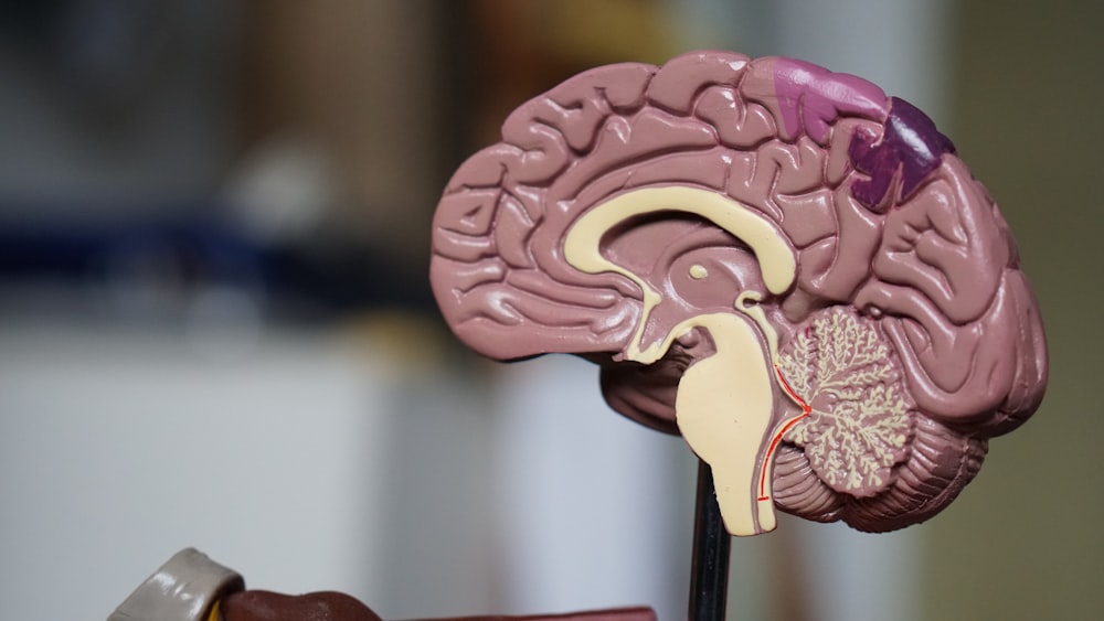
Imagine you could look at a slice of a human brain and not just see its structure, but also map out thousands of different molecules—the fats, the sugars, the proteins, the drugs—precisely where they are located. It would be like having a Google Maps for the molecular world, revealing the intricate chemical landscapes that define health, disease, and life itself. This is not science fiction; it is the power of Mass Spectrometry Imaging (MSI).
For centuries, scientists have had to grind up tissue to analyze its chemistry, losing all information about where those molecules came from. MSI shatters this limitation, allowing us to see the "where" with stunning clarity.
This technology is transforming our understanding of everything from cancer biology to drug development, painting a picture of life in vibrant molecular detail.
How Does It Work? From Snapshots to Maps
At its core, Mass Spectrometry Imaging combines the spatial detail of microscopy with the chemical identification power of a mass spectrometer. The process turns a tissue sample into a detailed chemical map.
Preparation
A thin slice of tissue—from a leaf, a tumor, or even a dinosaur bone—is mounted on a slide.
Ionization
This is the crucial imaging step. A focused beam (like a laser or ion stream) is rastered across the sample, spot by spot, row by row. At each spot, it desorbs and ionizes the molecules, effectively turning them into a charged gas. Different techniques exist for this, such as MALDI (Matrix-Assisted Laser Desorption/Ionization), which is particularly gentle and good for larger molecules like proteins and lipids .
Analysis
The cloud of ionized molecules from each spot is sucked into the mass spectrometer. This instrument acts as a molecular weighing scale, measuring the mass-to-charge ratio of every ion .
Mapping
A computer records the precise location of the laser spot and the mass spectrum from that spot. After scanning the entire sample, the software can then generate an image for any specific molecule. Want to see where a certain lipid is? The software plots the intensity of its specific mass as a pixelated image, creating a map where brightness corresponds to abundance.
In essence, MSI builds a molecular picture, one pixel at a time.
A Closer Look: Mapping a Drug in a Tumor
To understand the power of MSI, let's walk through a hypothetical but representative experiment crucial to modern cancer drug development.
Objective
To determine if an experimental anti-cancer drug is effectively reaching its target—the core of a solid tumor—and to understand how it affects the tumor's metabolism.
Methodology: A Step-by-Step Guide
Laboratory mice with a specific type of human colon cancer tumor are used. One group is treated with the experimental drug, while a control group receives a placebo.
After a set time, the mice are euthanized, and the tumors are rapidly removed and frozen in a way that preserves their chemical integrity.
The frozen tumor is sliced into extremely thin sections using a cryostat. Adjacent sections are prepared for histological staining and MSI analysis.
A chemical "matrix" is uniformly sprayed onto the MSI sample slide to assist in the ionization process during MALDI-MSI .
Results and Analysis
The results are a series of images that tell a compelling story.
By selecting the mass of the experimental drug, we can generate an image showing exactly where it is located within the tumor. The analysis might reveal that the drug is concentrated in the well-vascularized outer regions of the tumor but fails to penetrate the oxygen-deprived, necrotic core .
By also mapping the natural molecules of the tumor, we can see the drug's effect. For instance, we can image specific phospholipids that are building blocks of cell membranes. A comparison might show a dramatic decrease in certain lipids, confirming the drug is disrupting cancer growth mechanisms .
Experimental Data Visualization
The following tables and visualizations represent data from our hypothetical MSI experiment on tumor tissue.
Key Molecules Identified in the Tumor Tissue
This table lists some of the crucial molecules detected and their hypothesized role in the experiment.
| Molecule Detected | Mass (m/z) | Role / Significance |
|---|---|---|
| Experimental Drug X | 455.2 | The administered anti-cancer compound. Distribution is the primary target of the study. |
| Phosphatidylcholine (PC 34:1) | 798.5 | A common phospholipid; high levels indicate active cell membrane synthesis in proliferating cells. |
| Sphingomyelin (d18:1/16:0) | 725.6 | Another key membrane lipid. Changes in its distribution can signal cell stress or death. |
| Adenosine Triphosphate (ATP) | 507.0 | The primary energy currency of the cell. Its localization can indicate regions of high metabolic activity. |
Relative Abundance of Key Lipids in Tumor Regions
This data quantifies the drug's effect on the tumor's chemistry, comparing a treated tumor to an untreated one.
| Lipid Species | % Change |
|---|---|
| Phosphatidylcholine (PC 34:1) | -66% |
| Sphingomyelin (d18:1/16:0) | +41% |
Drug Penetration Metrics
This visualization summarizes the spatial distribution data of the drug itself.
| Metric | Value | Interpretation |
|---|---|---|
| Avg. Drug Intensity (Viable) | 8,500 | The drug is effectively reaching the active cancer cells. |
| Avg. Drug Intensity (Necrotic) | 750 | The drug is largely absent from the tumor's core. |
| Penetration Ratio | 11.3 | Highlights a significant penetration issue. |
The Scientist's Toolkit: Key Reagents for MSI
Conducting a successful MSI experiment, especially using the common MALDI technique, requires a set of specialized tools and reagents.
| Tool / Reagent | Function in MSI |
|---|---|
| MALDI Matrix (e.g., DHB, CHCA) | A critical chemical applied to the sample. It absorbs laser energy, causing controlled desorption and ionization of molecules from the tissue surface without completely destroying them . |
| Organic Solvents (e.g., Acetonitrile, Ethanol) | Used for rapid tissue freezing and for washing slides to remove salts and other contaminants that can interfere with the mass spectrometry analysis. |
| Indium Tin Oxide (ITO) Coated Slides | Special microscope slides with a conductive coating. They are essential for certain MSI techniques as they allow for the necessary electrical contact during the ionization process. |
| Trypsin (Protease) | An enzyme sprayed onto tissue sections to digest proteins into smaller peptides. This allows for "bottom-up" proteomics imaging, identifying a wider range of proteins . |
| Calibration Standards | A mixture of molecules with known masses. They are spotted alongside the sample to calibrate the mass spectrometer, ensuring that the mass measurements for the unknown tissue molecules are accurate. |
The Future is Mapped
Mass Spectrometry Imaging has moved from a niche technique to a cornerstone of spatial biology. It is helping surgeons identify the exact margins of a tumor, allowing pharmacologists to watch a drug travel through an organ, and enabling botanists to see how plants distribute defensive compounds against pests .
Medical Diagnostics
Enabling precise tumor margin detection and personalized medicine approaches.
Drug Development
Visualizing drug distribution and metabolism within tissues for better therapeutics.
Plant Science
Mapping defense compounds and metabolic pathways in agricultural research.
The Age of Chemical Cartography
As the technology advances, becoming faster and able to detect even more molecules with higher resolution, the maps we create will only get richer and more detailed. We are no longer just looking at life; we are reading its complex chemical story, one pixel at a time. The age of chemical cartography has just begun.