SPR Running Buffer Mastery: A Complete Guide to Preparation, Degassing, and Troubleshooting for Flawless Data
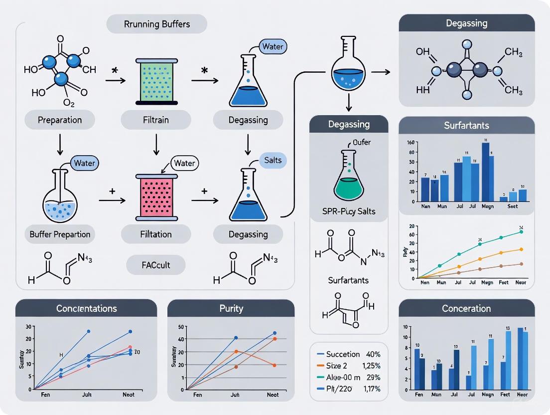
This comprehensive guide details the critical role of Surface Plasmon Resonance (SPR) running buffer in obtaining reliable, high-quality data.
SPR Running Buffer Mastery: A Complete Guide to Preparation, Degassing, and Troubleshooting for Flawless Data
Abstract
This comprehensive guide details the critical role of Surface Plasmon Resonance (SPR) running buffer in obtaining reliable, high-quality data. Tailored for researchers and drug development professionals, it covers foundational principles from buffer selection and component matching to advanced methodological protocols for degassing and vesicle preparation. The article provides a systematic troubleshooting framework for common issues like bulk shifts and air bubbles, and explores validation techniques and emerging technologies. By synthesizing established best practices with cutting-edge insights, this resource serves as an essential manual for optimizing SPR assays in biomedical and clinical research.
The Critical Role of SPR Running Buffer: Principles and Consequences
In Surface Plasmon Resonance (SPR) biosensing, the running buffer is not merely a carrier solution; it is a critical component of the experimental environment that directly influences every aspect of data quality. For researchers and drug development professionals, maintaining the integrity of biomolecular interactions—whether for kinetic characterization, affinity measurements, or off-target screening—demands uncompromising rigor in buffer preparation [1]. The running buffer provides the chemical matrix in which molecular recognition occurs, and its quality dictates the signal-to-noise ratio, baseline stability, and ultimately, the reliability of calculated kinetic parameters (ka, kd, KD) [2] [3]. This application note details the direct mechanistic links between buffer quality and data integrity, providing validated protocols to safeguard your SPR research outcomes.
The Direct Link: How Buffer Quality Dictates Data Integrity
The quality of SPR running buffer impacts data integrity through three primary mechanisms: the introduction of air bubbles, particulate contamination, and suboptimal chemical conditions.
Degassing: Preventing Air-Induced Artifacts
Improperly degassed buffers lead to the formation of air bubbles within the microfluidic path. These bubbles cause significant baseline spikes and drifts by altering the refractive index at the sensor surface in an uncontrolled manner [4]. This artifact directly obscures the true binding signal, making accurate quantification of binding events difficult or impossible. Furthermore, bubbles can disrupt laminar flow, leading to inconsistent analyte delivery and introducing variability in binding kinetics.
Filtration: Eliminating Particulate Contamination
Unfiltered buffers contain microscopic particulates that can clog the microfluidic channels [4]. This clogging manifests as a gradual increase in system pressure and unstable baseline. More critically, particulates can adhere to the sensor surface, creating sites for non-specific binding and permanently damaging the sensitive gold film. This not only compromises the current experiment but can also necessitate extensive system cleaning.
Preparation: Ensuring Chemical Consistency
The use of buffers that are not freshly prepared can lead to chemical degradation and microbial growth. This alters the pH and ionic strength of the solution, which in turn affects the activity and stability of immobilized ligands and injected analytes [4] [3]. Chemical inconsistency is a major source of poor reproducibility between experimental runs. Matching the buffer composition exactly between the running buffer and the sample buffer (after dilution) is also critical to prevent bulk shift effects, a common source of injection artifacts [4].
Table 1: Consequences of Poor Buffer Quality on SPR Data
| Buffer Defect | Direct Impact on System | Manifestation in Sensorgram | Effect on Data Integrity |
|---|---|---|---|
| Inadequate Degassing | Air bubble formation in microfluidics | Sharp spikes, irreversible baseline drift | Obscured binding response, inaccurate Rmax calculation |
| Incomplete Filtration | Particulate clogging of channels | Gradual baseline rise, increased noise | Poor signal-to-noise ratio, potential for surface damage |
| Non-Fresh Buffer | Chemical degradation, microbial growth | Baseline drift, altered binding kinetics | Poor reproducibility, inaccurate ka and kd values |
| Mismatched Sample/Running Buffer | Differences in refractive index | Bulk effect shifts at injection start/end | Inaccurate determination of binding onset and dissociation |
Essential Reagents and Materials
The following toolkit is essential for preparing high-quality SPR running buffers and maintaining system integrity.
Table 2: Research Reagent Solutions for SPR Buffer Preparation
| Reagent/Material | Function/Application | Key Specifications |
|---|---|---|
| Buffer Salts (e.g., PBS, HEPES) | Provides stable pH and ionic strength | Molecular biology grade, low UV absorbance |
| Detergent (e.g., Tween-20, P20) | Reduces non-specific binding to surfaces and tubing [4] [3] | Low fluorescence, sterile filtered |
| BSA (Bovine Serum Albumin) | Blocking agent to prevent non-specific binding to tubing and microfluidics [4] | Protease-free, low immunoglobulin content |
| DMSO (Dimethyl sulfoxide) | Increases solubility of small molecule analytes; must be matched in running and sample buffers [4] | High-purity, anhydrous |
| 0.2 µm Filter | Removes particulates to prevent microfluidic clogging [4] | Low protein binding, sterile |
| SDS (Sodium Dodecyl Sulfate) | Primary component of Desorb solution for deep system cleaning [5] [4] | High purity (≥99%) |
| Sodium Hypochlorite | Primary component of Sanitize solution (10% bleach) to remove biological contaminants [5] [4] | Fresh dilution from stock |
| Glycine-NaOH | Secondary component of Desorb solution (50 mM, pH 9.5) [5] [4] | High purity, pH-adjusted |
Validated Experimental Protocols
Standard Operating Procedure: Running Buffer Preparation
This protocol is adapted from established SPR maintenance guides and sample preparation tips [5] [4].
Principle: To consistently produce a high-quality, particle-free, and gas-free running buffer for SPR experiments to ensure stable baselines and reproducible binding data.
Materials:
- Buffer salts (e.g., for PBS or HEPES)
- Ultrapure water (18 MΩ·cm)
- Vacuum degassing apparatus or sonicator
- 0.2 µm vacuum filtration unit
- Optional additives: Tween-20 (0.005-0.05%), BSA (0.1 mg/mL), DMSO (1-5%)
Method:
- Solution Preparation: Dissolve buffer salts in ultrapure water to the desired concentration. Adjust the pH meticulously at the temperature the experiment will be performed (typically room temperature).
- Additive Introduction: If using detergents (e.g., Tween-20) or DMSO, add them after degassing and filter sterilizing the primary buffer solution. Pour gently to prevent reintroduction of gas [4].
- Filtration: Filter the buffer through a 0.2 µm filter into a clean, dedicated buffer bottle. This removes particulate matter that can clog microfluidics.
- Degassing: Degas the filtered buffer using a vacuum degasser (approximately 4 torr for 30 minutes) or by sonication under vacuum [4] [6]. Critical Step: Degassing immediately before use is most effective.
- Storage: Use the buffer immediately. Rule of thumb: Make fresh running buffer every day [4]. Do not store degassed buffers for extended periods.
Protocol: System Cleaning and Maintenance
This protocol is critical for data integrity after instrument idle time or when baseline drift is observed [5] [4].
Principle: To remove accumulated contaminants from the microfluidic system using a series of cleaning solutions, restoring a stable baseline and preventing carryover.
Materials:
- Desorb Solution 1: 0.5% (w/v) SDS in pure water [5] [4]
- Desorb Solution 2: 50 mM Glycine-NaOH, pH 9.5 [5] [4]
- Sanitize Solution: 10% bleach (0.5% sodium hypochlorite) [5] [4]
- ddH2O
- Blank or "Maintenance" sensor chip
Method:
- Chip Docking: Dock a blank or dedicated "Maintenance" sensor chip to avoid damaging an active experimental chip [5].
- Desorb Procedure:
- Place all active buffer and sample wash tubing into the Desorb 1 solution.
- Run the automated Desorb command. The system will typically wash with Desorb 1 for ~41 minutes, followed by Desorb 2 and ddH2O (total time ~2 hours) [4].
- Sanitize Procedure:
- Place all active tubing into the Sanitize solution.
- Run the automated Sanitize command. The system will wash with Sanitize, followed by ddH2O and running buffer (total time ~2 hours) [4].
- Equilibration: After cleaning, prime the system with freshly prepared, degassed running buffer and allow it to equilibrate on a continuous flow until a stable baseline is achieved.
Data Validation and Quality Control
Quantitative Benchmarks for Buffer-Derived Artifacts
Establishing expected performance benchmarks allows for the objective assessment of buffer-related problems.
Table 3: Quantitative Benchmarks for System Performance Related to Buffer Quality
| Performance Parameter | Acceptable Range | Indication of Buffer Problem |
|---|---|---|
| Baseline Noise Level | < 0.1-0.5 RU (instrument dependent) | High frequency noise > 1 RU suggests particulates or bubbles |
| Baseline Drift | < 5 RU/hour over 10 minutes | Consistent drift > 10 RU/hour suggests contamination or outgassing |
| Bulk Shift Magnitude | Minimal; should be fully reversible | Large, irreversible shifts indicate buffer mismatch |
| Chi² Value in Fitting | Square root of Chi² similar to instrument noise | High Chi² can indicate buffer-induced instability [2] |
| Rmax Consistency | < 5% decrease over multiple cycles | Progressive loss suggests harsh regeneration or surface fouling [2] |
Visual Inspection and Curve Validation
Always inspect sensorgrams and residuals for tell-tale signs of buffer issues [2].
- Check Residuals: After fitting a binding model, the residual plot (difference between fitted curve and actual data) should be randomly scattered around zero. Systematic deviations in the residuals can indicate a bulk effect or other buffer-related artifact that the model could not account for [2].
- Assess Dissociation: The dissociation phase should be monitored long enough to see at least 5% dissociation from the starting value. For very slow off-rates (kd < 1x10⁻⁵ s-1), this may require up to 90 minutes of dissociation time [2].
Workflow: Buffer Quality Impact on SPR Data
The following diagram illustrates the cascade of effects that poor buffer quality has on an SPR experiment, ultimately leading to compromised data integrity.
In SPR biosensing, there is no separation between sample integrity and buffer integrity. For researchers in drug development, where decisions are made based on nanomolar differences in affinity or subtle kinetic profiles, the quality of the running buffer is a primary determinant of success. As demonstrated, failures in buffer preparation—whether in degassing, filtration, or freshness—directly introduce noise, drift, and artifacts that corrupt the very data the instrument is designed to measure. By adhering to the rigorous protocols and validation checks outlined in this application note, scientists can ensure that their SPR data reflects biological truth rather than experimental artifact, thereby de-risking the critical path from discovery to development.
In Surface Plasmon Resonance (SPR) technology, the running buffer is a critical component that forms the hydrodynamic and biochemical environment for biomolecular interaction analysis. The choice of buffer directly influences the stability of the baseline, the specificity of binding events, and the overall quality of kinetic data. This application note details the core formulations of HBS-EP and HEPES-KCl buffers, along with essential additives, providing researchers with standardized protocols for reproducible SPR experimentation. Proper buffer preparation is fundamental to maintaining near-physiological conditions while minimizing non-specific binding and refractive index artifacts, thereby ensuring the accuracy of affinity and kinetic measurements [7] [8] [9].
Core Buffer Formulations and Composition
The selection of an appropriate running buffer is paramount for successful SPR experiments. The buffer must provide stable pH, maintain ionic strength, and include specific additives to reduce non-specific interactions. The following table summarizes the key components and their functions in two commonly used SPR running buffers.
Table 1: Core Components of Common SPR Running Buffers
| Component | Function | HBS-EP+ Buffer (10X) [7] | HEPES-KCl Buffer [8] |
|---|---|---|---|
| HEPES | Buffering capacity, pH stability | 100 mM | 10 mM |
| Sodium Chloride (NaCl) | Maintains ionic strength & osmolarity | 1.5 M | - |
| Potassium Chloride (KCl) | Maintains ionic strength & osmolarity | - | 150 mM |
| EDTA | Chelates divalent metal ions | 30 mM | - |
| Surfactant (P20/Tween-20) | Reduces non-specific binding | 0.50% (v/v) | - |
| Final pH | Optimal for biomolecular interactions | 7.4 ± 0.2 | 7.4 |
HBS-EP+ Buffer is a specialized formulation for SPR protocols. HEPES provides effective buffering in the physiological pH range, while 1.5 M NaCl (when diluted to 1X) creates a near-physiological salt environment. The inclusion of EDTA is crucial for chelating divalent cations that could promote non-specific aggregation or unwanted enzymatic activity. The surfactant P20, a proprietary formulation equivalent to Tween-20, is vital for coating fluidic paths and sensor surfaces to minimize nonspecific binding of analytes [7] [9]. This buffer is commercially available as a concentrated solution (e.g., 10X or 20X), requiring only dilution with sterile, ultrapure water to achieve the 1X working concentration [7] [9].
HEPES-KCl Buffer offers a simpler, detergent-free alternative, which is particularly advantageous when studying membrane proteins or lipid vesicles, as detergents can destabilize these structures [8]. Its lower ionic strength (150 mM KCl) may be preferable for studying electrostatic interactions. The absence of surfactant makes the baseline more susceptible to drift and non-specific binding, requiring exceptionally clean samples and surfaces. This buffer is typically prepared fresh in the laboratory from stock solutions.
Detailed Experimental Protocols
Buffer Preparation and Degassing
Proper preparation of running buffer is a critical step that directly impacts baseline stability and data quality.
Materials:
- Ultrapure water (18 MΩ resistivity at 25°C)
- HEPES, Sodium Chloride, EDTA Disodium Salt, Tween-20 (for HBS-EP+)
- HEPES, Potassium Chloride (for HEPES-KCl)
- 0.22 µm vacuum filtration unit
- Buffer degassing station or vacuum chamber
- Sterile storage bottles
Procedure for HBS-EP+ (1X Working Solution):
- Dilution: Aseptically dilute the 10X HBS-EP+ concentrate with ultrapure water to a 1X final concentration. For example, add 100 mL of 10X stock to 900 mL of water to make 1 L of 1X buffer [7].
- Homogenization: Invert the buffer bottle at least 8 times to ensure thorough mixing. Inadequate mixing can create concentration gradients, leading to a constantly drifting baseline due to refractive index mismatches [10].
- Degassing: Degas the buffer for approximately 30 minutes using a vacuum degassing system or by stirring under a mild vacuum. Degassing removes dissolved air that can form bubbles within the microfluidics, causing sharp "air-spikes" in the sensorgram [11] [4].
- Storage: Store the degassed buffer in a clean, sealed container at room temperature. Buffers stored at 4°C contain more dissolved air, which will outgas inside the warm SPR instrument, creating spikes [4].
Procedure for HEPES-KCl Buffer:
- Weighing and Dissolution: Weigh 2.38 g HEPES (10 mM final) and 11.18 g KCl (150 mM final). Dissolve in approximately 800 mL of ultrapure water.
- pH Adjustment: Adjust the pH to 7.4 using NaOH or KOH.
- Final Volume: Bring the final volume to 1 L with ultrapure water.
- Filtration and Degassing: Sterilize the buffer by filtering through a 0.22 µm filter, then degas as described above [8].
Note: If a detergent like Tween-20 is required for a HEPES-KCl buffer (typically at 0.005-0.05%), it should be added after the degassing step to prevent excessive foam formation [4].
System Equilibration and Baseline Stabilization
A stable baseline is a prerequisite for collecting high-quality binding data. The following workflow outlines the key steps to achieve system equilibrium.
Detailed Steps:
- System Priming: After preparing a fresh running buffer, prime the SPR system according to the manufacturer's instructions. This replaces the old buffer in the pumps and tubing, preventing buffer mixing that causes "waviness" in the baseline with each pump stroke [11].
- Chip Docking and Equilibration: Dock the appropriate sensor chip at least 12 hours prior to running the experiment. Flow running buffer continuously at the experimental flow rate. This prolonged equilibration hydrates the dextran matrix of the sensor chip and washes out preservatives, significantly reducing baseline drift [5] [11].
- Start-up Cycles: Incorporate at least three to five start-up cycles at the beginning of your experimental method. These cycles should mimic your sample injections but use running buffer instead of analyte. If a regeneration step is used, include it in these cycles. This "primes" the surface and stabilizes the system. These cycles should be excluded from the final analysis [11] [4].
- Baseline Assessment: Monitor the baseline response until it stabilizes. A stable baseline should have minimal drift and a low noise level (< 1 Resonance Unit (RU) is ideal) [11].
Troubleshooting Common Buffer-Related Issues
Even with careful preparation, buffer-related issues can arise. The following table outlines common problems, their likely causes, and recommended solutions.
Table 2: Troubleshooting Guide for Buffer-Related Issues in SPR
| Problem | Potential Cause | Solution |
|---|---|---|
| High Baseline Drift | Sensor surface not fully equilibrated; Buffer not freshly prepared; Buffer concentration gradient. | Equilibrate chip with running buffer for >12 hours [11]; Prepare fresh buffer daily [11] [4]; Invert buffer bottle 8+ times before degassing to ensure homogeneity [10]. |
| Air Spikes in Sensorgram | Dissolved air in buffer; Buffer stored at 4°C. | Ensure thorough degassing of buffer; Use buffer at room temperature [11] [4]. |
| High Noise Level | Contaminated buffers or fluidics; Inadequate filtration. | Prepare fresh, filtered (0.22 µm) buffer daily [11] [4]; Perform system cleaning (Desorb, Sanitize) [5] [4]. |
| Bulk Refractive Index Shifts | Mismatch between running buffer and sample buffer; Poorly mixed running buffer. | Dissolve/dilute analyte in the running buffer [8]; Ensure running buffer is thoroughly homogenized [10]. |
| Non-Specific Binding | Lack of surfactant in buffer; "Sticky" analyte. | Add detergent to running buffer (e.g., 0.005-0.05% Tween-20) after degassing [4]; Use a reference surface and apply double referencing during analysis [11]. |
The Scientist's Toolkit: Essential Research Reagent Solutions
A successful SPR experiment relies on more than just the running buffer. The following table details key reagents and materials essential for preparing and executing a robust SPR study.
Table 3: Essential Reagents and Materials for SPR Experiments
| Item | Function/Description | Example Application |
|---|---|---|
| L1 Sensor Chip | Sensor chip with lipophilic anchors for capturing lipid membranes. | Essential for studying protein-lipid interactions [8]. |
| CM5 Sensor Chip | Carboxymethylated dextran chip for covalent coupling. | General purpose chip for amine coupling of proteins, antibodies [12]. |
| Desorb Solutions | System cleaning solutions (0.5% SDS, 50 mM glycine pH 9.5). | Routine maintenance to remove contaminants from fluidics [5] [4]. |
| Sanitize Solution | Cleaning solution (0.5-10% sodium hypochlorite). | Sanitizes fluidics to remove biological contaminants [5] [4]. |
| Regeneration Solutions | Solutions to remove bound analyte without damaging ligand. | Acid (e.g., Glycine pH 2-3), base (e.g., 10-50 mM NaOH), detergents (0.01-0.5% SDS) [4]. |
| DMSO (Dimethyl Sulfoxide) | Solvent for small molecule analytes. | Increases compound solubility; match concentration in sample and running buffer (e.g., 1-5%) [4]. |
| BSA (Bovine Serum Albumin) | Additive to reduce non-specific binding. | Prevents non-specific binding to tubing and microfluidics (e.g., 0.1 mg/mL) [4]. |
The meticulous preparation of SPR running buffers is a foundational element of reproducible and high-quality biomolecular interaction data. Adherence to the detailed protocols for HBS-EP+ and HEPES-KCl buffer preparation, degassing, and system equilibration outlined in this document will minimize baseline drift, noise, and non-specific binding. Furthermore, integrating the recommended troubleshooting strategies and essential reagents into your SPR workflow will enhance experimental robustness. By standardizing these critical pre-experimental steps, researchers can ensure the reliability of their kinetic and affinity measurements, thereby strengthening the overall conclusions of their research.
Understanding Bulk Refractive Index Shifts and Their Impact on Sensorgrams
Surface Plasmon Resonance (SPR) is a powerful, label-free technology for investigating biomolecular interactions in real-time. The output of an SPR experiment is a sensorgram, a plot of the response (in Resonance Units, RU) against time, which provides a visual representation of binding events [13]. A critical, yet often confounding, factor in obtaining high-quality sensorgram data is the bulk refractive index (RI). A bulk RI shift is a change in the refractive index at the sensor surface that is not caused by a specific binding event between the ligand and analyte. Instead, it arises from a difference in composition between the running buffer and the sample solution [14]. This article, framed within a broader thesis on SPR running buffer preparation and degassing, details the sources and consequences of bulk effects and provides validated protocols for their mitigation.
The Principle of Bulk Refractive Index Shifts
The SPR signal is exquisitely sensitive to changes in the refractive index at the sensor surface. This principle is harnessed to measure the increase in mass from a binding event. However, any change in the solution's composition that alters its RI will also produce a signal. The analyte sample, often stored in a different buffer or containing stabilizing agents, will have a different RI than the running buffer flowing through the instrument.
When this sample is injected, the instrument detects the RI difference as a massive, instantaneous jump in the response—a bulk shift [14]. This shift can obscure the initial association phase of the binding event and complicate data analysis. The core of the problem is the mismatch between the running buffer and the analyte buffer [14]. Even small differences, such as variations in salt concentration or the presence of organic solvents, can cause significant responses. For example, a 1 mM difference in NaCl concentration can result in a bulk signal of approximately 10 RU [14].
Identifying and Troubleshooting Bulk Effects in Sensorgrams
Recognizing bulk shifts is the first step in troubleshooting. The table below summarizes common sensorgram artifacts and their primary causes.
Table 1: Common Sensorgram Artifacts and Their Identification
| Sensorgram Artifact | Description | Probable Cause |
|---|---|---|
| Bulk Shift Jumps | A sharp, vertical rise at the start of injection and a sharp drop at the end. The association and dissociation curves may appear normal but are offset. | Buffer mismatch between running buffer and analyte sample (e.g., differences in salt, DMSO, or glycerol concentration) [14]. |
| Spikes after Reference Subtraction | Sharp spikes only at the very beginning (1-4 seconds) and end of the injection after reference subtraction. | Slight "out-of-phase" arrival of the sample to the active and reference flow cells due to their serial arrangement, exacerbated by large bulk effects [14]. |
| Carry-over | A sudden buffer jump or spike at the beginning of an analyte injection. | Residual salt or viscous solution from a previous injection contaminating the next run [14]. |
| Air Spikes/Bubbles | Sudden, sharp spikes in the signal during an injection. | Formation of small air bubbles in the flow channels, often exacerbated by poorly degassed buffers or low flow rates [14]. |
The following diagram illustrates the logical workflow for diagnosing and resolving bulk RI issues based on observed sensorgram features.
Quantitative Impact of Common Solvents and Salts
Understanding the magnitude of the signal caused by common solutes is critical for experimental design. The following table provides a summary of the quantitative effects of typical solution components, illustrating why meticulous buffer matching is non-negotiable.
Table 2: Quantitative Impact of Common Solution Components on SPR Response
| Solution Component | Typical Context | Impact on SPR Signal & RI | Recommended Mitigation Strategy |
|---|---|---|---|
| DMSO | Solvent for small molecule compounds. | High refractive index causes large buffer jumps. Even small concentration differences (e.g., 0.5%) create significant RU shifts [14]. | Dialyze the analyte against the running buffer supplemented with the same DMSO concentration. Use this dialysate as the running and dilution buffer [14]. |
| Glycerol | Protein storage additive. | High refractive index, leading to bulk shifts. | Remove via dialysis or buffer exchange into the running buffer [14]. |
| NaCl (Salts) | Varying ionic strength in sample vs. running buffer. | ~10 RU signal per 1 mM NaCl concentration difference [14]. | Ensure precise buffer matching. Use the running buffer for the final sample dilution. |
| Evaporation | Sample storage in vials. | Increases analyte concentration and alters buffer composition, leading to jumps. | Always cap sample vials [14]. |
Experimental Protocols for Mitigating Bulk Effects
Protocol: Buffer Matching via Dialysis or Desalting
This protocol is the primary method for eliminating bulk shifts caused by differences in small molecule solutes and salts between the sample and running buffer.
- Preparation of Dialysis Buffer: Prepare a sufficient volume (e.g., 1 L) of the running buffer that will be used in the SPR experiment. If the analyte requires a component like DMSO for stability, add it to the running buffer at the exact final concentration that will be present in the sample.
- Sample Dialysis:
- Place the analyte sample in a dialysis tube or cassette with an appropriate molecular weight cutoff.
- Submerge the sealed dialysis unit in a large volume (e.g., 500x the sample volume) of the prepared running buffer.
- Dialyze at the recommended temperature (typically 4°C for proteins) with gentle stirring for several hours or overnight.
- Change the dialysis buffer at least once and continue dialysis for another few hours.
- Alternative: Buffer Exchange via Size Exclusion Chromatography:
- For smaller volumes, use a desalting or buffer exchange column (e.g., PD-10, Zeba Spin Columns) pre-equilibrated with the running buffer.
- Load the sample and elute according to the manufacturer's instructions, collecting the protein fraction in the new buffer.
- Final Buffer Usage: The buffer used in the final dialysis step or for column equilibration must be used as the running buffer in the SPR instrument and for any further dilution of the dialyzed analyte [14].
Protocol: Injection System and Buffer Integrity Test
This quality control procedure tests the overall health of the fluidics system and the quality of the buffer matching before running valuable samples.
- Chip and System Equilibration: Install a plain gold or dextran-coated sensor chip. Equilibrate the system with the degassed running buffer until a stable baseline is achieved.
- Preparation of Test Solutions: Create a solution with 50 mM extra NaCl dissolved in the running buffer. Then, prepare a dilution series of this solution in the running buffer (e.g., 50, 25, 12.5, 6.3, 3.1, 1.6, 0.8, 0 mM extra NaCl).
- Injection and Monitoring:
- Inject the solutions from low to high concentration (single cycle kinetics), ending with an injection of running buffer alone.
- Closely monitor the sensorgrams. The rise and fall of the curve should be smooth and immediate. The steady-state phase should be flat, without drift.
- The highest NaCl concentration will yield a signal of over 550 RU, confirming the ~10 RU/mM NaCl effect [14].
- Interpretation: The final running buffer injection checks for carry-over. A smooth, stepwise response confirms good system performance and well-matched buffers.
The Scientist's Toolkit: Essential Research Reagents and Materials
Table 3: Key Reagents and Materials for SPR Running Buffer Management
| Item | Function in Bulk RI Management |
|---|---|
| Degassing Apparatus | Removes dissolved air from buffers to prevent the formation of air bubbles, which cause spikes in the sensorgram [14]. |
| 0.22 µm Membrane Filters | Filters buffers to remove particulate matter that could clog the microfluidic system and introduces a source of contamination. |
| Dialysis Tubing/Cassettes | Allows for the equilibration of the analyte sample with the running buffer, eliminating differences in salt and small molecule composition [14]. |
| Size Exclusion Desalting Columns | Provides a rapid method for exchanging the buffer of small-volume analyte samples into the running buffer. |
| SPR Sensor Chips (e.g., CM5, C1, SA) | The sensor surface. Using a reference surface (a channel with no ligand immobilized) is crucial for subtracting residual bulk effects [14] [15]. |
Advanced Considerations and Buffer Preparation Best Practices
For a thesis focused on buffer preparation, it is critical to adhere to rigorous buffer hygiene. Fresh buffers should be prepared daily, 0.22 µm filtered, and degassed before use [14]. It is bad practice to top up old buffer with new, as this can introduce contaminants or biological growth.
Furthermore, the excluded volume effect can cause artifacts. Differences in ligand density between the active and reference surfaces can lead to them responding differently to changes in ionic strength or organic solvent, as they have different displaced volumes [14]. This can be diagnosed by injecting a control solution with the same RI as the analyte but no binding capability.
Modern SPR systems also offer advanced features like real-time bulk compensation (e.g., BioNavis's PureKinetics), which actively measures the bulk RI of the solution, allowing for the analysis of samples containing DMSO without requiring it in the running buffer [14]. The buffer itself is a key experimental variable; as studies on other optical biosensors like Whispering Gallery Modes (WGM) have shown, even minuscule changes in buffer RI can significantly impact the sensor's wavelength shift and apparent sensitivity [16].
Within surface plasmon resonance (SPR) research, the preparation of running buffer is a foundational step that directly influences data integrity and experimental success. A meticulously prepared buffer is paramount for maintaining stable baselines, minimizing non-specific binding, and achieving accurate kinetic measurements. The process of buffer degassing serves as a critical control point to prevent the formation of air bubbles, a common source of significant experimental artifact [11] [17]. In SPR systems, air bubbles introduce abrupt signal spikes, cause baseline instability and drift, and can potentially damage the sensitive hydrodynamic flow system or the functionalized sensor surface [11] [18]. These disruptions compromise the detection of true biomolecular interactions, leading to unreliable data and wasted resources.
The necessity of degassing is rooted in the physics of SPR instrumentation. SPR functions as a highly sensitive refractometer, detecting minute changes in the refractive index at the sensor surface [19] [20]. Air bubbles possess a refractive index vastly different from aqueous buffers, and their passage through the flow cell creates massive, anomalous signals that obscure legitimate binding events [20]. Furthermore, bubbles can become trapped in microfluidic channels, creating persistent baseline drift by altering flow dynamics and pressure [18]. Therefore, proper degassing is not merely a recommended best practice but an essential, non-negotiable step in any rigorous SPR running buffer preparation protocol designed to ensure the highest data quality.
Key Principles: How Bubbles Disrupt SPR Assays
Consequences of Air Bubbles in Microfluidic Systems
The integration of microfluidics with biosensors, while enabling automated and precise fluid handling, also increases susceptibility to disruptions from gaseous microbubbles [18]. The consequences of bubble formation are severe and multifaceted, directly impacting key performance metrics as shown in the table below.
Table 1: Impact of Air Bubbles on SPR System Performance
| Performance Metric | Impact of Air Bubbles |
|---|---|
| Baseline Stability | Induces significant drift and instability as bubbles traverse the flow cell or become lodged, altering local refractive index [11] [17]. |
| Signal Integrity | Causes sharp, unpredictable spikes in the sensorgram that can mask legitimate binding signals or be misinterpreted as binding events [11]. |
| Assay Replicability | Introduces random, uncontrolled variables, leading to high intra- and inter-assay variability and poor reproducibility [18]. |
| Sensor Surface Integrity | Can displace or damage the immobilized ligand layer upon contact, degrading surface binding activity and necessitating chip replacement [18]. |
| Operational Yield | A major cause of assay failure, requiring aborted runs and repetition, which wastes valuable samples, reagents, and time [18]. |
The Underlying Signaling Pathway of Bubble-Induced Artifacts
The disruptive effect of an air bubble follows a defined pathway from its formation to the final data artifact. The following diagram visualizes this cascade, which underpins the necessity of rigorous degassing protocols.
Diagram 1: Cascade of bubble-induced SPR artifacts.
Experimental Protocols: Standardized Degassing Procedures
Protocol 1: In-Laboratory Vacuum Degassing
This protocol describes a standardized method for preparing and degassing SPR running buffer using a vacuum filtration system, a common setup in most life science laboratories.
Principle: Applying a vacuum to the buffer solution reduces the partial pressure of dissolved gases, lowering their solubility and causing them to effervesce out of the solution, after which they are removed from the system.
Materials:
- Buffer Components: High-purity water and analytical grade reagents.
- Vacuum Source: Laboratory vacuum line or vacuum pump.
- Filtration Apparatus: Vacuum flask and a bottle-top vacuum filter with a 0.22 µm membrane [11].
- Storage Vessel: Clean, sterile glass bottle.
Step-by-Step Method:
- Solution Preparation: Completely dissolve all buffer constituents in high-purity water. Stir gently to minimize vortexing and shearing.
- Filtration and Degassing: Assemble the bottle-top vacuum filter (0.22 µm pore size) onto a clean vacuum flask. Pour the buffer into the filter unit, apply a vacuum for 10-15 minutes, or until vigorous effervescence ceases. Filtration simultaneously removes particulate matter and dissolved gases [11].
- Storage: Transfer the degassed buffer from the vacuum flask into a clean, sterile storage bottle. Seal the bottle to minimize reabsorption of atmospheric gases.
- Pre-Use Equilibration: Before starting the SPR experiment, bring the required volume of degassed buffer to the instrument's operating temperature. It is considered bad practice to add fresh buffer to old buffer remaining in the system, as this can introduce contaminants [11].
Protocol 2: On-Instrument Dynamic Degassing
Many modern SPR instruments are equipped with integrated, in-line degassers. This protocol outlines their use and verification.
Principle: These systems typically use gas-permeable membranes or create turbulent flow under negative pressure to strip dissolved gases from the buffer immediately before it enters the microfluidic cartridge.
Materials:
- SPR instrument with integrated dynamic degasser.
- Buffer prepared according to Protocol 1.
Step-by-Step Method:
- System Prime: Prime the entire fluidic system of the SPR instrument with the buffer. The priming process itself often routes the buffer through the internal degasser.
- Baseline Monitoring: After priming, flow the running buffer over the sensor surface and monitor the baseline response. A stable baseline with minimal noise indicates successful degassing and system equilibration [11].
- Start-Up Cycles: Incorporate at least three start-up cycles into the experimental method where buffer is injected instead of analyte. This helps to "prime" the surface and fluidics, stabilizing the system before actual data collection begins [11].
The Scientist's Toolkit: Essential Reagents and Materials
Successful degassing and buffer preparation rely on specific laboratory reagents and equipment. The table below details these essential items and their functions.
Table 2: Key Research Reagent Solutions for SPR Buffer Preparation
| Item | Function & Importance |
|---|---|
| 0.22 µm Membrane Filter | Critical for removing particulate matter that could nucleate bubble formation and clog microfluidic channels [11]. |
| Vacuum Pump / Degassing Unit | Provides the vacuum force required to remove dissolved gases from the buffer solution [19] [21]. |
| High-Purity Water | The solvent base; minimizes ionic and organic contaminants that contribute to background noise and nonspecific binding. |
| Non-ionic Surfactant (e.g., Tween-20) | Added to the buffer after degassing to reduce surface tension and wet the microfluidic channels and sensor surface, further inhibiting bubble formation [11] [22]. |
| Clean, Sterile Storage Bottles | Prevents bacterial growth and chemical contamination of the prepared buffer, which can cause drift and noise [11]. |
Advanced Strategies and Integrated Bubble Mitigation
For systems persistently plagued by bubbles, a multi-faceted approach beyond simple degassing is required. Research by Puumala et al. demonstrates that effective bubble mitigation is achieved by combining microfluidic device degassing, plasma treatment, and microchannel pre-wetting with a surfactant solution [18]. This combined strategy addresses the problem at multiple points: removing gases from the fluid, modifying the channel surface to be more hydrophilic, and reducing the fluid's surface tension.
The following workflow diagram integrates degassing into a comprehensive buffer preparation and system equilibration protocol, highlighting its role within a broader context.
Diagram 2: Integrated workflow for SPR buffer prep and equilibration.
Troubleshooting Guide: Resolving Persistent Baseline Issues
Despite degassing, baseline problems can occur. This guide helps diagnose and address common issues.
Table 3: Troubleshooting Baseline Drift and Bubble-Related Issues
| Observation | Potential Cause | Corrective Action |
|---|---|---|
| Gradual, continuous baseline drift | Buffer not fully equilibrated with sensor surface; insufficient degassing; buffer contamination [11] [17]. | Flow running buffer for longer to equilibrate (e.g., overnight for new surfaces). Re-degas buffer. Prepare fresh buffer in a clean container [11]. |
| Sudden, large spikes in sensorgram | Air bubbles entering the flow cell, often from a depleted buffer source or leak in the fluidic path [17]. | Check buffer supply for low volume. Inspect tubing and connections for leaks. Ensure degasser is functioning correctly. |
| High-frequency baseline noise | Electrical interference; mechanical vibrations; or very small, persistent microbubbles [17]. | Relocate instrument to minimize vibrations. Ensure proper grounding. Increase surfactant concentration slightly (e.g., 0.01% Tween-20) to suppress micro-bubbles. |
| Drift after buffer change | Inadequate priming after changing to a new buffer, leading to mixing of buffers with different refractive indices [11]. | Prime the system thoroughly after each buffer change. Allow sufficient time for the baseline to stabilize before starting injections. |
| Wavy or oscillating baseline | Mismatch between buffer temperature and instrument control, or a malfunctioning degasser. | Allow more time for the buffer to reach set temperature. Verify and service the instrument's degasser and temperature control unit. |
In Surface Plasmon Resonance (SPR) analysis, the principle of buffer matching is a critical precept for ensuring data accuracy and reliability. Buffer matching refers to the practice of minimizing differences in composition between the analyte sample and the running buffer, a process essential for reducing optical artifacts and obtaining true binding signals. Bulk shift, or solvent effect, occurs when a difference exists between the refractive index (RI) of the analyte solution and the running buffer [22]. This phenomenon creates a distinctive 'square' shape in sensorgrams due to large, rapid response changes at the start and end of analyte injection, potentially obscuring genuine binding events, particularly those with small responses or rapid kinetics [22]. This application note details systematic strategies and protocols for effective buffer matching within the broader context of SPR running buffer preparation, providing researchers with actionable methodologies to enhance data quality.
Understanding Bulk Shift Effects
Fundamental Principles and Consequences
Bulk shift effects originate from the core operating principle of SPR technology, which detects changes in the refractive index (RI) at the sensor surface [23]. While SPR instruments are exquisitely sensitive to binding-induced RI changes, they cannot intrinsically distinguish these from RI changes caused by variations in buffer composition between the sample and running buffer. Even minor differences in the concentration of salts, solvents, or other additives can generate significant bulk RI responses [22].
The primary consequence of uncompensated bulk shift is the distortion of binding sensorgrams, which complicates the differentiation of small binding-induced responses and other interactions with rapid kinetics from a high refractive index background [22]. Although reference subtraction can partially compensate for these effects, the correction may not always adequately account for the bulk effect, particularly with modern high-sensitivity SPR systems [22]. Therefore, preventive strategies through careful buffer matching are consistently more effective than post-acquisition correction.
Problematic Buffer Components
Certain buffer components are particularly prone to causing bulk shift effects, even when they are necessary for analyte or ligand stability. The table below summarizes common problematic components and recommended mitigation strategies:
Table 1: Common Buffer Components Causing Bulk Shift and Recommended Solutions
| Component | Typical Concentration Range | Primary Function | Bulk Shift Risk | Recommended Mitigation Strategy |
|---|---|---|---|---|
| DMSO | >1% | Solubilizing small molecules | High | Use the lowest possible concentration; ensure exact match between analyte and running buffer; employ calibration if necessary [22] [24] |
| Glycerol/Sucrose | 1-10% | Protein stabilization, cryoprotection | Moderate to High | Avoid unless absolutely necessary; use minimal required concentration with exact buffer matching [22] |
| High Salt Concentrations | >250 mM | Maintaining ionic strength | Moderate | Precisely match salt concentrations; use reference surface subtraction [22] [24] |
| Detergents | 0.01-0.1% | Reducing non-specific binding | Low to Moderate | Use consistent brand and concentration; Tween-20 is recommended at 0.01-0.1% [22] [24] |
Experimental Protocols for Buffer Matching
Standard Buffer Matching Procedure
This protocol establishes a baseline methodology for preparing matched running buffer and analyte solutions for SPR experiments.
3.1.1 Materials and Reagents
- HEPES Buffered Saline (HBS-PE): 10 mM HEPES pH 7.4, 150 mM NaCl, 3.4 mM EDTA, 0.01% surfactant P20 [24]
- Tris Buffered Saline (TBS-P): 50 mM TRIS-HCl pH 7.4, 150 mM NaCl, 0.01% P20 [24]
- Phosphate Buffered Saline (PBS-P): 10.1 mM Na₂PO₄, 1.8 mM KH₂PO₄, 137 mM NaCl, 2.7 mM KCl, pH 7.4, 0.01% P20 [24]
- Ultrapure water (18.2 MΩ·cm)
- Buffer additives as required (e.g., BSA, CaCl₂, CM-dextran)
- Analytical grade salts and pH adjustment solutions
3.1.2 Equipment
- pH meter with temperature compensation
- Vacuum filtration apparatus (0.22 µm membrane)
- Buffer degassing system (in-line or offline)
- Analytical balance (0.1 mg sensitivity)
- Volumetric flasks and pipettes
3.1.3 Procedure
- Prepare Running Buffer Stock Solution: Prepare a 2-5 liter stock of the selected running buffer (HBS-PE, TBS-P, or PBS-P) using ultrapure water. Precisely weigh all components to ensure consistency.
- Adjust pH and Osmolality: Adjust the buffer to the required pH at the temperature the experiment will be conducted. Verify osmolality if necessary.
- Filter and Degas: Filter the entire buffer volume through a 0.22 µm membrane to remove particulates [24]. Degas the buffer thoroughly to prevent micro-bubble formation in the fluidic system [5] [24].
- Prepare Analyte Dilutions: Using the freshly prepared and degassed running buffer, prepare all analyte dilution series. Perform serial dilutions to minimize cumulative pipetting errors [22].
- Equilibrate System: Before starting experiments, equilibrate the SPR system with running buffer until a stable baseline is achieved (typically 30-60 minutes).
3.1.4 Critical Steps Notes
- Always use the same buffer batch for running buffer and analyte dilution to minimize lot-to-lot variation.
- Avoid cooling degassed buffer as this can cause gas re-uptake and bubble formation [24].
- For multi-day experiments, store running buffer appropriately to prevent contamination or evaporation.
Specialized Buffer Matching with Additives
Some experimental systems require specific additives to maintain biomolecule stability or function. This protocol addresses matching strategies for these challenging systems.
3.2.1 DMSO-Containing Buffers
- Prepare Base Running Buffer: Prepare the standard running buffer without DMSO.
- Add DMSO to Running Buffer: Add the required percentage of DMSO directly to the running buffer. For small molecule studies, typically use 1-2% DMSO, never exceeding 5%.
- Calibrate System: Perform a calibration run if significant solvent effects are anticipated [24].
- Prepare Analyte Solutions: Dissolve analytes in the same DMSO-containing running buffer, not pure DMSO.
3.2.2 Stabilizing Additives (BSA, Carrier Proteins)
- Identify Minimal Effective Concentration: Determine the lowest concentration of additive (e.g., BSA) that prevents non-specific binding and surface adsorption.
- Add to Running Buffer Only: Incorporate the additive (typically 0.1% BSA) into the running buffer to minimize analyte adsorption to system vials and tubing [24].
- Exclude from Analyte Buffer: Do not add these blocking agents to analyte samples to prevent competition with binding interactions.
3.2.3 Ion-Dependent Interactions
- Identify Essential Ions: Determine which ions (e.g., Ca²⁺, Zn²⁺, Mg²⁺) are required for the interaction [24].
- Prepare Stock Solutions: Create concentrated stock solutions of these ions in ultrapure water.
- Add to Both Buffers: Precisely add the same concentration of ions to both running buffer and analyte dilution buffer.
The following workflow diagram illustrates the decision process for buffer matching strategies:
The Scientist's Toolkit: Essential Reagents and Materials
Successful buffer matching requires specific reagents and materials selected for their purity, consistency, and compatibility with SPR systems. The following table details the essential components of a buffer matching toolkit:
Table 2: Essential Research Reagent Solutions for SPR Buffer Matching
| Reagent/Material | Specifications | Function in Buffer Matching | Usage Notes |
|---|---|---|---|
| High-Purity Buffers | Molecular biology grade HEPES, TRIS, or phosphate salts | Provides consistent ionic background and pH control | Prefer single large batch to ensure consistency [24] |
| Surfactant P20 | 0.01% in final buffer (10% stock solution) | Reduces non-specific binding to fluidics and surfaces | Use consistent brand; concentration affects baseline [24] |
| Ultrapure Water | 18.2 MΩ·cm resistance, <5 ppb TOC | Prevents contamination and particulate introduction | Always use for all buffer preparations [5] |
| DMSO | Spectrophotometric grade, low UV absorbance | Solvent for small molecule analytes | Match concentration exactly between buffer and sample [24] |
| BSA | Protease-free, low immunoglobulin content | Blocks non-specific binding in running buffer | Add only to running buffer, not analyte samples [24] |
| Filter Membranes | 0.22 µm pore size, low protein binding | Removes particulates that could clog microfluidics | Always filter before degassing [5] [24] |
| Concentrated Salt Solutions | Analytical grade NaCl, KCl, etc. | Adjusts ionic strength to match physiological conditions | Precisely match concentrations in all solutions |
Quality Assessment and Validation
Bulk Shift Detection and Analysis
Validating successful buffer matching requires analytical methods to detect and quantify bulk shift effects. The primary assessment occurs during preliminary runs without immobilized ligand or using a reference surface.
5.1.1 Sensorgram Analysis
- Characteristic Signature: Look for the tell-tale 'square' shape in sensorgrams with large, rapid response changes at injection start and end points [22].
- Response Magnitude: Quantify the bulk response magnitude in Resonance Units (RU). While reference subtraction can correct for bulk effects, significant shifts (>10 RU) may indicate problematic mismatch.
- Kinetic Artifacts: Examine whether the bulk effect obscures the initial association or dissociation phases of binding events.
5.1.2 Buffer Scouting Approach For challenging systems with unavoidable additives, implement a systematic buffer scouting protocol:
- Run a dilution series of analyte in perfectly matched buffers (positive control).
- Introduce deliberate, known mismatches in specific components (e.g., ±1% DMSO, ±50 mM salt).
- Quantify the resulting bulk shift responses to establish acceptable tolerance ranges.
The following workflow illustrates the quality assessment process for buffer matching:
Documentation and Reporting Standards
Comprehensive documentation of buffer preparation is essential for experimental reproducibility and troubleshooting. The following elements should be recorded:
5.2.1 Buffer Composition Documentation
- Exact identities and sources of all buffer components
- Final concentrations of all salts, buffers, and additives
- pH measurement temperature and exact values
- Batch numbers and preparation dates
5.2.2 Preparation Parameters
- Filtration method and membrane specifications
- Degassing method and duration
- Storage conditions and duration before use
Effective buffer matching through meticulous preparation of running buffers and analyte solutions represents a foundational element of robust SPR experimentation. By systematically addressing potential refractive index mismatches at their source, researchers can significantly reduce bulk shift artifacts, thereby enhancing data quality and reliability. The protocols and strategies outlined in this application note provide a standardized approach to buffer matching that aligns with the broader objectives of SPR running buffer preparation research. Implementation of these methodologies will enable researchers to distinguish true molecular interactions from solvent-based artifacts, ultimately leading to more accurate kinetic and affinity measurements in drug development and basic research applications.
Step-by-Step Protocols: From Buffer Preparation to Liposome Handling
Within Surface Plasmon Resonance (SPR) research, the quality of the running buffer is a fundamental determinant of data reliability and instrument integrity. This Standard Operating Procedure (SOP) outlines the protocols for the preparation, filtration, and degassing of running buffers for SPR instrumentation. Consistent adherence to this procedure is critical for maintaining a stable baseline, preventing the introduction of air bubbles and particulate matter into the microfluidics, and ensuring the reproducibility of biomolecular interaction analyses [5] [1]. This document is framed within a broader thesis investigating the optimization of SPR running buffers to enhance the accuracy of kinetic and affinity measurements in drug development.
The Scientist's Toolkit: Essential Materials
The following table details the essential reagents and materials required for the successful execution of this protocol.
Table 1: Essential Materials and Reagents for SPR Buffer Preparation
| Item | Specification/Function |
|---|---|
| Buffer Salts | High-purity grade (e.g., USP, ACS) for consistent ionic strength and pH. |
| Ultrapure Water | Type I (18.2 MΩ·cm at 25°C) to minimize contaminant interference. |
| 0.22 µm Filters | Sterilization and removal of particulates and microorganisms [25] [26]. |
| Filter Membrane | PES: Recommended for aqueous buffers due to low protein-binding [25] [26]. |
| Vacuum Pump / Syringe | For driving the filtration process. |
| Degassing Unit | In-line degasser or sonication bath to remove dissolved gases [5]. |
| pH Meter | Calibrated instrument for accurate pH adjustment. |
| Sterile Storage Bottles | Chemical-resistant vessels (e.g., glass, PP) for storing prepared buffer. |
Quantitative Filter Selection Data
Selecting the appropriate 0.22 µm filter is crucial for both sterility and chemical compatibility. The following tables provide a quantitative guide for selection based on sample type and membrane properties.
Table 2: Filter Membrane Selection Guide by Chemical Compatibility
| Membrane Material | Hydrophilicity | Protein Binding | Strong Acid/Base | Organic Solvents | Recommended Use |
|---|---|---|---|---|---|
| Polyethersulfone (PES) | Hydrophilic | Low | Poor | Moderate | Aqueous SPR running buffers, cell culture media [25] [26]. |
| Polyvinylidene Fluoride (PVDF) | Hydrophilic | Medium | Moderate | Good | General purpose filtration, biopharma applications [26]. |
| Nylon | Hydrophilic | Medium-High | Moderate | Poor | Water samples, polar solvents (not for sensitive LC-MS) [26]. |
| Polytetrafluoroethylene (PTFE) | Hydrophobic | Very Low | Excellent | Excellent | Organic solvents, aggressive chemicals [25] [26]. |
Table 3: Performance Characteristics for Different Applications
| Application | Recommended Membrane | Key Rationale |
|---|---|---|
| Standard Aqueous SPR Buffer | PES | Low protein-binding preserves analyte integrity; high flow rate [26]. |
| Buffers with Organic Solvents | PTFE | Superior chemical resistance and compatibility [25] [26]. |
| Routine Filtration (aqueous) | PVDF | Good balance of flow rate and chemical resistance [26]. |
Detailed Experimental Protocol
Buffer Preparation
- Weighing: Using an analytical balance, weigh the required mass of high-purity buffer salts into a clean, sterile container.
- Reconstitution: Add the correct volume of Type I ultrapure water to achieve the desired buffer concentration. Stir vigorously until all salts are completely dissolved.
- pH Adjustment: Calibrate the pH meter with standard buffers. Adjust the pH of the solution to the target value (e.g., 7.4 for PBS) using concentrated acid (e.g., HCl) or base (e.g., NaOH). Note that the pH may shift slightly after filtration and degassing.
Filtration Protocol
The following workflow details the steps for sterilizing and clarifying the buffer solution using a 0.22 µm filter.
4.2.1 Pre-wetting (for hydrophilic membranes): Pre-wet the PES filter membrane by passing a small volume (e.g., 5-10 mL) of ultrapure water through it. This eliminates air bubbles within the membrane matrix and ensures a consistent, high flow rate during the main filtration process [25] [27].
4.2.2 Filtration Assembly and Execution: Aseptically assemble the filtration apparatus, connecting the vacuum pump to the filter flask. Pour the prepared buffer into the filter funnel. Apply a gentle vacuum to drive the solution through the membrane. Avoid using excessive pressure, as this can compromise the integrity of the membrane. The resulting filtrate is now sterile and ready for degassing.
Buffer Degassing
Dissolved gases in the running buffer are a primary cause of air bubble formation in SPR microfluidics, leading to significant signal noise and data artifacts.
4.3.1 Sonication Method:
- Transfer the filtered buffer into a glass bottle, leaving sufficient headspace.
- Place the bottle in a sonication bath and sonicate for 15-30 minutes. Gentle stirring or agitation during sonication enhances gas removal.
4.3.2 In-line Degassing (Preferred):
- For instruments equipped with an in-line degasser, this is the recommended method.
- It provides continuous and on-demand degassing immediately before the buffer enters the fluidic system, which is the most effective approach for preventing bubble formation [28].
Quality Control and Storage
- Inspection: Visually inspect the final buffer for any cloudiness or particulate matter. The solution should be clear.
- pH Verification: Re-check and record the pH of the degassed buffer.
- Storage: Store the prepared buffer in a tightly sealed, sterile container at room temperature or as required by the specific buffer formulation. It is recommended that freshly prepared buffer be used within a short period (e.g., 24-48 hours) to prevent microbial growth or chemical degradation.
Integration with SPR Instrument Maintenance
Proper buffer preparation is intrinsically linked to the long-term health of the SPR instrument. The use of freshly prepared, degassed, and detergent-free running buffer is explicitly recommended as part of routine instrument maintenance to prevent the accumulation of contaminants within the fluidic path [5]. Furthermore, a clean fluidic system supplied with high-quality buffer is a prerequisite for successful pre-concentration screening and other sensitive immobilization techniques that optimize the sensor surface for ligand binding studies [29].
Troubleshooting Guide
Table 4: Common Issues and Solutions
| Problem | Potential Cause | Solution |
|---|---|---|
| Low Flow Rate / Clogging | High particulate load in initial buffer. | Pre-filter the solution through a 0.45 µm or 1.0 µm filter before using the 0.22 µm filter [25] [27]. |
| Air Bubbles in Fluidics | Inadequate degassing. | Extend sonication time, ensure in-line degasser is functioning, or let buffer equilibrate to room temperature before use. |
| High Baseline Noise | Contaminated buffer or dirty fluidics. | Prepare fresh buffer, ensure all equipment is clean, and run instrument desorb and sanitize procedures [5]. |
| pH Drift | Buffer instability or CO₂ absorption. | Prepare buffer fresh, use tight-sealing containers, and consider the chemical stability of the buffer chosen. |
Within Surface Plasmon Resonance (SPR) biosensing, the quality of running buffer is a foundational element dictating data integrity. SPR detects minute changes in refractive index at a sensor surface, making it highly susceptible to artifacts caused by air bubbles or particulate matter in the buffer [14] [20]. Proper buffer preparation, specifically filtration and degassing, is therefore not a mere preliminary step but a critical experimental variable that directly influences baseline stability, signal-to-noise ratio, and the reliability of derived kinetic parameters [5] [14]. This application note provides detailed protocols and best practices for buffer degassing, framed within a broader research context on optimizing SPR running buffer preparation to enhance research reproducibility.
The Critical Role of Degassing in SPR
Air bubbles represent a primary failure mode in SPR experiments. Small air bubbles entrapped in buffer can form within the microfluidic flow channels of the instrument. At low flow rates (< 10 µL/min), these bubbles are not efficiently flushed out and can grow, causing sudden spikes in the sensorgram that obscure the binding data [14]. Furthermore, buffers stored at 4°C contain more dissolved air, which is released as the buffer warms to experimental temperature, creating a significant risk of bubble formation [11]. The consequences of inadequate degassing include:
- Data Corruption: Spikes and drifts can make kinetic analysis impossible [14].
- Experimental Halt: Severe bubbles can block fluidic paths, requiring manual intervention and cleaning.
- Reduced Reproducibility: Fluctuating baselines introduce variability, compromising the reliability of binding affinities (KD) and rate constants (ka, kd) [11].
Degassing mitigates these risks by reducing the dissolved air content, thereby minimizing the potential for bubble nucleation and growth under the controlled temperature and flow conditions of an SPR assay.
Core Principles and Protocols for Buffer Preparation
A robust buffer preparation routine is the first step toward superior SPR data. The following workflow integrates filtration and degassing into a single, cohesive protocol. Adherence to strict buffer hygiene is paramount; it is considered "bad practice to add fresh buffer to the old" as microbial growth or chemical contamination can occur [14] [11].
The following diagram outlines the logical sequence and decision points in the SPR running buffer preparation workflow:
Comprehensive Degassing Techniques
Vacuum Degassing
Vacuum degassing is a common laboratory method where a buffer solution is placed under a vacuum, which lowers the partial pressure of dissolved gases, encouraging them to come out of solution.
Procedure:
- Pour the filtered buffer into a clean, heat-resistant glass flask. Do not fill more than halfway to allow for vigorous bubbling.
- Place the flask on a magnetic stirrer and add a clean stir bar.
- Seal the flask with a stopper connected to a vacuum line. Incorporate a cold trap between the flask and the vacuum source to prevent buffer vapor from entering the pump.
- Begin stirring at a moderate rate. Gradually apply vacuum until the buffer begins to bubble vigorously as gases evolve.
- Maintain this vacuum for 20-30 minutes. The process is complete when the evolution of fine bubbles substantially decreases.
- Slowly release the vacuum to avoid violent bubbling that can re-introduce air.
Optimal Duration: While the exact duration can depend on buffer volume and composition, a period of 20-30 minutes is generally effective for standard aqueous buffers like PBS or HBS-EP [14] [30]. Continuously monitor the solution; extended degassing beyond this point typically offers diminishing returns.
In-line Degassing and Helium Sparging
Many modern SPR systems, such as the Octet SF3, feature in-line degassers that actively remove gases from the buffer immediately before it enters the fluidic system, preventing air bubble formation during operation [28]. This is highly effective for the running buffer but does not degas the sample.
For systems without in-line degassing or for samples, helium (He) sparging is an excellent alternative. Helium has very low solubility in aqueous solutions, and when bubbled through the buffer, it displaces more soluble gases like nitrogen and oxygen [30].
- Procedure:
- Use a disposable, sterile filter (0.22 µm) on the helium inlet line to maintain sterility.
- Submerge the tip of the helium line in the buffer and set the gas flow to a gentle, continuous stream of bubbles. Avoid a violent flow that could cause foaming, especially if detergents are present.
- Sparge intensively for an initial 5-minute period to remove the bulk of dissolved air [30].
- For storage, reduce the helium flow to a minimal rate to maintain a positive pressure of inert gas over the buffer, or simply maintain a "He blanket" [30].
Buffer Selection and Handling
The composition of the buffer itself influences degassing efficacy and baseline stability.
Table 1: Buffer Handling and Degassing Considerations
| Buffer Type | Key Characteristics | Degassing & Handling Notes | Common SPR Applications |
|---|---|---|---|
| Aqueous Buffers (e.g., PBS, HBS) | Low viscosity, high volatility for volatile additives. | Standard 20-30 min vacuum degassing. Volatile components may evaporate; sparge time may need optimization. | General protein-protein interactions, antibody-antigen binding. |
| DMSO-Containing Buffers | High refractive index, prone to evaporation. | Critical to match running and sample buffer DMSO concentration [14]. Evaporation changes concentration, causing large bulk shifts. | Small molecule screening where solubility requires DMSO. |
| High-Salt Buffers | Can increase solution viscosity. | Ensure complete dissolution before degassing. Risk of salt precipitation if stored cold; store at room temperature [11]. | Studies involving ionic strength dependence, DNA-protein interactions. |
Troubleshooting Common Buffer-Related Issues
Even with careful preparation, issues can arise. The table below outlines common symptoms, their likely causes, and corrective actions.
Table 2: Troubleshooting Guide for Buffer-Related SPR Issues
| Problem Symptom | Potential Cause | Solution |
|---|---|---|
| Frequent spikes in the sensorgram [14] | Air bubbles from inadequately degassed buffer; bubbles growing at low flow rates. | Re-degas buffer thoroughly. Increase flow rate temporarily to flush system. Ensure buffers stored at room temp, not 4°C [11]. |
| Baseline drift after docking chip or buffer change [11] | System not equilibrated; temperature difference; mixing of old and new buffer in lines. | Prime system multiple times after buffer change. Equilibrate a new sensor chip in running buffer for up to 12 hours [5]. |
| Bulk refractive index shift (jump at injection start/end) [14] | Mismatch between running buffer and analyte buffer (e.g., different salt, DMSO, glycerol content). | Dialyze analyte into running buffer. Use size-exclusion columns for buffer exchange. For DMSO, include the same concentration in the running buffer [14]. |
| Carry-over or contamination [14] | Old or contaminated buffer; microbial growth. | Prepare fresh buffers daily. Never top off old buffer with new. Use clean, sterile bottles. |
The Scientist's Toolkit: Essential Reagents and Materials
Table 3: Key Research Reagent Solutions for SPR Buffer Preparation
| Item | Function in SPR Buffer Prep |
|---|---|
| High-Purity Water | The solvent base for all buffers; must be of high purity (e.g., 18 MΩ·cm) to minimize contaminants. |
| Buffer Salts (e.g., HEPES, PBS components) | Maintain a stable pH and ionic strength to preserve biomolecule activity and interaction fidelity. |
| Detergent (e.g., Tween-20) | Added after filtration and degassing to reduce nonspecific binding to the sensor chip and fluidics. Pre-filtration addition can cause foam [14] [11]. |
| 0.22 µm Membrane Filter | Removes particulate matter that could clog the instrument's microfluidic channels. |
| Vacuum Pump & Filter Flask | Standard apparatus for vacuum degassing of buffer solutions prior to use. |
| Helium Gas Tank & Sparging Line | Used for helium sparging, an effective method for degassing and maintaining an inert atmosphere over the buffer. |
Meticulous preparation and degassing of running buffers are non-negotiable prerequisites for robust and reproducible SPR data. There is no universal "one-size-fits-all" vacuum duration; rather, 20-30 minutes of vacuum degassing serves as a reliable starting point for most aqueous buffers, while 5 minutes of initial helium sparging is effective for in-process degassing. The most critical factor is consistency—maintaining strict buffer hygiene, precisely matching the composition of running and sample buffers, and systematically implementing the described protocols. Integrating these degassing techniques as a core component of SPR experimental design will significantly minimize artifacts, reduce wasted time and reagents, and enhance the confidence in the derived kinetic and affinity constants that are vital to drug development and basic research.
In Surface Plasmon Resonance (SPR) analysis, the integrity of binding data is profoundly influenced by the chemical composition of the running buffer. Buffer mismatch between the analyte sample and the SPR running buffer can introduce significant refractive index changes, leading to bulk shift effects that obscure genuine binding signals and compromise kinetic measurements [31]. For researchers, scientists, and drug development professionals, achieving perfect buffer matching is not merely a preparatory step but a foundational requirement for generating publication-quality binding affinities and kinetics.
Buffer exchange via desalting columns offers a rapid and efficient method to transfer an analyte into the exact SPR running buffer, eliminating matrix-induced artifacts. This application note details a robust protocol for implementing gel filtration-based desalting columns into SPR sample preparation workflows. The content is framed within a broader research context on optimizing SPR running buffer preparation and degassing, emphasizing practical steps to ensure data accuracy and reliability.
The Critical Role of Buffer Matching in SPR
The principle of SPR detection is based on measuring changes in the refractive index at a sensor surface [8]. When an analyte is dissolved in a buffer that differs in composition from the SPR running buffer—even slightly in salt concentration, pH, or additives like glycerol or DMSO—the difference in refractive index between the two solutions causes a sharp, bulk response upon injection [31] [8]. This response is distinct from a specific binding event and can manifest as a large injection peak or an unstable baseline, complicating or even preventing accurate data interpretation.
- Minimizing Bulk Shift Effects: The primary goal of buffer exchange is to make the analyte's solution matrix (its buffer) identical to the SPR running buffer. This ensures that any change in the SPR signal (Response Units, RU) during analyte injection is due solely to molecular binding at the sensor surface, and not from a difference in the solution's composition [31].
- Preserving Analyte Activity: The use of size-exclusion based desalting columns is a gentle process that maintains the native structure and activity of proteins and other biomolecules, which is crucial for obtaining biologically relevant binding data [32] [33].
- Compatibility with Additives: Many small molecule analytes require DMSO for solubility. It is critical to match the concentration of DMSO in the running buffer and all analyte samples to prevent significant baseline distortions [31]. Desalting columns efficiently remove and exchange buffers while being compatible with a range of biological samples.
Technical Principles of Desalting Columns
Desalting columns, also known as spin desalting columns or gel filtration columns, operate on the principle of size exclusion chromatography (SEC) [33]. The column is packed with porous resin beads, and the separation process is based on molecular size:
- Macromolecules (e.g., proteins, peptides) are too large to enter the pores of the resin. They flow around the beads and elute from the column first, in the void volume.
- Small molecules (e.g., salts, detergents, impurities, old buffer constituents) can enter the pores, which increases their path length and slows their progress through the column. They elute later, separated from the macromolecules [33].
For buffer exchange, the column resin is pre-equilibrated with the desired destination buffer (e.g., the SPR running buffer). As the sample passes through the column, the larger analyte is collected in this new buffer, effectively achieving a rapid and efficient buffer exchange [33].
Workflow Diagram
The following diagram illustrates the logical workflow for integrating desalting columns into the SPR sample preparation process, from initial buffer preparation to the final SPR experiment.
Research Reagent Solutions: Essential Materials
The following table lists key materials and reagents required for successful buffer exchange and subsequent SPR analysis.
Table 1: Essential Research Reagents and Materials for SPR Analyte Preparation
| Item | Function/Description | Example Vendor/Product |
|---|---|---|
| Spin Desalting Columns | Gel filtration columns for rapid buffer exchange and desalting of protein/peptide samples. | AdvanceBio Spin Columns [32], Zeba Spin Desalting Columns [33] |
| SPR Running Buffer | The continuous phase for SPR analysis; must be matched by the analyte buffer. Common buffers include HBS-EP, PBS, or HEPES-KCl [19] [8]. | GE Healthcare (HBS-EP) [19], Laboratory preparation [8] |
| Ultrapure Water | Used for preparing buffer solutions; 18 MΩ resistivity at 25°C is recommended to minimize contaminants [8]. | Milli-Q Advantage A10 System [19] |
| Salts & Reagents | For buffer preparation (e.g., HEPES, NaCl, KCl). | Sigma-Aldrich [19] |
| Detergents | Added to running buffer (e.g., Tween-20) to reduce non-specific binding [19]. | Sigma-Aldrich [19] |
| DMSO | Organic solvent for dissolving small molecule analytes; concentration must be matched in all solutions [31]. | Sigma-Aldrich [19] |
Quantitative Performance Data
The performance of desalting columns is characterized by high protein recovery and efficient removal of small molecules. The following table summarizes typical performance metrics as established in the literature and commercial product data.
Table 2: Performance Metrics of Desalting Columns for Sample Preparation
| Parameter | Typical Performance | Experimental Context |
|---|---|---|
| Protein Recovery | >90% | Recovery of BSA demonstrated using Zeba Spin Desalting Columns across a concentration range of 0.04-1 mg/mL [33]. |
| Sample Volume | 2 µL to 4+ mL | Various commercial column formats are available to process a wide range of sample volumes [33]. |
| Molecular Weight Cutoff (MWCO) | ~7,000 Da | A common MWCO for spin columns, designed to retain proteins while excluding salts and small molecules [33]. |
| Processed Volume | 1.5 - 3.5 mL | Sample volumes of BSA successfully desalted with high recovery [33]. |
| Time Efficiency | Minutes per sample | Spin column procedures are designed for rapid processing, typically involving a short centrifugation step [32] [33]. |
Experimental Protocol: Buffer Exchange for SPR Analytes
This protocol describes the step-by-step procedure for exchanging the buffer of an analyte sample into an SPR running buffer using spin desalting columns.
Pre-Experiment Preparation
- Prepare and Degas Running Buffer: Prepare the SPR running buffer (e.g., 10 mM HEPES, 150 mM NaCl, 0.005% Tween-20, pH 7.4) [19] [8]. Degas the buffer thoroughly using a vacuum pump to prevent air bubble formation in the SPR microfluidic system during the experiment [19].
- Select the Desalting Column: Choose a spin desalting column with a molecular weight cutoff (MWCO) significantly lower than the molecular weight of your analyte to ensure its retention. Ensure the column size is appropriate for your sample volume [33].
Buffer Exchange Procedure
Column Equilibration:
- Resuspend the resin in the column by gently vortexing or tapping the tube.
- Remove the top cap and then the bottom cap of the column.
- Place the column in a clean 2 mL microcentrifuge tube (provided with most kits) and centrifuge at 1,000 × g for 1 minute to remove the storage solution.
- Discard the flow-through. Apply the degassed running buffer (approximately the same volume as the column's bed capacity) to the column.
- Centrifuge again at 1,000 × g for 1 minute and discard the flow-through.
- Repeat this equilibration step a second time. A properly equilibrated column is critical for effective buffer exchange.
Sample Application and Elution:
- Place the equilibrated column into a new, clean collection tube.
- Slowly and carefully apply your analyte sample directly onto the center of the compacted resin bed. Do not disturb the bed. The sample volume should not exceed the column's recommended capacity.
- Centrifuge the column at 1,000 × g for 2 minutes. The eluate collected in the tube contains your analyte in the new SPR running buffer.
- The collected analyte is now ready for SPR analysis. If necessary, the sample concentration can be determined post-exchange using an appropriate method.
Post-Exchange Verification
- Quality Control: Check the pH and conductivity of the eluted sample against the original SPR running buffer to confirm a successful exchange.
- SPR Signal Baseline: Before injecting the analyte, observe the SPR baseline with running buffer flowing. A stable baseline is a good indicator of proper buffer matching.
Integration with SPR Running Buffer Preparation
The use of desalting columns is an integral component of a comprehensive SPR running buffer preparation strategy. The entire workflow, from buffer formulation to analyte preparation, must be executed with precision to ensure the success of the SPR experiment. The diagram below integrates the buffer exchange protocol into the complete SPR experimental workflow.
Complete SPR Experiment Workflow
Concluding Remarks
Proper analyte preparation through buffer exchange is a critical, yet often overlooked, factor in obtaining reliable SPR data. The implementation of a standardized protocol using desalting columns, as outlined in this application note, provides researchers with a reliable method to achieve perfect buffer matching. This practice effectively minimizes bulk refractive index shifts, thereby enhancing the signal-to-noise ratio and the accuracy of kinetic and affinity measurements. Integrating this step into a rigorous SPR buffer preparation and degassing regimen is essential for any high-quality drug development or basic research program utilizing SPR technology.
Within the framework of a broader thesis on Surface Plasmon Resonance (SPR) running buffer preparation and degassing, the critical challenge of analyzing challenging sample types emerges as a significant research focus. SPR is a label-free, quantitative analytical technique that measures biomolecular interactions in real-time by detecting changes in the refractive index near a sensor surface [34]. The accuracy of these measurements is exceptionally sensitive to the composition of the running buffer and sample solutions. The presence of common sample additives—such as DMSO for solubilizing small molecules, glycerol for protein stability, and lipids for membrane protein studies—can introduce substantial refractive index artifacts, mass transport limitations, and non-specific binding if not properly managed [8] [31] [17]. This application note provides detailed, practical protocols for incorporating these challenging samples into SPR experiments, ensuring the generation of reliable, publication-quality binding data.
Guidelines for Specific Sample Types
DMSO (Dimethyl Sulfoxide)
Background and Challenge: DMSO is a ubiquitous solvent for small organic molecules, lipids, and other compounds with low aqueous solubility. However, even small differences in DMSO concentration between the sample and running buffer cause significant shifts in the refractive index, resulting in bulk refractive index effects that can obscure genuine binding signals [31].
Protocols and Best Practices:
- Concentration Matching: The highest concentration of DMSO required to dissolve the analyte should be determined first. The running buffer and all analyte dilutions must contain this same, precise percentage of DMSO to eliminate buffer mismatch [31].
- Sample Preparation Workflow: Prepare a stock solution of the analyte in 100% DMSO at the maximum solubility. Perform serial dilutions of this stock into running buffer that has been pre-mixed to contain the target percentage of DMSO. This ensures all samples and the running buffer have identical DMSO content.
- System Compatibility: Ensure that the SPR instrument's fluidic system is compatible with the planned DMSO concentration, as some materials may be degraded by high levels of organic solvents.
Glycerol
Background and Challenge: Glycerol is commonly added to protein storage buffers (often at 10-50%) to prevent aggregation and stabilize protein structure. Like DMSO, it significantly alters the solution's refractive index. A sudden injection of a glycerol-containing sample into a glycerol-free running buffer creates a large, positive spike in the sensorgram that can mask the initial association phase of binding [8].
Protocols and Best Practices:
- Buffer Matching Strategy: To minimize refractive index changes, the running buffer should ideally be the same as the analyte storage buffer. If the protein is stored in a buffer containing glycerol, the running buffer should also contain the same concentration of glycerol (e.g., 5%) [8].
- Desalting or Dialysis: As an alternative, the analyte can be transferred into a glycerol-free buffer using desalting columns or dialysis before the experiment. This is the preferred method if the running buffer composition is critical for the interaction being studied.
- Control Experiments: Always include a blank injection (zero analyte) of the glycerol-containing buffer to confirm that the refractive index matching is effective and that no significant bulk shift is observed.
Lipids and Liposomes
Background and Challenge: Lipid-protein interactions are a major application of SPR. The primary challenge is creating a stable, homogeneous lipid layer on the sensor chip while avoiding non-specific binding and maintaining protein functionality [8]. Sensor chips with a hydrophobic surface, such as the L1 chip, are designed to capture intact liposomes.
Protocols and Best Practices:
- Liposome Preparation:
- Composition: Start with control vesicles of 100 mol% POPC (phosphatidylcholine) or an 80:20 molar ratio of POPC:POPE (phosphatidylethanolamine). To study specific binding, create a second vesicle type by "spiking" in an anionic lipid of interest (e.g., phosphatidic acid, phosphoinositides) into the control mixture [8] [31].
- Extrusion: After resuspending the dried lipid film in SPR running buffer, extrude the mixture through a filter membrane (e.g., 0.1 µm pore size) 41 times to create large unilamellar vesicles (LUVs) of uniform size [8].
- Sensor Surface Capture: The L1 sensor chip surface is designed to capture lipid vesicles. A typical workflow involves cleaning the surface, capturing the liposomes to form a lipid monolayer, and then using a brief injection of a mild detergent (e.g., 40 mM Octyl-β-D-Glucopyranoside) to wash away excess material and create a stable bilayer [8].
- Non-Specific Binding Control: The use of control vesicles (lacking the specific lipid of interest) is essential to distinguish specific binding from non-specific adsorption of the protein to the lipid surface [8].
The following workflow diagram summarizes the core experimental process for handling these challenging samples in SPR, from buffer preparation to data acquisition.
Essential Experimental Protocols
Running Buffer Preparation and Degassing
The foundation of a stable SPR experiment is a properly prepared and degassed running buffer.
- Buffer Selection: Use a buffer that matches the analyte's storage conditions and maintains biological activity (e.g., HEPES-KCl, PBS, Tris). Avoid detergents if working with lipid vesicles [8].
- Degassing: Always degas buffers immediately before use to prevent the formation of air bubbles within the microfluidic system, which causes significant baseline drift and noise [17].
- Filtration: Sterile-filter the buffer through a 0.22 µm filter to remove particulate matter that could clog the instrument's fluidic channels.
Instrument Cleaning and Maintenance
Regular maintenance is crucial for preventing contamination and signal drift, especially when working with complex samples like lipids.
- Routine Desorb and Sanitize: If the instrument has been idle, run a desorb procedure using solutions like 0.5% SDS followed by 50 mM glycine-NaOH (pH 9.5). This should be followed by a sanitize cycle with a 10% bleach solution to remove any residual biomolecules. Perform these steps with a dedicated "maintenance chip" docked to avoid damaging a functional sensor chip [8].
- System Equilibration: After docking the experimental sensor chip, allow the system to equilibrate with the running buffer for at least 12 hours to achieve a stable baseline [8].
Critical Considerations for Binding Assays
The table below summarizes key parameters to optimize when designing SPR experiments with challenging samples.
Table 1: Key Experimental Parameters for SPR Binding Assays
| Parameter | Consideration | Guideline |
|---|---|---|
| Ligand Immobilization Level | Must be optimized for analyte size; low density minimizes mass transport effects. | For small molecules, higher density may be needed for detectable Rmax [31]. |
| Analyte Concentration | Should span a range above and below the expected KD. | A minimum of 3-5 concentrations in a 3-10-fold dilution series is standard [35]. |
| Flow Rate | Affects mass transport; higher rates reduce depletion at the surface. | Typically 30-50 µL/min; optimize to minimize bulk refractive effects and mass transport [17]. |
| Regeneration | Removes bound analyte without damaging the ligand. | Condition-specific; test mild (2 M NaCl) to harsh (10 mM Glycine pH 2.0) solutions [31]. |
The Scientist's Toolkit: Essential Research Reagents
Successful execution of SPR experiments with challenging samples requires the use of specific, high-quality reagents and materials. The following table details these essential components.
Table 2: Key Research Reagents and Materials for SPR
| Reagent/Material | Function and Application | Specific Example |
|---|---|---|
| L1 Sensor Chip | A sensor chip with a lipophilic surface designed for the capture of lipid vesicles and the study of lipid-protein interactions. | GE Healthcare Sensor Chip L1 [8]. |
| CM5 Sensor Chip | A general-purpose chip with a carboxymethylated dextran matrix for covalent immobilization of ligands via amine coupling. | GE Healthcare CM5 chip [31]. |
| NTA Sensor Chip | A chip coated with nitrilotriacetic acid for capturing His-tagged ligands in a defined orientation. | Ni-NTA chip for His6-Sec18 immobilization [31]. |
| CHAPS Detergent | A zwitterionic detergent used for cleaning sensor chips and in the preparation of lipid surfaces. | 20 mM solution for instrument maintenance [8]. |
| Octyl-β-D-Glucopyranoside | A non-ionic detergent used to create a stable lipid bilayer on the L1 sensor chip surface. | 40 mM solution for washing captured liposomes [8]. |
| Nanodiscs | Soluble, discrete patches of lipid bilayer encircled by a membrane scaffold protein (MSP), useful for studying membrane protein interactions. | MSP1D1 nanodiscs with incorporated PA [31]. |
| Regeneration Solutions | Chemical solutions used to remove strongly bound analyte from the ligand without causing permanent damage. | 2 M NaCl (mild) or 10 mM Glycine, pH 2.0 (acidic) [31]. |
Troubleshooting Common Issues
Even with careful preparation, issues can arise. The following troubleshooting guide addresses common problems linked to challenging samples.
Table 3: Troubleshooting Guide for Common Issues
| Problem | Potential Cause | Recommended Solution |
|---|---|---|
| Baseline Drift/Noise [17] | Improperly degassed buffer; buffer mismatch (DMSO/glycerol). | Degas buffer thoroughly; ensure exact DMSO/glycerol matching between running buffer and samples. |
| No Binding Signal [17] | Low ligand activity; analyte solubility issues; incorrect buffer. | Check ligand functionality; optimize sample buffer with additives to improve solubility. |
| High Non-Specific Binding [17] | Hydrophobic interactions with sensor chip or lipid surface. | Include a control surface; use a suitable blocking agent (e.g., BSA); adjust running buffer ionic strength. |
| Incomplete Regeneration [17] [31] | Analyte binds too strongly for gentle conditions. | Systematically test harsher regeneration solutions (e.g., low pH, high salt, detergent); ensure ligand stability to regeneration. |
| Irreproducible Data [17] | Inconsistent sample handling; ligand surface degradation. | Standardize sample prep; check for analyte precipitation; monitor surface stability and regenerate consistently. |
Large Unilamellar Vesicles (LUVs), with diameters typically ranging from 100 to 1000 nm, represent a crucial class of lipid-based nanoparticles widely employed in nanomedicine and biomedical applications [36]. Their structural composition—an aqueous core enclosed by a single phospholipid bilayer shell—makes them ideal candidates for drug delivery systems, enabling the encapsulation of both hydrophilic and lipophilic active compounds [36]. The preparation of LUVs with defined size, lamellarity, and stability is paramount for obtaining reliable and reproducible results in subsequent analytical assays, including those utilizing Surface Plasmon Resonance (SPR) to study interactions with biological targets [37]. This protocol details the formation and characterization of LUVs, framed within the context of preparing samples for SPR-based analysis, with particular attention to the critical factors that influence vesicle stability and performance in biosensing environments.
Theoretical Background
Structural and Physicochemical Properties of LUVs
Liposomes are classified based on their size and number of bilayers. LUVs are characterized by a single bilayer, distinguishing them from multilamellar vesicles (MLVs) and small unilamellar vesicles (SUVs) [36]. A key parameter governing the formation and stability of lipid bilayers is the critical packing parameter (CPP), which is a function of the lipid's head group area, tail volume, and length [36]. The self-assembly of phospholipids into vesicular structures is a metastable process that depends on the preparation method and the solution conditions, such as ionic strength, pH, and temperature [36].
The lipid composition directly determines the bilayer's fluidity and phase behavior. Phospholipids undergo a phase transition from a gel phase to a liquid-crystalline phase at a characteristic temperature (Tm). For instance, liposomes containing saturated phospholipids like DPPC or DSPC have a higher Tm, resulting in a more rigid and stable bilayer with reduced drug leakage. Conversely, unsaturated phospholipids produce less stable bilayers with higher permeability [36]. This property can be exploited to design temperature-sensitive liposomes that release their cargo near body temperature (37°C) [36].
The Role of LUVs in SPR Biosensing
SPR is a powerful, label-free technique for real-time monitoring of biomolecular interactions [1] [37]. In the context of drug delivery research, SPR has emerged as a versatile tool for studying the interactions between LUVs and biological targets, serum proteins, and components of the tumor extracellular matrix [37]. LUVs can be immobilized on sensor chips via hydrophobic attachment onto commercial lipid-capture surfaces, allowing for the direct analysis of their binding behavior [37]. The successful application of LUVs in SPR assays depends on a well-controlled preparation process to ensure colloidal stability and uniform size distribution, which are critical for minimizing non-specific signals and obtaining high-quality kinetic data [36].
Materials and Reagents
Research Reagent Solutions
The following table details the essential materials required for the preparation and characterization of LUVs.
Table 1: Key Reagents and Materials for LUV Preparation
| Item | Function/Description |
|---|---|
| Phospholipids (e.g., DPPC, DSPC, Egg PC) | Primary building blocks of the lipid bilayer. Choice influences membrane fluidity, permeability, and stability [36]. |
| Running Buffer (e.g., HEPES, PBS) | Aqueous medium for lipid hydration and subsequent vesicle dispersion. Must be degassed and filtered for SPR applications [5]. |
| Organic Solvent (e.g., Chloroform) | Solvent for dissolving lipids to create a thin, uniform film during the initial preparation step. |
| Extrusion Apparatus | Device used to force the lipid suspension through polycarbonate membranes to achieve a uniform size distribution [36]. |
| Polycarbonate Membranes (various pore sizes) | Filters used in extrusion to define the final size of the LUVs (e.g., 100 nm membranes for ~100 nm LUVs) [36]. |
| Degassing System | Prevents introduction of air bubbles into the SPR instrument, which can disrupt measurements and cause baseline instability [38] [39]. |
Methodology
Workflow for LUV Preparation and Analysis
The following diagram illustrates the comprehensive workflow for preparing LUVs and integrating them into an SPR assay.
Detailed Experimental Protocol
Lipid Film Formation and Hydration
- Dissolve Lipids: Dissolve the selected phospholipid(s) in an organic solvent such as chloroform in a glass vial to achieve a desired final lipid concentration (e.g., 10 mg/mL).
- Form Thin Film: Evaporate the solvent under a stream of inert gas (e.g., nitrogen or argon) while gently rotating the vial to form a uniform thin lipid film on the walls of the vial.
- Remove Solvent Traces: Place the vial under vacuum for several hours (or overnight) to ensure complete removal of any residual organic solvent.
- Hydrate with Buffer: Add the degassed running buffer (e.g., PBS or HEPES) to the vial above the lipid transition temperature (Tm). Vigorously agitate the mixture using a vortex mixer for several minutes to hydrate the lipid film and form a heterogeneous suspension of large multilamellar vesicles (MLVs) [36].
Size Reduction and Extrusion to Form LUVs
- Pre-equilibrate Extruder: Assemble the extrusion apparatus with a polycarbonate membrane of the desired pore size (e.g., 100 nm) and pre-equilibrate it with warm buffer.
- Extrude Lipid Suspension: Pass the MLV suspension through the membrane repeatedly (typically 11-21 times) using syringes. The process should be performed at a temperature above the Tm of the lipids to ensure uniformity.
- Collect LUVs: The final extruded suspension will contain a homogeneous population of LUVs with a diameter close to the pore size of the membrane used [36].
LUV Characterization Parameters
A thorough characterization of the prepared LUVs is essential before proceeding to SPR experiments. The key parameters to analyze are summarized in the table below.
Table 2: Key Characterization Parameters for LUVs
| Parameter | Description | Recommended Method(s) |
|---|---|---|
| Size & Polydispersity | Mean hydrodynamic diameter and distribution width. Preferably between 100-150 nm for optimal cell uptake and to avoid immune clearance [36]. | Dynamic Light Scattering (DLS) |
| Surface Charge | Zeta potential, indicating surface charge and colloidal stability. | Electrophoretic Light Scattering |
| Lamellarity | Number of lipid bilayers. The goal is unilamellar structure. | Electron Microscopy, NMR |
| Concentration | Lipid concentration in the final preparation. | Spectrophotometric assays (e.g., phosphate analysis) |
| Phase Behavior | Gel to liquid-crystalline phase transition temperature (Tm). | Differential Scanning Calorimetry (DSC) |
Critical Factors for SPR Integration
The following diagram outlines the key structural and experimental factors that critically influence the success of LUVs in an SPR assay.
- Buffer Preparation and Degassing: All buffers used for LUV preparation and as running buffers in SPR must be freshly prepared, detergent-free, and thoroughly degassed to prevent the introduction of air bubbles into the microfluidic system, which can disrupt measurements and cause baseline instability [5] [38] [39]. Homogenize the buffer by inverting the bottle at least 8 times before degassing to prevent refractive index mismatch [10].
- Sensor Chip Selection: The choice of sensor chip is critical. For studying LUV interactions, hydrophobic capture sensor chips (e.g., L1 chips) are often used as they facilitate the immobilization of intact lipid vesicles via the hydrophobic effect [37]. It is recommended to dock the sensor chip at least 12 hours prior to the experiment for proper equilibration with the running buffer [5].
- Use of a Reference Cell: Incorporating a reference flow cell is essential to compensate for refractive index changes and non-specific binding. The reference surface should closely match the active surface, for example, by using an inactive ligand or a non-related protein like BSA [39].
- System Maintenance: Prior to experiments, especially after the instrument has been unused, perform a cleaning and maintenance procedure using a blank sensor chip to avoid damaging a functional chip. This typically involves running desorb and sanitize protocols as per the instrument manufacturer's guidelines [5].
The preparation of well-defined LUVs is a foundational step for generating reliable data in liposome-based assays, particularly when coupled with sensitive analytical techniques like SPR. By adhering to the detailed protocols outlined herein—emphasizing controlled extrusion, rigorous characterization, and SPR-specific buffer preparation—researchers can consistently produce LUV formulations with the desired structural properties. Attention to critical factors such as lipid composition, size control, and the use of degassed buffers is paramount for ensuring the colloidal stability of the vesicles and the success of subsequent interaction studies. This methodology provides a robust framework for advancing research in drug delivery and biomolecular interactions.
Diagnosing and Solving Common Buffer-Related SPR Problems
In Surface Plasmon Resonance (SPR) analysis, a stable baseline is the fundamental prerequisite for obtaining reliable, publication-quality kinetic data. Baseline instability, manifesting as drift, noise, or sudden jumps, can obscure genuine binding events, lead to inaccurate calculation of kinetic parameters, and compromise experimental reproducibility. Within the broader context of SPR running buffer preparation and degassing research, it is critical to recognize that the buffer is not merely a carrier for the analyte but an integral component of the sensor's microenvironment. This application note provides a systematic framework for diagnosing and resolving common baseline issues, with a particular emphasis on the pivotal role of proper buffer management and experimental design.
Understanding and Diagnosing Baseline Anomalies
Before attempting to correct baseline issues, accurate diagnosis of their root causes is essential. The following table summarizes the characteristic signatures of common baseline problems, their likely causes, and initial diagnostic steps.
Table 1: Diagnostic Guide to Common Baseline Issues
| Issue Type | Characteristic Signature | Common Causes | Initial Diagnostic Steps |
|---|---|---|---|
| Baseline Drift [11] [17] | A continuous, slow change in Resonance Units (RU) over time, either upward or downward. | - Improperly equilibrated sensor chip or buffer.- Temperature fluctuations.- Buffer evaporation or degradation.- Rehydration of a newly docked sensor chip. | - Monitor baseline for 30+ minutes after startup or buffer change.- Check for adequate system equilibration and degassing. |
| Excessive Noise [17] | High-frequency fluctuations or a "fuzzy" appearance of the baseline signal. | - Air bubbles in the fluidic system.- Electrical or environmental noise (vibrations).- Contaminated sensor surface or buffer.- Inadequate buffer degassing. | - Inspect fluidic lines for bubbles.- Ensure instrument is on a stable, vibration-free surface.- Check buffer for clarity and particulates. |
| Bulk Refractive Index Shift [22] | A sharp, square-shaped signal jump at the start and end of an injection that returns to baseline. | - Mismatch between the refractive index of the running buffer and the analyte sample buffer. | - Compare the composition of the running buffer and sample buffer.- Use a reference flow cell for subtraction. |
| Spikes [11] | Sudden, abrupt peaks or dips in the signal. | - Air bubbles passing through the flow cell.- Pressure fluctuations from pump strokes or leaks. | - Observe if spikes correlate with pump strokes.- Check the microfluidic system for leaks or obstructions. |
The logical relationship between a symptom, its cause, and the appropriate corrective action is outlined below.
Protocols for Resolution and Prevention
Protocol: Running Buffer Preparation and Degassing
Proper buffer preparation is the most critical step in preventing baseline issues [11].
- Preparation: Prepare at least 2 liters of buffer using high-purity water and reagents. Fresh buffer should be prepared daily to prevent microbial growth or chemical degradation. Avoid adding fresh buffer to old stock.
- Filtration: Filter the buffer through a 0.22 µm filter to remove particulate matter that could cause clogging or noise.
- Degassing: Degas the filtered buffer thoroughly using an in-line degasser or by stirring under vacuum for approximately 20 minutes. Buffers stored at 4°C must be warmed to room temperature and degassed immediately before use, as cold liquid holds more dissolved air, which can form destructive air-spikes in the sensorgram [11].
- Additive Introduction: If detergents (e.g., Tween 20) or carrier proteins like BSA are required, add them after the degassing step to prevent foam formation [11] [22].
- Storage: Store the prepared, degassed buffer in a clean, sterile bottle at room temperature.
Protocol: System Startup and Equilibration
A well-equilibrated system is key to a stable baseline [11].
- Prime the System: After any buffer change or at daily startup, prime the fluidic system according to the instrument manufacturer's guidelines. This ensures the previous buffer is completely purged from the lines and IFC (Integrated Fluidic Cartridge).
- Flow Buffer: Initiate a continuous flow of running buffer at the experimental flow rate. For a new sensor chip or after an immobilization procedure, the system may require extended equilibration (30 minutes to several hours, sometimes overnight) to level out drift caused by surface rehydration or wash-out of chemicals [11].
- Stabilization Check: Monitor the baseline until the drift rate falls below an acceptable threshold (e.g., < 1 RU/min).
- Start-up Cycles: Incorporate at least three "start-up" or "dummy" cycles into your experimental method. These cycles should mimic the experimental cycle exactly but inject running buffer instead of analyte. This further stabilizes the surface and the fluidics. These cycles should not be used for data analysis [11].
Advanced Technique: Double Referencing
Double referencing is a powerful data processing technique to compensate for residual drift, bulk effects, and channel differences [11] [22].
- Reference Channel Subtraction: First, subtract the signal from a reference flow cell (which should have no specific binding activity) from the signal of the active flow cell. This compensates for the majority of the bulk refractive index shift and systemic drift.
- Blank Injection Subtraction: Second, subtract the average response from several blank injections (injections of running buffer) from the analyte injections. This step compensates for any remaining differences between the reference and active channels and for injection artifacts. Blank cycles should be spaced evenly throughout the experiment.
Advanced Noise Reduction: Algorithmic Approaches
For high-resolution applications like live-cell imaging or trace molecular detection, instrumental noise can be a limiting factor. Recent algorithmic advances demonstrate significant improvements. Researchers have developed a Polarization Pair, Block Matching, and 4D Filtering (PPBM4D) algorithm for phase-sensitive SPR imaging [40] [41].
This algorithm extends the BM3D framework by leveraging inter-polarization correlations in a quad-polarization filter array camera system. It generates virtual measurements for each polarization channel, enabling more effective collaborative filtering. This approach has been shown to achieve a 57% reduction in instrumental noise and a refractive index resolution of 1.51 × 10⁻⁶ RIU, establishing a robust framework for high-resolution SPR applications across a broad dynamic range [41].
The Scientist's Toolkit: Essential Research Reagents
The following table details key reagents and materials crucial for troubleshooting and preventing baseline issues in SPR experiments.
Table 2: Key Research Reagent Solutions for Baseline Stabilization
| Reagent/Material | Function & Application | Key Considerations |
|---|---|---|
| High-Purity Water | Solvent for all running buffers and sample preparations. | Use high-resistivity (e.g., 18.2 MΩ·cm) water to minimize ionic contaminants and background noise. |
| Buffering Salts (HEPES, Acetate) | Maintains pH stability during the experiment. | Choose a buffer with pKa near your desired experimental pH. Ensure adequate buffering capacity. |
| Salts (e.g., NaCl) | Adjusts ionic strength and shields charge-based non-specific interactions [22]. | Concentration must be optimized; too little can cause NSB, too much may precipitate proteins. |
| Non-ionic Detergents (e.g., Tween 20) | Reduces hydrophobic non-specific binding (NSB) to the sensor surface [42] [22]. | Typically used at low concentrations (0.005-0.05% v/v). Add after degassing to prevent foam. |
| Carrier Proteins (e.g., BSA) | Blocks non-specific binding sites on the sensor surface [22]. | Use at ~1% concentration. Add to running buffer during analyte runs only to avoid coating the surface prematurely. |
| Regeneration Solutions | Removes bound analyte between cycles without damaging the ligand [22]. | Common solutions: low pH (Glycine-HCl), high salt, or mild surfactants. Must be empirically optimized for each interaction. |
| 11-MUA (11-mercaptoundecanoic acid) | Forms a self-assembled monolayer (SAM) on gold sensors for covalent coupling [43]. | Provides a carboxyl-terminated surface for activation with EDC/NHS. |
| Protein G | Enables oriented immobilization of antibodies via their Fc region, maximizing antigen-binding site availability and improving sensitivity [43]. | Leads to higher binding affinity and lower limits of detection compared to random covalent immobilization. |
The following workflow integrates these reagents and protocols into a logical, step-by-step procedure for diagnosing and resolving baseline instability.
Within the broader research on Surface Plasmon Resonance (SPR) running buffer preparation and degassing, the elimination of instrumental artifacts is paramount for obtaining high-quality, publication-ready data. Spikes and sudden jumps in sensorgrams are frequent challenges that can obscure true binding kinetics, leading to erroneous interpretation of biomolecular interactions. These artifacts predominantly originate from two key areas: the formation of air bubbles within the microfluidic system and the operational cycles of the instrument's liquid handling pumps [14]. This application note provides a detailed experimental framework for researchers and drug development professionals to identify, mitigate, and troubleshoot these issues, thereby enhancing the reliability of kinetic and affinity measurements in drug discovery and development.
Theoretical Background and Impact on Data Quality
Air bubbles and pump refills introduce noise through distinct physical mechanisms. Air bubbles, which can form if buffers are not properly degassed or due to temperature fluctuations, disrupt the consistent flow of liquid and the optical path of the incident light. This causes sharp, transient spikes in the sensorgram [14]. The refractive index (RI) of air is significantly different from that of aqueous buffers, and its presence in the flow cell scatters light and creates local RI anomalies.
Similarly, modern SPR instruments utilize sophisticated pumping systems to maintain a stable flow rate. When these systems engage in a refill cycle, the flow can momentarily halt or fluctuate. This interruption in flow manifests as a distinctive spike or a small step-change in the baseline [14]. For interactions with fast kinetics, these artifacts can be mistaken for or mask a real binding event, compromising the accuracy of derived rate constants (ka and kd) and the equilibrium dissociation constant (KD). The following diagram illustrates the cause-and-effect relationship of these artifacts and the primary mitigation strategies.
Protocols for Artifact Elimination
Comprehensive Buffer Preparation and Degassing Protocol
The first line of defense against air bubbles is meticulous buffer preparation. The quality of the running buffer directly influences baseline stability.
Materials:
- High-purity water (e.g., 18 MΩ·cm)
- Buffer salts and additives (e.g., HEPES, NaCl, detergents)
- Vacuum filter unit (0.22 µm pore size)
- Source bottle (e.g., 1-2 L, clean and sterile)
- Laboratory vacuum degassing system or sonicator
Methodology:
- Fresh Buffer Preparation: Prepare the running buffer solution on the same day of use. Do not add new buffer to old stock, as microbial growth or chemical changes can exacerbate bubble formation and cause baseline drift [14].
- Filtration: Filter the buffer through a 0.22 µm membrane into a clean bottle. This step removes particulate matter that can act as nucleation sites for bubbles.
- Degassing: Degas the filtered buffer for 20-30 minutes using a vacuum degasser while stirring with a magnetic stir bar. Alternatively, sonicate for 15-20 minutes. This removes dissolved air, significantly reducing the risk of bubble formation in the flow cell, especially at higher operating temperatures (e.g., 37°C) [14].
- Homogenization: Before attaching the buffer to the instrument, invert the sealed bottle at least 8 times to ensure it is thoroughly mixed. A gradient in the buffer bottle can cause a constantly drifting baseline [10].
Systematic Instrument Prime and Flush Protocol
Before beginning an experiment, especially after system storage or buffer changes, a high-flow-rate flush is critical to clear the fluidic path of any residual air.
Materials:
- Freshly prepared and degassed running buffer
- New or freshly cleaned sensor chip
Methodology:
- Prime the instrument's fluidic system with the degassed buffer according to the manufacturer's instructions.
- Install the sensor chip.
- Set the flow rate to a high value (e.g., 100-150 µL/min) and allow the system to flush for at least 10-15 minutes. High flow rates help to drive out small, trapped air bubbles from the microfluidic channels [14] [10].
- Monitor the baseline signal in real-time. A stable, low-drift baseline indicates a well-primed system. Continue flushing until the baseline stabilizes.
Strategic Run Design to Manage Pump Refills
Pump refill spikes are managed through intelligent experimental design and scheduling.
- Methodology:
- Identify Refill Triggers: Consult your instrument's user manual to understand the pump refill volume or time triggers.
- Schedule Wash Steps: Program wash steps or buffer blanks to coincide with anticipated pump refill events. This prevents the refill spike from occurring during a critical analyte injection or dissociation phase [14].
- Place Report Points Carefully: When defining report points for data analysis, ensure they do not overlap with the timing of pump refills or washing sequences to avoid capturing artifact-laden data points [14].
- Utilize Standby Flow: For instruments like the Carterra LSA, if there is a delay between the end of a run and the next steps, enable the "Standby Flow" function. This ensures a continuous, slow flow of buffer over the chip surface, preventing drying and maintaining system stability for up to 72 hours [10].
Troubleshooting and Artifact Analysis
Even with preventative measures, artifacts may occur. The table below summarizes diagnostic features and solutions for common problems.
Table 1: Troubleshooting Guide for Spikes, Jumps, and Drift
| Artifact | Diagnostic Features | Recommended Corrective Actions |
|---|---|---|
| Air Bubbles [14] | Sharp, random, positive-going spikes. May cause a permanent baseline shift if large. | 1. Re-degas running buffer thoroughly.2. Perform a high-flow rate flush (100 µL/min) between cycles.3. Ensure all buffer lines are securely connected and submerged. |
| Pump Refill Spikes [14] | Small, periodic spikes or jumps occurring at regular intervals corresponding to pump cycle. | 1. Reschedule method commands so wash steps align with refills.2. Adjust report point placement to avoid the spike.3. Verify pump is functioning correctly. |
| Bulk Refractive Index Jumps [22] [14] | Large, square-wave-like response at the start and end of injection. | 1. Dialyze analyte into running buffer.2. Use buffer exchange columns for small volumes.3. Match DMSO concentrations exactly if used. |
| Baseline Drift [39] [10] | Continuous, slow rise or fall of the baseline before/after injections. | 1. Prepare fresh, homogenized buffer (invert bottle 8x).2. Ensure temperature is equilibrated.3. Equilibrate system overnight after immobilization if chemicals cause drift. |
| Carry-over Spikes [14] | Spikes at the very beginning of an injection. | Add extra wash steps with running buffer between sample injections. |
The Scientist's Toolkit: Essential Research Reagents and Materials
The following table lists key materials and their specific functions in ensuring a stable, artifact-free SPR experiment.
Table 2: Essential Reagents and Materials for SPR Running Buffer Management
| Item | Function in Protocol | Specific Example or Note |
|---|---|---|
| Vacuum Degasser | Removes dissolved air from running buffer to prevent bubble formation in the flow cell. | Integral to HPLC systems or available as standalone units. Sonication is an alternative. |
| 0.22 µm Filter | Removes particulates that can nucleate bubbles or clog microfluidic channels. | Use sterile, low-protein-binding filters compatible with your buffer. |
| High-Flow Rate System | Flushes small, trapped air bubbles from microchannels. | A flow rate of 100-150 µL/min is often effective [14]. |
| Non-corroding Buffer Bottles | Storage for degassed running buffer. | Clean, sterile bottles prevent contamination and buffer degradation. |
| Tween-20 (or similar) | A non-ionic surfactant added to running buffer to reduce hydrophobic non-specific binding and potentially lower surface tension. | Typical concentration: 0.05% v/v [44]. |
| Bovine Serum Albumin (BSA) | A blocking agent used in sample and running buffers to minimize non-specific binding to the sensor surface and fluidics. | Typical concentration: 0.1-1% w/v [22] [44]. |
Mitigating spikes and jumps from air bubbles and pump refills is achievable through a disciplined approach to buffer management and experimental design. The core principles for high-quality data are the use of fresh, filtered, and thoroughly degassed buffers, an initial high-flow rate system flush, and strategic scheduling of instrument operations to minimize the impact of inherent pump cycles. Adherence to these detailed protocols will significantly reduce experimental noise, thereby increasing confidence in kinetic and affinity data critical for pharmaceutical development and basic research.
Bulk shift, also known as solvent effect, is a common phenomenon in Surface Plasmon Resonance (SPR) biosensing that occurs when there is a difference between the refractive index (RI) of the analyte solution and the running buffer [22]. This discrepancy creates a distinctive 'square' shape in sensorgrams due to large, rapid response changes at both the start and end of analyte injection [22]. While bulk shift does not alter the inherent kinetics of binding partners, it significantly complicates the differentiation of small binding-induced responses and can obscure interactions with rapid kinetics against a high refractive index background [22]. For researchers, scientists, and drug development professionals, accurately identifying and mitigating bulk shift is crucial for generating reliable binding data, particularly when working with compounds dissolved in solvents such as DMSO or glycerol [45] [22].
Within the broader context of SPR running buffer preparation and degassing research, proper buffer management serves as the first line of defense against bulk shift artifacts. The precision required in buffer matching and preparation directly impacts data quality, especially in high-stakes applications like therapeutic antibody characterization and off-target binding screening where transient interactions might otherwise be missed [1].
Understanding and Identifying Bulk Shift
Fundamental Principles and Impact on Data Quality
Bulk shift arises from non-specific changes in the refractive index at the sensor surface that are unrelated to the biomolecular binding event of interest. These artifacts can be positive or negative depending on the direction of the RI difference between the analyte solution and running buffer [22]. The primary challenge lies in distinguishing genuine binding signals, especially for small molecules or interactions with fast kinetics, from the bulk background effect [1] [22]. While reference subtraction can partially compensate for these effects, improper correction may lead to spikes at the start and end of analyte injections or residual artifacts that skew kinetic calculations [45].
Visual Identification in Sensorgrams
The tell-tale signature of bulk shift is an immediate, sharp response jump at the injection start point that maintains a relatively constant level throughout the injection phase, followed by an equally abrupt return to baseline at injection end [22]. This creates a rectangular or square-shaped signal superimposed on the binding curve. The magnitude of this shift depends on the extent of the refractive index mismatch, with more significant differences producing larger artifacts.
Table 1: Characteristic Signs of Bulk Shift in SPR Sensorgrams
| Feature | Appearance | Distinguishing from Specific Binding |
|---|---|---|
| Injection Start | Immediate, vertical response jump | Lacks the gradual curvature of association kinetics |
| During Injection | Sustained plateau level | No exponential approach to equilibrium |
| Injection End | Abrupt, vertical return to baseline | Dissociates immediately rather than gradually |
| Curvature | Square or rectangular shape | Lacks the sigmoidal quality of true binding events |
Preventive Strategies: Buffer System Design
Buffer Component Matching
The most effective approach to bulk shift management is prevention through careful buffer matching. All buffer components in the analyte sample should precisely match the running buffer composition to minimize refractive index differences [22]. However, certain additives necessary for analyte stability or solubility often cannot be omitted, creating inevitable RI mismatches.
Table 2: Common Bulk Shift Contributors and Mitigation Strategies
| Buffer Component | Typical Concentration | Bulk Shift Risk | Recommended Mitigation |
|---|---|---|---|
| DMSO | >1% | High | Reference subtraction; limit concentration where possible [45] [22] |
| Glycerol | >5% | High | Reference subtraction; avoid unless necessary for stability [45] [22] |
| Salts (NaCl) | >500 mM | Moderate | Precise buffer matching; reference subtraction [22] |
| Sucrose | >5% | Moderate | Precise buffer matching; reference subtraction [22] |
| Detergents | >0.1% | Low-Moderate | Use at consistent concentrations; reference subtraction [46] |
Practical Buffer Preparation Protocol
Running Buffer Preparation: Prepare a sufficient volume (≥1L) of running buffer for entire experiment to ensure consistency [28]. Filter through a 0.22µm membrane and degas under vacuum (approximately 4 torr) for at least 30 minutes prior to use [5] [6].
Analyte Sample Preparation: Dilute analyte samples directly in running buffer whenever possible. For stock solutions prepared in DMSO or other solvents, use the minimal necessary concentration and ensure the final solvent concentration matches between all analyte samples and the running buffer [22].
Buffer Degassing: Consistent degassing of all buffers prevents bubble formation in fluidics during experiments. Use fresh, degassed, detergent-free running buffer for all intake lines [5]. The Octet SF3 system utilizes in-line buffer degassing to prevent air bubble formation, which is particularly important when working at physiological temperatures [28].
System Equilibration: Dock the sensor chip at least 12 hours prior to running experiments to allow equilibration with running buffer [5]. Passivate surfaces with BSA-T (1% BSA with 0.05% Tween-20) for 30 minutes and equilibrate in appropriate running buffer prior to binding studies [6].
Corrective Approaches: Reference Subtraction Techniques
Principles of Reference Subtraction
Reference subtraction serves as the primary corrective method for bulk shift artifacts after preventive measures have been implemented. This technique uses a reference channel to measure and subtract non-specific responses, including those caused by refractive index differences [45]. The reference surface should closely match the active surface in all properties except for the specific ligand immobilization. For carbohydrate arrays, this might involve using a self-assembled monolayer (SAM) without the bioactive glycan [6], while for protein interactions, it could be a surface with inactivated ligand or an unrelated protein.
Implementation Protocol: Double Referencing
Double referencing combines reference surface subtraction with blank injection subtraction to compensate for both bulk effects and instrument artifacts like drift [45]. The following protocol outlines this essential technique:
Surface Preparation: Prepare matched active and reference surfaces using appropriate chemistry. For carboxyl sensors, immobilize ligand on active surface only, leaving reference surface activated and blocked [22]. For capture surfaces like NTA, leave reference surface without captured ligand [22].
Blank Injection: Include zero-concentration analyte injections (buffer blanks) throughout the experiment cycle. These are assigned a concentration of '0' or 'b' in processing software [45].
Data Processing Workflow:
- Zero in Y-axis: Select a small timeframe just before injection start and zero all curves to this baseline. Avoid ranges with spikes or dips [45].
- Cropping: Remove stabilization periods and washing/regeneration steps from the dataset [45].
- Zero in X-axis (aligning): Align curves with injection start at t=0, particularly important for flow channels in series [45].
- Reference subtraction: Subtract reference channel data from active channel data to compensate for bulk refractive index differences [45].
- Blank subtraction: Subtract blank injection responses from analyte injections to compensate for drift and small differences between reference and active channels [45].
Validation: Examine subtracted sensorgrams for residual artifacts. Spikes at injection start/end may indicate poor alignment—return to the alignment step to improve curve synchronization [45].
Advanced Correction: Excluded Volume Correction
For analytes dissolved in high refractive index solutions like DMSO or glycerol, an additional excluded volume correction may be necessary [45]. This method accounts for differences between immobilized ligand density on active versus reference surfaces:
- Calibration Curve: Inject several DMSO concentrations at the end of the experiment to construct a calibration curve [45].
- Correction Application: Use the calibration curve to correct for differences in recorded response due to varying immobilized ligand levels [45].
- Validation: Ensure the DMSO concentration range in calibration adequately covers the range present in analyte injections. If the range is too narrow (as indicated by red circles outside the calibration range), the correction may not be effective [45].
Experimental Design for Minimizing Artifacts
Strategic Assay Development
Effective bulk shift management begins with thoughtful experimental design that anticipates and minimizes potential artifacts. The ligand-analyte pairing strategy significantly impacts susceptibility to bulk effects. When designing SPR experiments, select the smaller binding partner as the ligand to maximize response signal relative to bulk artifacts [22]. For multivalent analytes, ensure they are in the solution phase rather than immobilized to prevent artificially low signals [22]. Utilize tagged binding partners when available to facilitate proper orientation and binding site accessibility [22].
Instrument parameter optimization also plays a crucial role in reducing bulk shift interference. Higher flow rates minimize mass transport limitations and can reduce bulk artifact duration [46] [22]. When using multi-channel instruments with reference subtraction capability, implement automatic reference subtraction during data recording rather than relying solely on post-processing correction, as electronic alignment of curves is typically superior [45].
Quality Control and Validation
Rigorous quality control measures ensure the reliability of bulk shift corrections. System suitability tests should be performed regularly using well-characterized binding pairs. Include replicates, blanks, and multiple sample concentrations (at least 4-5 concentrations between 0.1 to 10 times the expected KD value) to validate data quality [46] [22]. Visually inspect raw sensorgrams before any correction to identify bulk shift characteristics, then compare pre- and post-correction data to verify effective artifact removal without distortion of genuine binding signals.
The Scientist's Toolkit: Essential Research Reagents
Table 3: Key Research Reagent Solutions for Bulk Shift Management
| Reagent/Chemical | Function in Bulk Shift Management | Example Application/Concentration |
|---|---|---|
| BSA (Bovine Serum Albumin) | Blocking agent to reduce non-specific binding | 1% in PBST for surface passivation [6] |
| Tween-20 | Non-ionic surfactant to disrupt hydrophobic interactions | 0.05% in running buffer [6] |
| HEPES Buffer | Buffer system for protein interactions | 0.1 M HEPES, 1.5 M NaCl, pH 7.4 [6] |
| PBS Buffer | Physiological buffer for biomolecular interactions | 27 mM KCl, 1.37 M NaCl, 0.1 M phosphate, pH 7.4 [6] |
| DMSO | Common solvent for small molecule compounds | Limit concentration where possible; match exactly between samples and running buffer [45] [22] |
| Sodium Chloride (NaCl) | Salt for shielding charged interactions | Varying concentrations to prevent charge-based NSB [22] |
| Carbonic Anhydrase II | Model system for method validation | Interaction with inhibitor Acetazolamide [45] |
| Concanavalin A | Lectin for carbohydrate binding studies | 10 mg/mL in HEPES buffer [6] |
Effective management of bulk shift artifacts requires an integrated approach combining preventive buffer matching strategies with sophisticated reference subtraction techniques. By implementing the protocols outlined in this guide—including careful buffer preparation, systematic double referencing, and excluded volume correction when necessary—researchers can significantly improve data quality and reliability. These methods are particularly valuable in drug development applications where detecting transient interactions with fast kinetics is essential for comprehensive off-target profiling [1]. As SPR technology continues to evolve with innovations like the SPOC platform and high-throughput systems such as the Octet SF3, the fundamental principles of rigorous buffer management and appropriate reference controls remain essential for generating meaningful kinetic data [1] [28].
Within the framework of research on Surface Plasmon Resonance (SPR) running buffer preparation and degassing, the strategic optimization of the regeneration phase is a critical determinant for successful, reproducible binding kinetics studies. Regeneration involves the removal of tightly bound analyte from the immobilized ligand on the sensor chip surface between binding cycles, enabling the same ligand surface to be reused for multiple analyte injections. The central challenge lies in identifying conditions that are sufficiently harsh to completely remove the analyte, yet sufficiently gentle to preserve the ligand's biological activity and binding capacity over many cycles. An ineffective regeneration strategy can lead to two primary failure modes: a gradual accumulation of residual analyte on the surface if the conditions are too mild, or an irreversible denaturation of the ligand if the conditions are too harsh. This application note provides a detailed, systematic protocol for scouting and optimizing regeneration conditions to achieve this balance, thereby ensuring the integrity of kinetic data and the cost-effective operation of SPR biosensors.
The Critical Role of Regeneration in SPR
The necessity for a regeneration step is intrinsically linked to the dissociation kinetics of the ligand-analyte complex under study. For interactions with a high off-rate, where the analyte dissociates from the ligand within a short timeframe (e.g., a few minutes), a regeneration step may be superfluous as the surface will naturally return to baseline between injections. Conversely, for interactions with a very low off-rate, which could take many hours to dissociate fully, a regeneration step is essential to make multiple analyte injections within a practical experimental timeframe [47].
The principal goal of regeneration is to disrupt the specific interactions between the ligand and analyte without compromising the ligand's activity. Achieving this allows researchers to reuse a single sensor chip for numerous experimental cycles, making SPR a highly cost-effective technique. The success of regeneration is judged by two key criteria: First, the baseline response unit (RU) must return to its original level after regeneration, indicating complete analyte removal. Second, the binding response (RU) for a fixed analyte concentration must remain consistent across multiple cycles, confirming that the ligand's binding capacity is intact [47].
A Systematic Approach to Regeneration Scouting
Guiding Principles for Buffer Selection
The most effective regeneration buffer is highly specific to the chemical nature of the molecular interaction being studied. A strategic approach begins with the mildest possible conditions and progressively increases the intensity (e.g., lower pH, higher ionic strength, or adding denaturants) until the surface is fully regenerated. This progressive strategy helps to minimize unnecessary stress on the ligand [47].
Table 1: Common Regeneration Buffers for Various Molecular Interactions
| Interaction Type | Example Regeneration Buffers | Typical Concentration Range | Key Considerations |
|---|---|---|---|
| Proteins / Antibodies | Acid (e.g., Glycine-HCl) | 5 - 150 mM | A common starting point; pH and buffer type can be varied. |
| Peptides / Proteins with Nucleic Acids | Sodium Dodecyl Sulfate (SDS) | 0.01% - 0.5% | Effective for hydrophobic interactions; requires thorough washing. |
| Nucleic Acids / Nucleic Acids | Sodium Hydroxide (NaOH) | 10 mM | High pH disrupts hydrogen bonding; can be too harsh for some proteins. |
| Lipids | Isopropanol-HCl (IPA:HCl) | 1:1 ratio | Effective for very hydrophobic surfaces and lipid-based interactions. |
In many cases, a single solution may be insufficient. Researchers may need to employ multiple cycles of different solutions or even use a cocktail of different components to achieve complete regeneration without damaging the ligand [47].
Essential Reagents and Materials
A successful regeneration scouting experiment requires careful preparation of reagents and the SPR instrument.
The Scientist's Toolkit: Key Research Reagent Solutions
| Item | Function in Regeneration Scouting |
|---|---|
| Maintenance Chip | A blank or sacrificial sensor chip used during system cleaning procedures to avoid damaging a functional experimental chip [5]. |
| Degassed, Detergent-Free Running Buffer | Freshly prepared buffer for system equilibration and as a diluent; degassing prevents air bubble formation [5] [11]. |
| BIAdesorb Solutions | Commercial cleaning solutions (e.g., 0.5% SDS, 50 mM glycine-NaOH pH 9.5) for systematic decontamination of the fluidics [5]. |
| Sodium Hypochlorite (Bleach) | A sanitizing agent (e.g., 10% solution) used to remove biological contaminants from the fluidic system [5]. |
| Series of Regeneration Buffers | A pre-prepared scouting set covering a range of conditions (e.g., pH 3.0, 4.0, 5.0; low to high salt; with/without additives) [47]. |
| High Concentration Analyte | Used to condition the ligand surface and for testing the robustness of regeneration conditions over multiple cycles [47]. |
Prior to docking the experimental sensor chip, it is crucial to perform a thorough cleaning of the SPR instrument's fluidic path. This is done by running desorb and sanitize procedures with a dedicated maintenance chip docked to prevent permanent damage to a valuable sensor surface. The system should then be equilibrated with a freshly prepared, degassed, and filtered running buffer [5].
Experimental Protocol: Scouting for Optimal Regeneration
The following diagram outlines the logical workflow for a systematic regeneration scouting experiment, from surface preparation to final condition validation.
Detailed Step-by-Step Methodology
Step 1: Surface Conditioning Once your ligand is immobilized, condition the surface by performing 1 to 3 injections of your chosen regeneration buffer. Alternatively, inject a high concentration of analyte followed by a regeneration injection, repeating this cycle 1 to 3 times. This process stabilizes the ligand surface, making it less susceptible to drift or loss of activity during subsequent experimental cycles [47].
Step 2: Incorporate Start-Up Cycles Before beginning data collection, add at least three "start-up" cycles to your experimental method. These cycles should be identical to your sample cycles but inject running buffer instead of analyte. Include the regeneration step in these cycles. The purpose is to prime the surface and fluidics, allowing the system to stabilize. The data from these start-up cycles should be excluded from the final analysis [11].
Step 3: Execute Regeneration Scouting
- Establish a stable baseline with a continuous flow of running buffer.
- Inject a middle-range concentration of your analyte (Analyte A) to obtain a robust binding response. Allow for a short dissociation period.
- Inject the first, mildest regeneration candidate buffer from your scouting set for a contact time of 15-60 seconds.
- Monitor the response. A successful regeneration will show a rapid return of the signal to the original baseline level.
- Once the baseline is stable, re-inject the same concentration of Analyte A. Compare the maximum binding response (Rmax) to that of the previous cycle.
- Ideal Regeneration: The baseline returns to its original level, and the binding response for the second analyte injection is identical to the first. This indicates complete analyte removal and full preservation of ligand activity [47].
- Too Harsh: The baseline returns, but the subsequent analyte binding response is significantly lower. This indicates that the regeneration buffer has partially denatured or inactivated the ligand.
- Too Mild: The baseline does not return to its original level, and the subsequent analyte binding response may be higher. This indicates incomplete removal of the analyte, leading to a buildup on the surface.
Step 4: Validate the Optimal Condition Once a candidate buffer demonstrates ideal regeneration in initial tests, validate its robustness by performing a series of 5-10 repeated cycles of analyte injection and regeneration. Monitor the binding responses and baseline stability throughout. A high-quality regeneration condition will show minimal drift in the baseline and highly consistent analyte binding responses across all cycles, confirming that the ligand activity is preserved over time [47].
Data Interpretation and Troubleshooting
Correctly interpreting the sensorgram output is vital for diagnosing the quality of your regeneration.
Diagnosing Regeneration Quality from Sensorgrams
The following diagram illustrates the key diagnostic features to look for in your sensorgrams when evaluating regeneration success.
Advanced Optimization and Troubleshooting
- Addressing Baseline Drift: Significant baseline drift after regeneration can indicate that the surface is not fully equilibrated or that the regeneration buffer itself is causing a slow conformational change in the ligand. Ensure the system is thoroughly equilibrated with running buffer before starting. If drift persists, consider a different regeneration buffer with a pH and composition closer to your running buffer [11].
- Double Referencing: To compensate for minor baseline drift, bulk refractive index effects, and differences between flow channels, employ double referencing in your data analysis. This involves subtracting the signal from a reference flow cell and also subtracting the response from blank (buffer) injections collected throughout the experiment [11].
- Ligand Integrity Test: To definitively confirm ligand integrity after regeneration, periodically inject a standardized, high-concentration analyte sample throughout a long series of cycles. A consistent, unchanging maximum response (Rmax) confirms that the binding capacity of the ligand remains intact.
A meticulously optimized regeneration protocol is not merely a technical step but a cornerstone of robust and cost-effective SPR analysis. By adhering to a systematic scouting strategy—starting with mild conditions and progressively increasing stringency—researchers can identify the precise buffer that completely removes the analyte while faithfully preserving ligand activity. This process, integrated with thorough system preparation and careful data interpretation, ensures the generation of high-quality, reproducible binding kinetics data, thereby maximizing the return on investment from both sensor chips and instrument time.
Preventing Carry-over and Sample Dispersion with Improved Wash Steps
In Surface Plasmon Resonance (SPR) biosensing, the accuracy of biomolecular interaction analysis is heavily dependent on the integrity of the fluidic system. Carry-over (the transfer of analyte between samples) and sample dispersion (the broadening of analyte concentration profiles) represent two critical challenges that can compromise data quality, leading to inaccurate kinetic measurements and affinity constants [48]. These issues are particularly problematic in high-throughput screening and low-concentration analyte detection, where signal fidelity is paramount [1] [49].
This application note, framed within broader research on SPR running buffer preparation and degassing, details optimized protocols to mitigate carry-over and dispersion. We provide validated wash procedures, reagent specifications, and system maintenance schedules designed to ensure data reproducibility and instrument reliability for researchers and drug development professionals.
Key Concepts and Challenges
Carry-over occurs when sample material remains in the fluidic path after an injection and appears as a contaminating signal in subsequent runs [48]. In SPR systems, this can manifest as elevated baseline signals or ghost peaks, potentially leading to false-positive interactions or overestimation of binding affinity. Primary sources include:
- Ineffective wash solvents that fail to fully resolubilize residual analytes.
- Poorly seated fluidic connections creating microscopic reservoirs that trap sample [48].
- Interaction of "sticky" analytes with specific components of the fluidic path, such as PVC peristaltic pump tubing [50].
Sample Dispersion: Causes and Consequences
Sample dispersion refers to the broadening of the injected analyte bolus as it travels through the fluidic system. Excessive dispersion blurs the sharp boundaries of concentration gradients, which is especially detrimental for advanced injection technologies like OneStep or NeXtStep that rely on precise concentration profiles for accurate single-injection kinetics [28]. Causes include:
- Suboptimal fluidic path design (e.g., excessive tubing length, dead volumes).
- Inadequate buffer degassing, leading to bubble formation and flow instability [5] [28].
- Obstructed or worn fluidic components like nebulizers or valves [50].
Optimized Protocols for Prevention and Maintenance
Routine Cleaning and Desorb Procedure
This procedure is recommended as routine maintenance before starting new experiments, especially after the instrument has been unused or after analyzing high-concentration samples [5].
Materials:
- BIAdesorb Solution 1 (0.5% w/v SDS in pure water)
- BIAdesorb Solution 2 (50 mM glycine-NaOH, pH 9.5)
- Sanitize Solution (10% bleach)
- Freshly prepared, degassed, detergent-free running buffer
- A blank or "Maintenance" sensor chip
Method:
- Preparation: Dock the maintenance sensor chip. Ensure the buffer intake is placed in fresh running buffer [5].
- Desorb: Run the instrument's Desorb procedure as per its prompts, sequentially using BIAdesorb Solution 1 and BIAdesorb Solution 2 [5].
- Sanitize: Follow the Desorb procedure with the Sanitize procedure using 10% bleach solution, according to the instrument handbook [5].
- Equilibration: After cleaning, allow the instrument to run on a "Continue" setting or at a low, continuous flow rate with standard running buffer until the next experiment. For a proper experiment, dock the desired sensor chip at least 12 hours prior to use for equilibration [5].
Systematic Fluidic Maintenance Checklist
Adapted from best practices in analytical chemistry, this checklist helps minimize carry-over sources [50].
| Component | Maintenance Action | Frequency | Notes |
|---|---|---|---|
| Peristaltic Pump Tubing | Inspect for elasticity; replace if worn or stretched. | Ideally after 12-24 hours of use. | "Sticky" elements (e.g., Hg, Se, Th) have strong affinity to PVC tubing [50]. |
| All Fluidic Connections | Ensure all fittings are properly seated and tightened. | Before each run. | Poorly seated connections create sample-retaining reservoirs [48]. |
| Nebulizer/Spray Chamber | Inspect for blockages; clean via back-flushing. | Regularly, per manufacturer's schedule. | Optimize gas flow rates to prevent performance decay [50]. |
| Rinse Station & Reservoir | Clean the autosampler rinse station and replace feeding tubing. | Periodically. | Prevents introduction of contaminants [50]. |
| Needle Guide | Inspect for sample residue or debris; clean or replace. | As needed. | A contaminated guide is a direct source of carry-over [48]. |
Selection and Use of Specialized Rinse Solutions
The choice of wash solvent is critical for removing specific, hard-to-clean analytes [50] [48].
Table 1: Specialized Rinse Solutions for Sticky Analytes
| Analyte Category | Recommended Rinse Solution | Mechanism of Action |
|---|---|---|
| General Use / Precipitated Proteins | Dilute SDS (0.5%) [5] | Surfactant action disrupts protein-protein interactions and solubilizes residues. |
| Sticky Elements (B, I, Hg, Os) | 1-5% v/v NH₄OH [50] | Alkaline conditions help solubilize specific elemental residues. |
| Particularly Sticky Elements (Hg, Os, Bi) | HCl/Thiourea mixture [50] | Chelation and reduction of elemental ions for improved removal. |
| Organic Residues / General Decontamination | 2.5% v/v RBS-25 solution [50] | Powerful detergent for deep cleaning of spray chambers and fluidic paths. |
| Salt Crystallization & Precipitation | Dedicated water line [28] | Flushing with water minimizes buffer salt precipitation and blockages. |
Procedure for Method Rinse Optimization:
- Diagnose: Run a calibration blank before and after your highest concentration standard. An elevated signal in the second blank indicates insufficient rinsing and potential carry-over [50].
- Select: Choose a rinse solution from Table 1 appropriate for your analyte.
- Implement: Incorporate the selected rinse as the method rinse between samples during analysis. Ensure the rinse time is long enough for the signal to return to baseline.
- Validate: Continuously monitor blank signals throughout your run to confirm the absence of carry-over.
The Scientist's Toolkit: Essential Research Reagent Solutions
Table 2: Key Reagents for SPR Fluidic Maintenance and Assay Development
| Reagent / Material | Function | Application Note |
|---|---|---|
| BIAdesorb Solutions | Ready-to-use solutions for systematic instrument desorption. | Used sequentially (Solution 1: SDS, Solution 2: high-pH glycine) to remove a broad range of bio-contaminants [5]. |
| Acetate Buffer Optimization Kit | Buffers at pH 4.0, 4.5, 5.0, and 5.5 for preconcentration screening. | Used to determine optimal pH for electrostatic preconcentration of ligands on carboxyl sensors, maximizing immobilization efficiency [29]. |
| Amine Coupling Kit | Contains chemicals (EDC, NHS) for activating carboxylated sensor surfaces. | Essential for covalent immobilization of ligands containing primary amines. Preconcentration is applied during this process [29]. |
| HCl/Thiourea Rinse (e.g., ICP-TRUE-RINSE) | Specialty rinse for "sticky" elements like Hg and Os. | Effective for removing analytes that persistently bind to fluidics, used as an end-of-batch clean [50]. |
| Degassed, Detergent-Free Running Buffer | The primary solvent and carrier for all SPR experiments. | Must be freshly prepared and filtered to prevent microbial growth, particle introduction, and bubble formation [5] [28]. |
Experimental Data and Validation
Preconcentration to Minimize Sample Usage
Preconcentration is an immobilization technique that electrostatically accumulates ligand on the sensor surface prior to covalent coupling, dramatically increasing immobilization density from a dilute ligand solution [29].
Protocol for Preconcentration Screening:
- Prepare: Dissolve ligand at a low concentration (5-25 µg/mL) in a series of 10 mM acetate buffers ranging from pH 4.0 to 5.5.
- Load: Place a non-activated carboxyl sensor into the SPR instrument.
- Test & Regenerate: For each buffer pH, inject the ligand solution, then regenerate the surface with a 10 mM HCl injection to remove the non-covalently bound ligand.
- Analyze: Plot the response for each injection. The optimal pH is the highest pH that produces a large signal increase, balancing good electrostatic accumulation with efficient covalent coupling [29].
Table 3: Sample Preconcentration Data for IgG Immobilization
| Acetate Buffer pH | Immobilization Response (RU) | Recommended for Use? |
|---|---|---|
| 4.0 | Low | No |
| 4.5 | Low | No |
| 5.0 | Medium | No |
| 5.5 | High | Yes (if pH 6 is not available) |
| 6.0 | High | Yes (Optimal) |
Validating Wash Efficiency with Blank Measurements
Quantifying carry-over is essential for method validation.
Method:
- Establish Baseline: Inject a calibration blank and record the stable baseline signal.
- Run High Sample: Inject a high-concentration sample of your analyte.
- Test for Carry-over: Immediately follow with a second injection of the calibration blank.
- Analyze: Compare the signals of the two blank injections. A significant signal in the second blank indicates carry-over from the high-concentration sample. The system and wash method are validated when no significant difference is observed between the two blanks [50].
Integrated Workflow for Carry-over and Dispersion Prevention
The following diagram illustrates the logical workflow integrating the protocols and checks described in this note to prevent carry-over and sample dispersion in SPR experiments.
Preventing carry-over and sample dispersion is not a single action but a continuous practice integrated into the entire SPR workflow. By adhering to the systematic maintenance schedules, employing specialized rinse solutions for challenging analytes, and validating wash efficiency through blank measurements, researchers can significantly enhance the quality and reliability of their kinetic data. These protocols, rooted in robust buffer management and fluidic system care, form a critical foundation for successful drug discovery and development programs.
Ensuring Assay Robustness: QC, System Suitability, and Tech Comparisons
Within the framework of a comprehensive thesis on Surface Plasmon Resonance (SPR) biosensor utilization, this application note addresses a critical foundational element: the implementation of a robust quality control (QC) routine for buffer preparation and instrument performance. SPR features include real-time monitoring of biomolecular interactions, label-free detection, and high sensitivity, making it a valuable tool in pharmaceutical development [51]. However, the generation of reliable, publication-quality data is highly dependent on meticulous experimental preparation, with running buffer quality being a paramount factor. Even minor inconsistencies in buffer composition, pH, or degassing can introduce significant artifacts, compromising kinetic measurements and experimental reproducibility.
This protocol details the implementation of a standardized QC routine using NaCl series injection tests. This procedure serves as a straightforward yet powerful diagnostic tool to verify system stability, confirm fluidic path integrity, and establish a performance baseline before committing precious samples to experimental runs. By integrating this QC check into regular SPR practice, researchers can enhance data quality, streamline troubleshooting processes, and improve overall research reproducibility.
Theoretical Background
The Critical Role of Running Buffers in SPR
The running buffer in an SPR experiment is far more than a mere carrier fluid; it constitutes the chemical environment in which molecular interactions occur. Its properties directly influence every aspect of the binding event and its detection. Refractive index (RI) is a key property, as the SPR signal is sensitive to changes in RI near the sensor surface. A perfectly matched buffer and sample matrix will minimize bulk shift effects, which manifest as large, rapid response changes at the start and end of an injection, complicating data analysis [22]. Furthermore, buffer ionic strength, primarily controlled by salt concentration, modulates electrostatic interactions. High salt concentrations can shield charged groups, potentially reducing non-specific binding (NSB) that occurs when the analyte interacts with non-target sites on the sensor surface [22] [3].
Rationale for NaCl Series Injection Tests
A NaCl injection series provides a controlled method to probe the SPR system's response to a well-characterized change in ionic strength and refractive index. Since NaCl solutions are stable, easy to prepare with high accuracy, and generate a predictable, concentration-dependent response, they are an ideal candidate for QC tests. By injecting a series of NaCl solutions in a defined sequence and concentration range, researchers can:
- Verify Fluidic and System Stability: A stable, predictable response to the NaCl series indicates a well-functioning fluidic system free from blockages or major contamination.
- Establish a Performance Baseline: The response magnitude for a given NaCl concentration can be documented over time, serving as a benchmark for instrument performance.
- Identify Bulk Refractive Index Issues: The test helps characterize the system's inherent RI response, aiding in the differentiation of specific binding from non-specific solvent effects in actual assays.
Materials and Equipment
Research Reagent Solutions
The following table details the essential materials required for the execution of the NaCl series buffer injection test.
Table 1: Essential Materials for NaCl Series QC Testing
| Item | Specification | Function in the Protocol |
|---|---|---|
| Sodium Chloride (NaCl) | High-purity (e.g., ACS grade or higher) | The active component used to create solutions of defined ionic strength and refractive index for system testing. |
| Running Buffer | Detergent-free, freshly prepared (e.g., HBS-EP, PBS) | Serves as the base solution and the 0 M NaCl reference point. Must be compatible with the SPR system. |
| Deionized Water | High-resistance (e.g., 18.2 MΩ·cm) | Used for preparing all aqueous solutions to prevent contamination. |
| SPR Instrument | e.g., Biacore series, Octet SF3 | The platform on which the QC test is performed. |
| Sensor Chip | Blank or "Maintenance" Chip (e.g., a dedicated C1 or undeactivated chip) | Docked in the instrument to protect the fluidics and avoid damaging an active experimental sensor chip during cleaning or QC procedures [5]. |
Preparation of NaCl Solutions
All solutions for SPR analysis must be freshly prepared, filter-sterilized (using a 0.22 µm filter), and thoroughly degassed prior to use to prevent the formation of air bubbles within the microfluidics, which can cause baseline drift and signal artifacts [5] [3].
- Prepare the Running Buffer: Accurately prepare the chosen running buffer (e.g., 10 mM HEPES, 150 mM NaCl, pH 7.4) in a clean vessel using degassed, deionized water.
- Prepare NaCl Stock Solution: Using the same degassed water and running buffer as a diluent, prepare a 2.0 M NaCl stock solution. Filter-sterilize this solution.
- Generate NaCl Dilution Series: Perform serial dilutions of the 2.0 M NaCl stock using the running buffer as the diluent to create the final test series. A recommended 5-point series is shown in the table below.
Table 2: Example NaCl Series for QC Injection Test
| Solution | NaCl Concentration (M) | Preparation Method (from 2.0 M stock) |
|---|---|---|
| 1 | 0.00 (Running Buffer) | Pure running buffer |
| 2 | 0.05 | 1 part stock + 39 parts running buffer |
| 3 | 0.25 | 1 part stock + 7 parts running buffer |
| 4 | 0.50 | 1 part stock + 3 parts running buffer |
| 5 | 1.00 | 1 part stock + 1 part running buffer |
Experimental Protocol
Pre-Test System Preparation
A properly prepared instrument is crucial for obtaining meaningful QC data.
- System Cleaning: If the instrument has been idle, begin with a cleaning procedure. Dock a blank or maintenance sensor chip to avoid damaging a functional one. Run a desorb procedure using solutions like 0.5% (w/v) SDS followed by 50 mM glycine-NaOH (pH 9.5), and optionally a sanitize step with 10% bleach solution, as per the instrument manufacturer's guidelines [5].
- Buffer Equilibration: Place the instrument's buffer lines in freshly prepared, filtered, and degassed running buffer. Prime the system thoroughly according to the manufacturer's instructions to ensure all fluidic lines are filled with the new buffer and to remove any air bubbles.
- Baseline Stabilization: Allow the instrument to run at a continuous, low flow rate (e.g., 10-30 µL/min) until a stable baseline is achieved. This may take 30-60 minutes or, as recommended in some protocols, even longer (e.g., overnight) to ensure complete equilibration after chemical exposure [39].
QC Test Procedure
The following workflow outlines the steps for executing the NaCl series injection test.
Detailed Injection Steps:
- Method Programming: Using the instrument control software, program an automated method. A typical cycle should include:
- An initial 5-minute stabilization period with running buffer.
- A 2-minute injection of the NaCl test solution at a constant flow rate (e.g., 30 µL/min).
- A 5-minute dissociation period with running buffer.
- Sample Injection: Execute the method, injecting the NaCl solutions from Table 2 in ascending order of concentration (from 0.05 M to 1.00 M). Inject each solution in triplicate to assess reproducibility.
- Data Collection: Record the sensorgrams for all channels and injections, ensuring that the response units (RU) for each injection are documented.
Data Analysis and Interpretation
The primary goal of analysis is to confirm a linear, stable, and reproducible response to the increasing NaCl concentration.
- Response Measurement: For each injection, measure the steady-state response level during the injection plateau.
- Standard Curve Generation: Plot the average steady-state response (in RU) against the NaCl concentration (in M). The data should fit a linear regression model (R² > 0.98), confirming the system's proportional response to refractive index changes.
- Stability Assessment: Overlay the sensorgrams from the replicate injections for each concentration. The curves should superimpose closely, indicating excellent run-to-run reproducibility and system stability. A stable baseline before and after each injection, with a complete return to baseline after the injection ends, is a key indicator of a clean system.
Troubleshooting and Optimization
Even a straightforward QC test can reveal underlying issues. The following table guides the interpretation of common aberrant results and suggests corrective actions.
Table 3: Troubleshooting Guide for NaCl Series QC Test
| Observed Problem | Potential Cause | Recommended Solution |
|---|---|---|
| Non-Linear Response | - Inaccurate solution preparation- Sensor chip surface fouling | - Confirm pipette calibration and solution preparation methods.- Perform a more rigorous system cleaning and desorb procedure [5]. |
| High Replicate Variability | - Air bubbles in fluidic path- Unstable flow rate- Incomplete buffer degassing | - Prime the system thoroughly to clear bubbles.- Check instrument for fluidic errors.- Ensure all buffers are freshly degassed for >20 minutes [39] [3]. |
| Irreversible Signal Drift | - Contamination of fluidics or sensor chip- Strong non-specific binding to the surface | - Execute a sanitization wash with 10% bleach solution [5].- Use a different, more inert maintenance chip. |
| Unexpectedly Low Response | - Fluidic blockage- Incorrect buffer composition | - Check instrument fluidics for partial blockages and run a high-flow-rate wash if needed.- Verify the salt concentration in the running buffer and stock solutions. |
Integration in Broader Research Context
A rigorous QC routine is not an isolated task but a foundational practice that supports all subsequent SPR experimentation.
- Pre-Experiment Validation: This NaCl injection test should be performed as part of routine maintenance before starting a new experiment, especially if the instrument has been unused [5]. A passing QC test provides confidence that the fluidics and detection system are performing optimally before injecting valuable biological samples.
- Troubleshooting Complex Data: When anomalous binding data is observed (e.g., high noise, drift, or unexpected binding profiles), re-running the NaCl QC test can help isolate the problem. If the QC test fails, the issue is likely instrumental or buffer-related. If it passes, the problem may lie in the assay chemistry (e.g., ligand activity, specific buffer components).
- Supporting Research Reproducibility: Documenting the results of regular QC tests creates a performance log for the instrument. This record is invaluable for identifying long-term performance drift, validating the integrity of published data, and ensuring that experiments can be reliably reproduced over time, a critical concern in academic and drug development research.
The implementation of a standardized quality control routine using NaCl series injection tests is a simple, rapid, and highly effective strategy for ensuring the integrity of SPR data. By systematically verifying instrument and buffer performance, researchers can prevent costly experimental failures, save time on troubleshooting, and build a stronger, more reproducible foundation for their kinetic and affinity analyses. Integrating this protocol into the standard operating procedures of any SPR laboratory is a best practice that directly contributes to research quality and reliability.
The reliability of data generated by Surface Plasmon Resonance (SPR) biosensors is fundamentally dependent on the proper functioning of its fluidic system. This system, responsible for delivering analyte samples with precision to the sensor surface, must be meticulously validated to ensure the integrity of binding kinetics and affinity measurements. Carry-over, the unintended transfer of a residual analyte from one sample injection to the next, can lead to significant cross-contamination and inaccurate data. Similarly, inadequate sample separation within the fluidic path can cause sample mixing, compromising concentration-dependent analyses. This application note details standardized protocols for assessing these critical parameters, providing researchers with a framework to validate their SPR fluidic system performance, thereby enhancing data quality and reproducibility in drug discovery and development [51].
Key Concepts and Importance of Validation
The fluidic system is the circulatory system of any SPR instrument, and its validation is a prerequisite for high-quality, label-free analysis. Carry-over effects artificially inflate response signals and can lead to erroneous conclusions about binding affinity and kinetics, particularly in high-throughput screening where samples of vastly different concentrations are run sequentially. Similarly, poor sample separation undermines the core principle of concentration-gradient analyses, which are essential for determining accurate kinetic rate constants and equilibrium dissociation constants (KD) [2] [28].
The Octet SF3 SPR system addresses some sample separation challenges with its OneStep Injection Technology, which eliminates the need to prepare concentration dilution series for each analyte, saving on time, reagents and plate space and diffuses a single concentration of analyte into a stream of buffer to create an analyte concentration gradient of at least three orders of magnitude [28]. Furthermore, proper fluidic maintenance, including system desorb, clean and decontamination protocols, are critical to ensure maximum system up-time and minimize potential sources of blockage that can exacerbate carry-over and separation issues [28]. The following sections provide a detailed experimental framework to quantitatively assess and mitigate these risks.
Experimental Protocols
Protocol 1: Assessing System Carry-over
This protocol is designed to quantify the degree of analyte carry-over between sample injections.
3.1.1 Research Reagent Solutions
Table 1: Essential Reagents for Carry-over Assessment
| Reagent/Solution | Function |
|---|---|
| Running Buffer (e.g., HBS-EP) | Serves as the liquid medium for sample dilution and system priming; establishes a stable baseline [52]. |
| High-Concentration Analyte | A concentrated sample of a stable protein (e.g., BSA, 1 mg/mL) used to saturate the fluidic path and sensor surface. |
| Blank Running Buffer | Used in the subsequent injection to detect any residual signal from the high-concentration analyte. |
3.1.2 Procedure
- System Preparation: Prime the entire fluidic system and degas the running buffer thoroughly. In-line buffer degassing prevents air bubble formation which can disrupt flow and contribute to carry-over [28]. Ensure the instrument's Standby Flow option is activated if there are any delays before the run to prevent the sensor chip from drying out [10].
- Baseline Stabilization: Flow running buffer until a stable baseline is achieved in the sensorgram.
- High-Concentration Injection: Inject the high-concentration analyte sample (e.g., 1 mg/mL BSA) using a standard injection time and flow rate (e.g., 180 seconds at 30 µL/min). Do not use a regeneration solution after this injection.
- Blank Buffer Injection: Immediately following the dissociation phase of the first injection, inject blank running buffer using the same injection parameters (180 seconds at 30 µL/min).
- Data Analysis: The response (in Resonance Units, RU) observed during the blank buffer injection is a direct measure of carry-over. Calculate the percentage carry-over as follows:
- % Carry-over = (RU during blank injection / Max RU from sample injection) × 100
Protocol 2: Evaluating Sample Separation
This protocol evaluates the fluidic system's ability to maintain a sharp interface between a sample and the running buffer, which is critical for accurate sample delivery.
3.2.1 Research Reagent Solutions
Table 2: Essential Reagents for Sample Separation Evaluation
| Reagent/Solution | Function |
|---|---|
| Running Buffer | The primary buffer for system equilibration and as a low-refractive-index control. |
| High-Refractive-Index Solution | A solution like 10% (v/v) glycerol in running buffer, which produces a significant bulk refractive index shift without binding to the sensor surface. |
3.2.2 Procedure
- System Preparation: Prime and equilibrate the system with running buffer as in Protocol 1.
- Buffer Blank Injection: Perform a short injection (e.g., 60 seconds) of the running buffer onto a blank sensor channel. This serves as a negative control and should yield a minimal refractive index (RI) shift.
- High-RI Solution Injection: Inject the high-refractive-index solution (10% glycerol) using the same parameters. A sharp, square-shaped RI peak should be observed.
- Data Analysis: Inspect the sensorgram for both injections. A well-functioning fluidic system will show:
- A sharp rise and fall of the RI signal at the beginning and end of the injection.
- A stable signal plateau during the injection.
- A minimal signal for the buffer blank injection. Drifting baselines or distorted peak shapes during the high-RI injection indicate poor sample separation, often caused by poorly mixed running buffer or issues within the fluidic path [10]. To avoid buffer-related issues, ensure that running buffer is thoroughly homogenized by inverting the bottle at least 8 times before degassing [10].
The following workflow diagram illustrates the logical sequence for a comprehensive fluidic system validation, integrating both carry-over and sample separation assessments:
Troubleshooting and Data Interpretation
Even with a carefully executed protocol, issues can arise. The table below outlines common problems, their potential causes, and recommended solutions.
Table 3: Troubleshooting Guide for Fluidic System Validation
| Observation | Potential Cause | Recommended Solution |
|---|---|---|
| High % Carry-over | - Contaminated sample loop or tubing.- Incomplete washing between injections.- Adsorption of analyte to fluidic components. | - Perform a more aggressive system cleaning regimen (e.g., with 0.5% SDS or 50 mM NaOH) [53] [52].- Increase wash volume or flow rate between injections.- Include a surfactant (e.g., 0.005% P20) in the running buffer [52]. |
| Drifting Baseline / Poor Sample Separation | - Poorly mixed or inconsistently prepared running buffer [10].- Air bubbles in the fluidic path.- Mismatch between sample and running buffer composition. | - Thoroughly homogenize running buffer by inverting the bottle >8 times before degassing [10].- Ensure in-line degasser is functioning and check for bubbles.- Use the same batch of buffer for sample dilution and running buffer. |
| Negative Binding Signals | - Buffer mismatch between sample and running buffer [53].- Bulk refractive index effects. | - Dialyze the sample into the running buffer to perfect buffer matching.- Improve reference channel subtraction by using a well-designed reference surface [2]. |
| Non-specific Binding | - Analyte sticking to the sensor chip matrix or fluidics. | - Add additives like BSA or surfactants to the running buffer [53].- Change the type of sensor chip to one with a different chemistry [2]. |
Rigorous validation of the SPR fluidic system is a critical yet often overlooked component of robust assay development. The systematic assessment of carry-over and sample separation, as detailed in this application note, provides researchers with a clear methodology to confirm the integrity of their sample delivery system. By adhering to these protocols, scientists can identify and mitigate potential sources of error early in their experimental workflow, leading to more reliable kinetic and affinity data. This is paramount for making informed decisions in critical applications such as therapeutic antibody characterization, off-target binding screening, and hit-to-lead optimization in drug discovery [1] [51]. Incorporating these validation checks as a standard practice in any SPR-based research ensures the generation of high-quality, reproducible data, thereby strengthening the scientific conclusions drawn from SPR biosensing.
Surface Plasmon Resonance (SPR) is a label-free analytical technique that has become a gold standard for quantitatively measuring biomolecular interactions in real time, providing critical data on affinity and kinetics for drug discovery and development [54] [1]. The core principle involves detecting changes in the refractive index at a sensor surface, occurring when a ligand immobilized on the surface binds to an analyte in solution [54]. However, the method by which fluids are handled and delivered to this sensor surface fundamentally influences the performance, application, and accessibility of the technology.
This application note provides a detailed comparative analysis of two distinct fluidic paradigms: conventional flow-based SPR, which relies on networks of pumps, valves, and tubing for continuous liquid flow, and emerging digital microfluidics (DMF) SPR, which uses electrowetting to manipulate discrete, nanoliter-sized droplets on an array of electrodes [54] [55]. Framed within broader research on SPR running buffer preparation and degassing, this document delivers structured quantitative data, detailed experimental protocols, and actionable insights to guide researchers in selecting and implementing the optimal system for their needs.
Key System Comparisons and Performance Data
The choice between conventional and DMF-based SPR systems has significant implications for resource consumption, operational efficiency, and data quality. The table below summarizes a direct comparative study of the two platforms performing an equivalent assay—the selection of antibodies from Human Combinatorial Antibody Libraries (HuCAL) [54] [56].
Table 1: Quantitative Workflow Comparison: Conventional vs. DMF SPR (HuCAL Assay)
| Performance Metric | Conventional Flow-Based SPR | Digital Microfluidics (DMF) SPR (Alto) |
|---|---|---|
| Total Sample Consumption | 100 - 500 µL [54] | ~1% of conventional system volume [56] |
| Hands-On Time | Benchmark (~60 minutes) | ~70% reduction (approx. 18 minutes) [54] |
| Total Assay Time | Benchmark | >1 hour saved [54] |
| Fluidic System | Complex tubing, pumps, valves [54] | Fluidics-free instrument; disposable cartridge [54] |
| Kinetics & Affinity Data | Equivalent accuracy and comparable standard errors achieved by the DMF system [54] | Equivalent accuracy and comparable standard errors [54] |
| Throughput | Limited | High; 16-channel system allowing simultaneous analysis of up to 8 unique ligands [54] |
The data demonstrates that DMF SPR can achieve equivalent analytical accuracy while delivering dramatic efficiencies in sample consumption and researcher time [54] [56]. The miniaturization and automation inherent to DMF address key bottlenecks in early-stage biologics discovery where sample volumes are often precious and screening throughput is critical.
Experimental Protocols
The following protocols outline the core procedures for operating conventional and DMF SPR systems, with special emphasis on running buffer preparation—a critical step for robust assay performance.
Universal Running Buffer Preparation and Degassing Protocol
Proper buffer preparation is fundamental to the success of any SPR experiment, as it minimizes bulk refractive index shifts and prevents the formation of air bubbles, which can disrupt measurements and damage the sensor surface [24] [14].
- Reagent Solutions: The most common running buffers are HBS-PE (10 mM HEPES pH 7.4, 150 mM NaCl, 3.4 mM EDTA, 0.01% surfactant P20), TBS-P, and PBS-P [24].
- Step-by-Step Procedure:
- Preparation: Ideally, prepare 2 liters of buffer fresh daily using high-purity water and reagents [14].
- Filtration: Filter the buffer through a 0.22 µm membrane to remove particulates that could clog fluidic lines [14].
- Degassing: Degas the filtered buffer thoroughly before use. This is a critical step to remove dissolved air that can form micro-bubbles within the fluidic system, especially at higher operating temperatures (e.g., 37°C) [24] [14]. Note: Buffers stored at 4°C contain more dissolved air and must be degassed after warming [14].
- Additives: After degassing, add detergent (e.g., 0.01% P20) to suppress non-specific binding. For some challenging samples, adding 0.1% BSA can minimize analyte adsorption to system tubing and vials [24].
- Troubleshooting: A sudden, sharp spike in the sensorgram is a classic indicator of an air bubble passing over the sensor surface [14]. Consistently using properly degassed buffers is the primary mitigation strategy. For conventional systems, using higher flow rates between sample injections can help flush out any nascent bubbles [14].
Protocol for Conventional Flow-Based SPR Kinetics Analysis
This protocol describes a standard kinetics experiment to determine association (ka) and dissociation (kd) rate constants.
- Research Reagent Solutions:
- Running Buffer: As prepared in Section 3.1.
- Ligand: The molecule to be immobilized on the sensor chip.
- Analyte: The binding partner in solution, serially diluted in running buffer.
- Step-by-Step Procedure:
- System Startup: Power on the SPR instrument and degasser. Place running buffer and all solutions needed for immobilization on the system.
- Chip Priming: Install a suitable sensor chip and prime the system with running buffer to stabilize the baseline.
- Ligand Immobilization: Immobilize the ligand onto the sensor surface using a standard coupling chemistry (e.g., amine coupling). A reference surface should be prepared in parallel for background subtraction.
- Analyte Injection Series: Dilute the analyte to a minimum of five different concentrations in running buffer. Using the automated fluidics, inject each analyte concentration over the ligand and reference surfaces for a set time (e.g., 3 minutes) at a constant flow rate (e.g., 30 µL/min).
- Dissociation Monitoring: After each injection, switch back to running buffer to monitor the dissociation of the bound complex for a set time (e.g., 5-10 minutes).
- Surface Regeneration: If necessary, inject a regeneration solution (e.g., low pH or high salt) to remove bound analyte without damaging the immobilized ligand.
- Data Processing: Subtract the signal from the reference surface from the active ligand surface. Fit the resulting sensorgrams to a suitable binding model (e.g., 1:1 Langmuir) to calculate
ka,kd, and the equilibrium dissociation constantKD.
Protocol for DMF SPR (Alto) Affinity and Kinetics Analysis
The DMF SPR workflow automates several manual steps, significantly streamlining the process [54].
- Research Reagent Solutions:
- Running Buffer: As prepared in Section 3.1.
- Ligand & Analyte: Prepared in standard tubes at a defined starting concentration.
- Step-by-Step Procedure:
- Cartridge Loading: Load a disposable DMF cartridge with running buffer, ligand solution, and analyte stock solution into designated wells.
- Assay Script Selection: Select or design an automated assay script on the instrument software. This script defines droplet movements, mixing, dilution ratios, and contact times.
- Automated Ligand Immobilization: The system uses electrowetting to transport a droplet of ligand solution to specific sensor spots on the cartridge for immobilization.
- Automated Serial Dilution & Injection: The instrument automatically performs serial dilutions of the analyte stock to create a concentration series. Each dilution is then transported as a discrete droplet to the sensor surface, where it is held for the association phase, before being moved away for the dissociation phase.
- Data Collection and Analysis: The system collects binding data in real-time across all active sensor channels. Software automatically processes the data and can generate kinetic or affinity constants.
System Workflow and Fluidic Path Diagrams
The fundamental difference between the two systems lies in their approach to fluid handling. The diagrams below illustrate the distinct workflows and fluidic paths.
The Scientist's Toolkit: Essential Research Reagent Solutions
Successful SPR analysis, regardless of the platform, relies on the quality and preparation of key reagents. The following table details these essential components.
Table 2: Key Research Reagent Solutions for SPR Assays
| Reagent / Material | Function / Purpose | Key Considerations & Examples |
|---|---|---|
| Running Buffer | Serves as the carrier fluid for the analyte; maintains stable pH and ionic strength. | HBS-PE, PBS-P, TBS-P. Must be particle-free and properly degassed to prevent bubble formation [24] [14]. |
| Surfactant (e.g., P20) | Added to running buffer (typically at 0.01%) to reduce non-specific binding and prevent sample adsorption to fluidic components [24]. | Concentration can be increased (e.g., up to 0.1%) to further suppress non-specific binding for challenging samples [24]. |
| Ligand | The molecule immobilized on the sensor surface to capture the analyte. | Can be a protein, antibody, DNA, etc. Purity and activity are critical for meaningful results. |
| Analyte | The binding partner in solution, flowed over the ligand surface. | Should be diluted in running buffer to match the refractive index and minimize bulk shifts [24]. |
| Regeneration Solution | (Primarily Conventional SPR) Removes bound analyte from the ligand to reuse the sensor surface. | Must be strong enough to disrupt binding but not damage the immobilized ligand (e.g., low pH, high salt). |
| DMF Disposable Cartridge | (DMF SPR only) Integrated device containing the sensor surface and electrodes for droplet manipulation. | Eliminates the need for permanent, complex fluidics and minimizes cross-contamination between experiments [54]. |
The evolution from conventional flow-based SPR to DMF SPR represents a significant advancement in making detailed biomolecular interaction analysis more accessible, efficient, and robust. Conventional SPR systems remain powerful and versatile but require more extensive infrastructure, sample volume, and operator skill to manage complex fluidics. In contrast, DMF SPR systems offer a compelling alternative by miniaturizing and automating fluid handling, which drastically reduces sample consumption, hands-on time, and operational challenges like bubble formation and system clogging, especially when working with crude samples [54].
The choice between these platforms should be guided by specific application needs. For high-volume, established assays where sample is plentiful, conventional systems are highly effective. For applications in early-stage drug discovery, where sample is precious and throughput is key, or in environments requiring ease-of-use and minimal maintenance, DMF SPR provides a powerful and transformative solution. By understanding the comparative strengths, workflows, and technical requirements outlined in this application note, researchers can make an informed decision to best accelerate their biologics discovery and development pipeline.
Surface Plasmon Resonance (SPR) has established itself as a gold-standard technique in biomedical research and drug discovery for the real-time, label-free analysis of biomolecular interactions [1] [46]. While much emphasis is rightly placed on immobilization chemistry and analyte quality, the critical role of the running buffer is sometimes underestimated. The running buffer is not merely a carrier fluid; it constitutes the chemical environment in which interactions occur, directly influencing complex formation, stability, and detection. This application note examines, through specific case studies and quantitative data, how buffer composition, preparation methodology, and quality control directly impact the kinetic and affinity parameters derived from SPR experiments, framed within a broader thesis on SPR running buffer preparation and degassing research.
Theoretical Impact of Buffer Composition on SPR Data Quality
The running buffer in an SPR experiment serves multiple essential functions: it hydrates the sensor chip dextran matrix, provides a constant pH and ionic environment for interacting biomolecules, and suppresses non-specific binding to the sensor surface [24]. Variations in the buffer's physicochemical properties can therefore introduce significant artifacts in the sensorgram data.
Refractive Index (RI) and Bulk Effects: Any mismatch between the RI of the running buffer and the sample buffer creates a shift in the baseline response, potentially obscuring the initial association phase and complicating data interpretation [10]. This is particularly critical for samples containing solvents like DMSO.
Non-Specific Binding (NSB): Inadequately formulated buffers can fail to shield the sensor chip surface, leading to analyte adsorption to the dextran matrix or other non-target surfaces. This manifests as a drifting baseline and an overestimation of binding response [39] [24].
Biomolecular Function and Stability: The biological activity of proteins, nucleic acids, and other ligands is inherently dependent on their environment. Suboptimal pH, ionic strength, or missing essential co-factors (e.g., Ca²⁺, Zn²⁺) can alter conformational states and binding pockets, leading to inaccurate measurements of association ((ka)) and dissociation ((kd)) rates, and consequently, the equilibrium dissociation constant ((K_D)) [1] [24].
Table 1: Common SPR Running Buffers and Their Typical Applications
| Buffer Name | Key Components | Typical Application Areas |
|---|---|---|
| HBS-PE | 10 mM HEPES pH 7.4, 150 mM NaCl, 3.4 mM EDTA, 0.01% Surfactant P20 [24] | General protein-protein/interaction studies; standard benchmark buffer. |
| PBS-P | 10 mM Phosphate, 2.7 mM KCl, 137 mM NaCl, 0.01% Surfactant P20 [24] | Immunoassays, antibody-antigen interactions, biological contexts mimicking serum. |
| TBS-P | 50 mM Tris-HCl pH 7.4, 150 mM NaCl, 0.01% Surfactant P20 [24] | Interactions where phosphate may interfere; enzymatic assays. |
The following diagram illustrates the logical relationship between buffer properties, their direct effects on the SPR system, and the ultimate impact on the kinetic data.
Case Studies and Experimental Data
Case Study 1: Buffer Homogenization and Baseline Drift
Background: A research team observed consistently drifting baselines and anomalous refractive index shifts during the dissociation phase of their screening campaign, leading to unreliable fitting of dissociation rates.
Investigation & Findings: The support team identified the cause as inadequate mixing of running buffer components, leading to a concentration gradient within the buffer bottle [10]. The buffer was not homogenous, causing a constantly changing refractive index during the experiment.
Experimental Protocol:
- Buffer Preparation: A standard HBS-EP+ buffer was prepared. For the "problem" condition, the buffer bottle was not mixed after preparation. For the "optimized" condition, the buffer was inverted at least 8 times before degassing to ensure complete homogenization [10].
- SPR Analysis: Both buffers were degassed and used in the same instrument method. A blank sensor chip was docked, and the system was allowed to equilibrate. Multiple buffer blanks were injected to monitor the baseline stability and the response unit (RU) shift upon injection.
Results: The following table quantifies the observed differences:
Table 2: Quantitative Impact of Buffer Homogenization on Baseline Stability
| Buffer Condition | Average Baseline Drift (RU/min) | RU Shift in Buffer Blank Injection | Impact on Dissociation Phase Fitting |
|---|---|---|---|
| Poorly Mixed Buffer | > 1.5 | High (≥ 5-10 RU), inconsistent | Poor fit, high chi² values, unreliable kd |
| Thoroughly Mixed Buffer | < 0.5 | Low (≤ 2 RU), consistent | Excellent fit, low chi² values, robust kd |
Conclusion: This case demonstrates that a simple procedural oversight in buffer preparation—inadequate homogenization—can directly compromise the quality of kinetic data, particularly for the accurate determination of dissociation rates.
Case Study 2: Additives for Suppressing Non-Specific Binding
Background: In a project to characterize the binding of a small molecule candidate to a membrane protein target, researchers encountered high background signal on the reference flow cell, suggesting prevalent non-specific binding (NSB).
Investigation & Findings: Systematic testing of buffer additives was performed to shield the sensor chip surface without affecting the specific interaction.
Experimental Protocol:
- Surface Preparation: A capture-based sensor chip was used to immobilize the target GPCR. A reference surface was prepared identically but without the receptor.
- Buffer Formulation: The running buffer (HBS-P) was supplemented with various additives:
- Condition A: Base HBS-P (0.01% P20).
- Condition B: HBS-P + 0.1% BSA.
- Condition C: HBS-P + increased salt (250 mM NaCl).
- Condition D: HBS-P + 0.1% CM-dextran [24].
- SPR Analysis: The small molecule analyte was injected over both target and reference surfaces under each buffer condition. The response from the reference surface was subtracted from the target surface to yield specific binding.
Results:
Table 3: Efficacy of Different Buffer Additives in Reducing NSB
| Buffer Condition | Observed NSB (RU on Reference Cell) | Signal-to-Noise Ratio (Specific/NSB) | Measured KD (nM) |
|---|---|---|---|
| A: Base HBS-P | 45 RU | 3:1 | 15.2 ± 5.1 |
| B: + 0.1% BSA | 8 RU | 12:1 | 12.8 ± 1.2 |
| C: + 250 mM NaCl | 15 RU | 9:1 | 13.5 ± 1.5 |
| D: + 0.1% CM-Dextran | 12 RU | 10:1 | 13.9 ± 1.8 |
Conclusion: The addition of BSA at 0.1% provided the most effective suppression of NSB, resulting in a cleaner signal and a more precise and reliable measurement of the affinity constant (reduced standard deviation). This highlights that optimizing buffer additives is crucial for studying challenging interactions like those involving small molecules or membrane proteins [57] [24].
Detailed Protocol for SPR Running Buffer Preparation
This protocol provides a standardized procedure for the preparation and quality control of running buffer to ensure reproducibility and high-quality data in SPR kinetics and affinity studies.
Materials and Reagents
Table 4: Research Reagent Solutions for SPR Running Buffer Preparation
| Item | Specification / Function |
|---|---|
| High-Purity Water | USP Purified Water, 18 MΩ·cm resistivity, particle-free. Serves as the solvent. |
| Buffer Salts | Molecular biology grade (e.g., HEPES, Tris, NaCl). Maintains pH and ionic strength. |
| Detergent | Surfactant P20 or Tween-20, 10% stock solution. Redines non-specific binding. |
| Chelating Agent | EDTA, 0.5 M stock solution, pH 8.0. Chelates divalent cations to inhibit metalloproteases. |
| Carrier Protein | Fatty-acid free BSA. Blocks non-specific binding to surfaces and fluidic lines. |
| Bacterial Inhibitor | 0.22 µm syringe filter. Sterilizes and removes particulates from the final buffer. |
| Sterilization Filter | 0.22 µm syringe filter. Sterilizes and removes particulates from final buffer. |
Step-by-Step Procedure
Formulation:
- Select an appropriate buffer recipe based on the biomolecular system under investigation (refer to Table 1). HBS-EP (10 mM HEPES pH 7.4, 150 mM NaCl, 3 mM EDTA, 0.005% v/v Surfactant P20) is a recommended starting point [24].
- Weigh all buffer components accurately and dissolve them in approximately 90% of the final volume of high-purity water.
pH Adjustment:
- Use a calibrated pH meter to adjust the buffer to the desired pH at room temperature (20-25°C). Note that the pH of Tris-based buffers is highly temperature-dependent.
- Bring the buffer to the final volume with water.
Homogenization and Filtration:
- Invert the buffer container vigorously at least 8 times to ensure complete mixing and homogeneity [10].
- Filter the buffer through a 0.22 µm membrane filter into a clean, dedicated buffer bottle. This step removes particulate matter that can clog the instrument's microfluidics.
Degassing:
- Degas the filtered buffer using an in-line degasser on the SPR instrument or by following the manufacturer's recommended offline degassing procedure.
- Critical Note: Buffers must be degassed to prevent the formation of micro-bubbles during the experiment, which cause spikes and instabilities in the baseline signal [5] [46].
System Equilibration:
- Dock the sensor chip and prime the system with the freshly prepared, degassed running buffer.
- Allow the system to equilibrate with a continuous flow until a stable baseline is achieved (drift < 1-2 RU/min). For new surfaces, overnight equilibration is recommended to ensure full hydration of the matrix and wash-out of preservatives [5] [39].
The following workflow diagram summarizes the key stages of the buffer preparation protocol.
The evidence presented in this application note unequivocally demonstrates that the running buffer is a critical experimental variable in SPR studies, not a mere supporting component. As shown in the case studies, improper buffer homogenization can lead to significant baseline drift and erroneous kinetic fitting, while the strategic inclusion of additives like BSA is highly effective in mitigating non-specific binding, thereby improving data accuracy and precision. Adherence to a rigorous, standardized protocol for buffer preparation—emphasizing precise formulation, thorough homogenization, strict degassing, and adequate system equilibration—is fundamental to achieving reproducible, high-quality kinetic and affinity data. This practice is essential for advancing research reproducibility and accelerating decision-making in critical fields like drug discovery and diagnostics.
Surface Plasmon Resonance (SPR) technology has established itself as a cornerstone technique in biomolecular interaction analysis, particularly within drug discovery and development. Its label-free, real-time monitoring capabilities provide significant advantages over traditional endpoint assays, which risk false-negative results for transient interactions with fast dissociation rates [1]. The core principle of SPR involves immobilizing a ligand on a sensor chip and injecting an analyte over this surface; binding events are monitored in real-time as changes in the refractive index at the sensor surface [53]. As the demand for higher throughput and more sensitive biosensing grows, SPR technology continues to evolve, with implications for both high-throughput screening (HTS) in pharmaceutical development and the creation of robust clinical assays. This progress is multifaceted, encompassing innovations in instrumentation, sensing materials, and experimental protocols, all aimed at improving the consistency, accuracy, and cost-effectiveness of biomolecular interaction data [51] [58] [59]. This application note explores these future directions, providing detailed protocols and contextualizing them within a broader research framework focused on SPR running buffer optimization.
Current Applications and Advantages in Drug Discovery
SPR biosensors are renowned for their real-time monitoring of biomolecular interactions, label-free detection, high sensitivity, and ability to analyze multiple samples simultaneously [51]. These characteristics make them invaluable in the pharmaceutical industry for ensuring drug efficacy and safety. A critical application is in off-target binding screening, a necessary step in secondary pharmacological profiling required by regulatory guidelines for investigational new drugs [1]. It is estimated that small molecule drugs interact with ~6–11 unintended targets in the human body, and approximately 75% of adverse drug reactions (ADRs) are due to dose-limiting toxicity from such off-target interactions, contributing to an estimated 30% of drug failures [1]. Traditional endpoint assays, which rely on fluorescent or radioligand-based detection after incubation and wash steps, can miss these transient but clinically significant interactions because the bound complexes may dissociate rapidly before detection [1]. SPR addresses this limitation by monitoring interactions as they form and disassemble, thereby reducing the risk of false negatives and enabling the detection of weak, transient off-target binding [1].
Furthermore, SPR's ability to provide detailed kinetic parameters (association rate, k_on; dissociation rate, k_off; and equilibrium dissociation constant, K_D) is crucial for optimizing therapeutic modalities where binding affinity must be precisely tuned. This is particularly important for emerging therapies like chimeric antigen receptor T-cell (CAR-T) therapy, antibody drug conjugates (ADCs), and targeted protein degradation (TPD) [1]. For instance, in CAR-T therapies, moderate affinity (K_D ≈ 50.0–100 nM) of the antigen-binding domain correlates with antitumor efficacy, while in ADCs, reducing target binding affinity has been explored as a strategy to improve tumoral diffusion and reduce on-target, off-site toxicity [1].
Table 1: Key Kinetic Parameters Measurable by SPR and Their Significance in Drug Discovery
| Kinetic Parameter | Symbol | Description | Significance in Drug Discovery |
|---|---|---|---|
| Association Rate Constant | k_on (k_a) |
Rate at which the analyte binds to the immobilized ligand. | A high k_on can be beneficial for drugs requiring rapid target engagement. |
| Dissociation Rate Constant | k_off (k_d) |
Rate at which the analyte-ligand complex dissociates. | A low k_off (long complex half-life) is often linked to prolonged efficacy and duration of action. |
| Equilibrium Dissociation Constant | K_D |
Ratio of k_off/k_on; concentration of analyte needed for half-maximal binding. |
Measures overall binding affinity; critical for dose-response relationships and optimizing therapeutic windows. |
| Half-Life | t_1/2 |
Time for half of the analyte-ligand complex to dissociate. Calculated as ln(2)/k_off. |
Provides an intuitive measure of how long the drug remains bound to its target. |
Advanced SPR Methodologies and Protocols
Detailed SPR Experimental Procedure
The following protocol outlines a standard SPR procedure for kinetic characterization, which can be adapted for high-throughput screening and clinical assay development.
Stage 1: Ligand and Analyte Preparation
- Ligand Immobilization: Express and purify the ligand (e.g., protein, antibody). Check purity and stability prior to immobilization [53].
- Analyte Preparation: Measure the concentration of the analyte (e.g., drug candidate, small molecule, protein). Dilute the analyte to a range of concentrations (e.g., 5-1100 µM, depending on the expected affinity) using the running buffer to minimize matrix effects [60] [53].
Stage 2: Sensor Chip Selection and Ligand Immobilization
- Chip Selection: Select a suitable sensor chip. CM5 chips, which have a carboxymethylated dextran surface, are commonly used for immobilizing proteins via amine coupling [60] [53].
- Surface Activation: Perform conditioning and activation of the sensor chip surface. For amine coupling, this typically involves injecting a solution like a mixture of N-ethyl-N'-(3-dimethylaminopropyl)carbodiimide (EDC) and N-hydroxysuccinimide (NHS) to form reactive esters on the sensor chip surface [53].
- Ligand Injection: Inject the ligand protein over the activated surface at a specified concentration and flow rate (e.g., 100 µg/mL at 2 µL/min) until the desired immobilization level is reached (e.g., ~400 Response Units, RU) [60].
- Surface Blocking: Inject a solution such as ethanolamine to deactivate and block any remaining reactive groups on the sensor chip surface [53].
Stage 3: Analyte Binding and Kinetic Measurement
- Sample Introduction: Place the diluted analyte samples in the instrument's sample holder. The use of an autosampler is essential for high-throughput screening [53].
- Binding Cycle: Inject the analyte over the ligand surface at a constant flow rate (e.g., 20-30 µL/min). Monitor the association phase for a set time (e.g., 270 s), followed by a dissociation phase where only running buffer flows over the surface (e.g., 300 s) [60].
- Surface Regeneration: After each cycle, regenerate the sensor chip surface to remove any bound analyte without denaturing the immobilized ligand. This is often achieved with a brief injection (e.g., 30 s) of a regeneration solution, such as 10 mM NaOH or glycine-HCl at low pH [60] [53]. The addition of 10% glycerol can aid in target stability during regeneration [53].
Stage 4: Data Analysis
- Reference Subtraction: Subtract the signal from a reference flow cell (which lacks the ligand or has an inert protein) from the sample sensorgram to account for bulk refractive index changes and non-specific binding [60].
- Kinetic Modeling: Fit the processed sensorgram data to an appropriate binding model (e.g., a 1:1 Langmuir binding model) using the instrument's evaluation software (e.g., Biacore Evaluation Software) to determine the kinetic rate constants (
k_on,k_off) and the equilibrium dissociation constant (K_D) [60].
Diagram 1: SPR Experimental Workflow for HTS.
The Scientist's Toolkit: Essential Research Reagent Solutions
A successful SPR assay relies on a suite of carefully selected reagents and materials. The following table details key components and their functions.
Table 2: Essential Reagents and Materials for SPR Experiments
| Reagent/Material | Function | Application Notes |
|---|---|---|
| Running Buffer | Continuous phase for analyte injection; establishes baseline refractive index. | Phosphate-buffered saline (PBS) or HBS-EP (with surfactant) is common. Must be degassed and matched to analyte dilution buffer to avoid bulk shift [60] [1]. |
| Sensor Chips (e.g., CM5) | Platform for ligand immobilization via a dextran matrix. | A CM5 chip is versatile for amine coupling. Chip choice depends on ligand properties and application [60] [53]. |
| Activation Reagents | Chemicals that create reactive groups on the sensor surface for covalent ligand attachment. | EDC and NHS are standard for amine coupling chemistry [53]. |
| Regeneration Solution | Removes bound analyte without damaging the immobilized ligand, enabling chip re-use. | Conditions vary (e.g., low pH, high salt). Common solutions include 10 mM NaOH or glycine-HCl (pH 1.5-3.0) [60] [53]. |
| Ligand | The molecule immobilized on the sensor chip (e.g., protein, antibody, peptide). | Must be pure and stable. Concentration and immobilization level are optimized for each system [60] [53]. |
| Analyte | The molecule in solution whose binding to the ligand is measured (e.g., drug, small molecule). | Serially diluted in running buffer. Purity and monodispersity are critical for accurate kinetic analysis [53]. |
Technological Advancements and Future Outlook
Enhancing SPR Performance and Throughput
Recent technological advancements are pushing the boundaries of SPR performance, making it more suitable for high-throughput screening and clinical assay development. A significant innovation is the sensor-integrated proteome on chip (SPOC) technology. This next-generation platform leverages in vitro transcription and translation (IVTT) to synthesize proteins of interest directly onto SPR biosensors. This allows for cost-efficient, high-density protein array production, with one study demonstrating a capacity of ~864 protein ligand spots—a 2.2-fold increase over standard 384-spot commercial instruments [1]. This multiplexing capability is poised to revolutionize kinetic evaluation of therapeutics and off-target screening.
Another key area of development is in improving the signal-to-noise ratio (SNR) and measurement consistency of SPR systems. A 2025 study demonstrated a simple and cost-effective spectral shaping method using a multi-field-of-view spectrometer combined with a mask. This technique controls the amount of light received by the sensor, creating uniform spectral intensity across different SPR wavelengths. The results showed a 70% reduction in the difference of SNR at various resonance wavelengths and an 85% reduction in the difference of measurement accuracy [59]. Such enhancements facilitate the wider application of SPR in fields requiring high precision.
Concurrently, research into novel sensing materials and nanostructures for both SPR and Localized Surface Plasmon Resonance (LSPR) is ongoing. These innovations aim to maximize sensor performance for real-time chemical analyte detection, with a focus on applying these technologies across diverse fields including environmental monitoring, food safety, and medical diagnostics [58].
Diagram 2: Key Drivers for Future SPR Development.
Troubleshooting Common SPR Challenges
Even with robust protocols, researchers may encounter technical challenges. The table below outlines common issues and their solutions.
Table 3: Troubleshooting Guide for SPR Experiments
| Problem | Potential Causes | Recommended Solutions |
|---|---|---|
| Inactive Targets | Protein denaturation or inactivation; low binding activity of the sensor chip surface. | Check protein quality and stability before analysis. Try alternative coupling methods to orient the ligand favorably [53]. |
| Non-Specific Binding | Analyte binding to the sensor chip matrix or other non-target sites. | Add surfactants (e.g., Tween-20) or BSA to the running buffer. Use a well-designed reference surface. Change sensor chip type if necessary [53]. |
| Negative Binding Signals | Buffer mismatch between analyte sample and running buffer; issues with reference channel subtraction. | Ensure the analyte is diluted in running buffer. Check the suitability of the reference surface [53]. |
| Poor Regeneration | Inadequate regeneration solution strength or duration; ligand instability. | Test a range of regeneration solutions (acidic, alkaline, high salt). Add 10% glycerol to the storage buffer to improve ligand stability [53]. |
SPR technology is dynamically evolving to meet the increasing demands of high-throughput screening and clinical assay development. The convergence of innovative platforms like SPOC, which enable highly multiplexed real-time kinetic analysis, alongside engineering improvements in signal consistency and cost-effectiveness, is expanding the frontiers of what is possible in biomolecular interaction analysis [1] [59]. The detailed protocols and troubleshooting guides provided here offer a foundation for researchers to implement and optimize SPR assays. As these technologies mature and become more accessible, they will undoubtedly accelerate the drug discovery pipeline, enhance the critical assessment of off-target effects, and contribute to the development of safer and more effective therapeutics. Future work will continue to focus on integrating these advanced biosensing capabilities with automated, data-rich workflows to further empower researchers and drug development professionals.
Conclusion
The preparation and degassing of SPR running buffer is far from a routine preliminary task; it is a foundational element that dictates the success or failure of an entire interaction study. A meticulously prepared buffer, perfectly matched to the analyte and thoroughly degassed, is the most effective safeguard against pervasive artifacts like bulk shifts, baseline drift, and air spikes. By integrating the core principles of refractive index matching, adopting rigorous methodological protocols, and employing systematic troubleshooting, researchers can transform their SPR data from noisy and unreliable to publication-quality. As SPR technology evolves towards digital microfluidics and higher-throughput applications, these fundamental practices in buffer management will become even more critical in accelerating drug discovery and enhancing the reproducibility of biomedical research.
References
- 1. Real-Time SPR Biosensing to Detect and Characterize ... [https://pmc.ncbi.nlm.nih.gov/articles/PMC12190930/]
- 2. Validation [https://www.sprpages.nl/data-fitting/validation]
- 3. Troubleshooting and Optimization Tips for SPR Experiments [https://www.creative-proteomics.com/resource/troubleshooting-optimization-tips-for-spr-experiments.htm]
- 4. 2 – Tips on Sample Preparation and Experimental Setup [https://support.brukerspr.com/2-tips-on-sample-preparation-and-experimental-setup/]
- 5. 3.1 Preparation of SPR Instrument [https://bio-protocol.org/exchange/minidetail?id=1855596&type=30]
- 6. Glycosylated Self-Assembled Monolayers for Arrays and ... [https://pmc.ncbi.nlm.nih.gov/articles/PMC3832357/]
- 7. HBS-EP+ Buffer (10X, pH 7.4) [https://www.bostonbioproducts.com/products/hbs-ep-buffer-10x-ph-7-4]
- 8. Using Surface Plasmon Resonance to Quantitatively ... [https://pmc.ncbi.nlm.nih.gov/articles/PMC5964981/]
- 9. 20X HBS-EP+ Buffer, pH 7.6.1000mL, Sterile. [https://www.teknova.com/en/products/product-page.html/h8022-20x-hbs-ep-buffer-ph-7-6-1000ml.html?srsltid=AfmBOoqmntLJkNjJURYERyuqOe_mvCcjdoSyeYxkouuh8tpmdwa-EbrJ]
- 10. Technical Support Update: November 17, 2022 [https://carterra-bio.com/technical-support-updates/november-17-2022/]
- 11. Baseline drift [https://www.sprpages.nl/troubleshooting/baseline-drift]
- 12. Surface Plasmon Resonance (SPR) [https://bio-protocol.org/exchange/minidetail?id=18188209&type=30]
- 13. SPR Sensorgram Explained [https://www.affiniteinstruments.com/post/spr-sensorgram-explained]
- 14. Bulk and Spikes [https://www.sprpages.nl/troubleshooting/bulk-and-spikes]
- 15. Principle and Protocol of Surface Plasmon Resonance (SPR) [https://www.creativebiomart.net/resource/principle-protocol-principle-and-protocol-of-surface-plasmon-resonance-spr-361.htm]
- 16. Ultra-sensitive optical biosensor based on whispering gallery ... [https://pmc.ncbi.nlm.nih.gov/articles/PMC4204496/]
- 17. Surface Plasmon Resonance (SPR) Troubleshooting Guide [https://knowledge.kactusbio.com/trouble-shooting-guide-for-surface-plasmon-resonance-spr]
- 18. factors shaping assay yield and variability in microfluidics ... [https://pubs.rsc.org/en/content/articlehtml/2025/lc/d5lc00575b]
- 19. A surface plasmon resonance-based method for ... [https://pmc.ncbi.nlm.nih.gov/articles/PMC6706161/]
- 20. Surface Plasmon Resonance: A Comprehensive Review of ... [https://www.ovid.com/journals/jpdts/pdf/10.4103/jpdtsm.jpdtsm_40_25~surface-plasmon-resonance-a-comprehensive-review-of]
- 21. From Experimental Design to Validated Hits: A ... [https://www.sciencedirect.com/science/article/abs/pii/B978012381274200008X]
- 22. Top 10 tips for high quality SPR data | SPR Guide [https://nicoyalife.com/blog/spr-tips/top-10-tips-for-high-quality-spr-data/]
- 23. On the Use of Surface Plasmon Resonance-Based ... [https://www.mdpi.com/2227-9717/9/11/1996]
- 24. Experiments [https://www.sprpages.nl/experiments]
- 25. The Ultimate Guide to 0.22um and 0.45um Filters [https://www.genfollower.com/the-ultimate-guide-to-syringe-filters/]
- 26. The Complete Guide to 0.22 Micron Filters: Everything You ... [https://www.hplcvials.com/faq/the-complete-guide-to-022-micron-filters-everything-you-need-to-know.html]
- 27. What Is A 0.22 Micron Filter Used For? - Blogs - News [https://www.alwsci.com/news/what-is-a-0-22-micron-filter-used-for-82082546.html]
- 28. Octet® SF3 SPR System - Surface Plasmon Resonance [https://www.sartorius.com/en/products/surface-plasmon-resonance/octet-sf3-spr]
- 29. How Preconcentration can save your Protein Samples in ... [https://nicoyalife.com/blog/spr-tips/save-protein-samples-preconcentration-surface-plasmon-resonance/]
- 30. Buffers and Degassing [http://www.chromforum.org/viewtopic.php?t=2072]
- 31. Use of Surface Plasmon Resonance (SPR) to Determine ... [https://pmc.ncbi.nlm.nih.gov/articles/PMC8489108/]
- 32. Spin Columns | Desalting | Buffer Exchange | Agilent [https://www.agilent.com/en/product/biopharma-hplc-analysis/biomolecule-sample-preparation/advancebio-spin-columns]
- 33. Overview of dialysis, desalting, buffer exchange and protein concentration | Thermo Fisher Scientific - US [https://www.thermofisher.com/us/en/home/life-science/protein-biology/protein-biology-learning-center/protein-biology-resource-library/pierce-protein-methods/overview-buffer-exchange.html]
- 34. Surface plasmon resonance technology: Recent advances ... [https://www.sciencedirect.com/science/article/pii/S0165993623001668]
- 35. 4 Types of Binding Assays you can do with SPR [https://nicoyalife.com/blog/types-of-binding-assays-with-spr/]
- 36. Methods of Liposomes Preparation: Formation and Control ... [https://pmc.ncbi.nlm.nih.gov/articles/PMC8955843/]
- 37. Surface Plasmon Resonance as a Characterization Tool ... [https://www.frontiersin.org/journals/chemistry/articles/10.3389/fchem.2020.605307/full]
- 38. Enhancing the Performance of SPR Instruments [https://www.lab-worldwide.com/enhancing-the-performance-of-spr-instruments-a-e849c98e22dd927c5c959b26d6f4d076/]
- 39. How to make a reference cell? [https://www.sprpages.nl/spr-howto-faq/how-to]
- 40. Denoising Algorithm for High-Resolution and Large-Range ... [https://pubmed.ncbi.nlm.nih.gov/40807808/]
- 41. Denoising Algorithm for High-Resolution and Large-Range ... [https://www.mdpi.com/1424-8220/25/15/4641]
- 42. Improving SPR Data Quality: Controls, Design and ... [https://nicoyalife.com/blog/improving-spr-data-quality/]
- 43. Enhancing Shiga toxin detection using surface plasmon ... [https://www.nature.com/articles/s41598-025-01686-9]
- 44. A New Approach to Examine Cell–Antibody Avidity with ... [https://www.mdpi.com/2079-6374/15/9/559]
- 45. Data processing [https://www.sprpages.nl/experiments/data-processing]
- 46. Tips for Your Surface Plasmon Resonance Experiments [https://bitesizebio.com/31306/surface-plasmon-resonance-experiment-tips/]
- 47. Master the Art of Regeneration in SPR Experiments [https://nicoyalife.com/blog/regeneration-buffer-spr-experiment/]
- 48. Reducing carryover - Waters Help Center [https://help.waters.com/help/en/product-support/alliance-is-system-support/715008450/110D520.html]
- 49. Algorithm assisted comprehensive optimization of SPR ... [https://www.sciencedirect.com/science/article/abs/pii/S0956566325007225]
- 50. Checklist to Minimize Washout and Carryover during ICP ... [https://www.inorganicventures.com/blog/news/checklist-to-minimize-washout-and-carryover-during-icp-analysis]
- 51. Optimizing drug discovery: Surface plasmon resonance ... [https://www.sciencedirect.com/science/article/pii/S2667022423002530]
- 52. Surface Plasmon Resonance (SPR) Analysis of Binding ... [https://pmc.ncbi.nlm.nih.gov/articles/PMC2864718/]
- 53. Surface Plasmon Resonance Protocol & Troubleshooting [https://www.creativebiolabs.net/surface-plasmon-resonance.htm]
- 54. Conventional vs Digital SPR: Comparing label-free tools ... [https://nicoyalife.com/comparison-study/conventional-vs-digital-spr-for-biologics-discovery/]
- 55. Emerging Trends in Integrated Digital Microfluidic ... [https://www.mdpi.com/2072-666X/15/11/1358]
- 56. Conventional vs Digital SPR | Comparison Study [https://nicoyalife.com/notes/conventional-vs-digital-spr-comparison-study/]
- 57. Latest surface plasmon resonance advances for G protein- ... [https://www.sciencedirect.com/science/article/pii/S2095177925001984]
- 58. Advancements in surface plasmon resonance sensors for ... [https://pubs.rsc.org/en/content/articlelanding/2025/tc/d4tc04890c]
- 59. Cost-effective enhancement of SPR performance via ... [https://opg.optica.org/ao/abstract.cfm?uri=ao-64-13-3494]
- 60. 2.5. Surface plasmon resonance (SPR) experiments [https://bio-protocol.org/exchange/minidetail?id=8583838&type=30]